Letters to the Editor
Radiol Bras. 2016 Mar/Abr;49(2):126–132
131
Ultrasound guided injection of botulinum toxin into the salivary glands of children with neurological disorders
Dear Editor,
Here, we report the case of a 2-year-old male patient with corpus callosum atrophy who was under investigation for genetic syndrome. The patient had a gastrostomy and a permanent tra-cheostomy. He had sialorrhea (drooling) that had not responded to clinical treatment with sublingual atropine and had been hos-pitalized for pneumonia on multiple occasions. He was referred for ultrasound-guided injection of botulinum toxin—recom-mended for therapeutic use since 1822(1–7)—into the parotid and submandibular glands.
Ultrasound studies of the parotid and submandibular glands, all conducted by the same physician (with 15 years of experience in ultrasound), revealed that the glands were normal in appear-ance. Prior to, 30 days after, and 60 days after injection of the botulinum toxin, the glands were measured and their volumes were calculated. Ultrasound guidance allowed the best site for
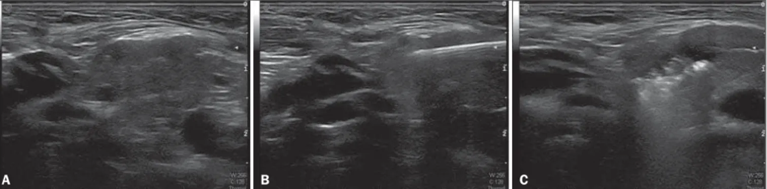
in-jection of the botulinum toxin to be identified, which prevented the toxin affecting structures adjacent to the salivary glands, such as the muscles involved in swallowing and vascular structures (Fig-ure 1).
In follow-up visits, the mother reported that there was a sig-nificant decrease in the number of pads used for cleaning drool and a 50% reduction in the number of tracheal aspirations, with-out any complaints suggesting that the botulinum toxin had pro-voked an inflammatory process. The patient had no episodes of bronchopneumonia during the two-months observation period. The ultrasound studies of the parotid and submandibular glands showed no parenchymal changes subsequent to injection of the botulinum toxin.
The use of ultrasound to guide botulinum toxin injections is important in pediatric patients, especially because the small size of the salivary glands makes them difficult to palpate in such pa-tients. In neurologically impaired children, the use of the ultra-sound guidance is even more relevant, because they can present with increased muscle tone and often have a tracheostomy in an
Luiz de Abreu Junior1, Henrique T. Martucci2, Paulo Tarso
Reck de Mendonça3, Gustavo Garcia Marques1, Célia
Rodrigues1
1. Universidade São Camilo, São Paulo, SP, Brazil. 2. Clínica São Camilo, Sinop, MT, Brazil. 3. Instituto Neurocirúrgico de Sinop, Sinop, MT, Brazil. Mailing address: Dr. Luiz de Abreu Junior. Rua Baturité, 200, ap. 32B, Aclimação. São Paulo, SP, Brazil, 01530-030. E-mail: [email protected]. Remote cerebellar hemorrhage has been defined as
bleed-ing within the cerebellar parenchyma, a rare complication that can occur after neurosurgical intervention. The entity was first described in the 1970s, by Yasargil et al.(1). The reported inci-dence of remote cerebellar hemorrhage after supratentorial in-terventions ranges from 0.08% to 0.6%(2). However, it has been reported to occur after various other surgical procedures involving the cranium or spinal cord(2–7).
Several hypotheses have been suggested to explain the ap-pearance of bleeding in the cerebellum away from the primary (supratentorial or spinal) surgical site. One such hypothesis is that resection of a supratentorial lesion creates a pressure gradient, resulting in suction on the cerebellar veins, particularly in the upper portion of the vermis(8). However, there is another hypothesis that might explain the two findings in the case reported here. That hypothesis is based on the supposition that opening the cisterns or the ventricular system promotes intracranial hypotension, trig-gering the process that culminates in the distension and rupture of cerebellar veins, resulting in cerebellar hemorrhage (9).
Various neurosurgical procedures have been associated with the occurrence of remote cerebellar hemorrhage, including the clipping of aneurysms (ruptured or otherwise), tumor resection, drainage of parenchymal or extra-axial hematomas, and spinal sur-gery(2–7,9). In imaging examinations, remote cerebellar hemor-rhage has a characteristic presentation, with a tendency for the blood to be distributed among the cerebellar folia with a curvilin-ear configuration. This aspect results in the pattern known as the zebra sign(8).
The symptoms of intracranial hypotension syndrome include headache that is orthostatic in presentation, tending to improve in the recumbent position. In imaging studies of the brains of patients with intracranial hypotension(10), findings include dural thickening and diffuse dural enhancement; engorgement and dilatation of venous structures; subdural fluid collections; down-ward displacement of the midbrain; and herniation of the cerebellar tonsils.
The case presented here demonstrates a chain of events that could have collectively resulted in the two central nervous system complications observed. The supratentorial surgical manipulation and the placement of the ventricular shunt could have caused intracranial hypotension, resulting in the traction, distension, and
consequent rupture of cerebellar veins, as well as hemorrhage in the cerebellar parenchyma.
Radiologist knowledge of these entities is relevant, because their proper, early characterization can promote interventions aimed at their correction and at alleviating the associated symp-toms.
REFERENCES
1. Yasargil MG, Yonekawa Y. Results of microsurgical extra-intracranial arterial bypass in the treatment of cerebral ischemia. Neurosurgery. 1977;1:22–4.
2. Bokhari R, Baeesa S. Remote cerebellar hemorrhage due to ventriculo-peritoneal shunt in an infant: a case report. J Med Case Rep. 2012; 6:222.
3. Honegger J, Zentner J, Spreer J, et al. Cerebellar hemorrhage arising postoperatively as a complication of supratentorial surgery: a retrospec-tive study. J Neurosurg. 2002;96:248–54.
4. Smith R, Kebriaei M, Gard A, et al. Remote cerebellar hemorrhage following supratentorial cerebrovascular surgery. J Clin Neurosci. 2014; 21:673–6.
5. Suzuki M, Kobayashi T, Miyakoshi N, et al. Remote cerebellar hemor-rhage following thoracic spinal surgery of an intradural extramedullary tumor: a case report. J Med Case Rep. 2015;9:68.
6. Biasi PR, Mallmann AB, Crusius PS, et al. Hemorragia cerebelar re-mota como complicação de cirurgia de coluna vertebral. Relato de dois casos e revisão da literatura. J Bras Neurocir. 2011;22:66–71. 7. Paola L, Troiano AR, Germiniani FMB, et al. Cerebellar hemorrhage
as a complication of temporal lobectomy for refractory medial tempo-ral epilepsy: report of three cases. Arq Neuropsiquiatr. 2004;62:519– 22.
8. Amini A, Osborn AG, McCall TD, et al. Remote cerebellar hemorrhage. AJNR Am J Neuroradiol. 2006;27:387–90.
9. Chalela JA, Monroe T, Kelley M, et al. Cerebellar hemorrhage caused by remote neurological surgery. Neurocrit Care. 2006;5:30–4. 10. Savoiardo M, Minati L, Farina L, et al. Spontaneous intracranial
hy-potension with deep brain swelling. Brain. 2007;130(Pt 7):1884–93.
Letters to the Editor
Radiol Bras. 2016 Mar/Abr;49(2):126–132
132
Figure 1.A: Normal right submandibular gland. B: Needle inserted into the gland. C: Botulinum toxin within the gland.
A B C
http://dx.doi.org/10.1590/0100-3984.2015.0056
anatomically narrow location, as well as showing anatomical ab-normalities(1). In addition, the injection of botulinum toxin into adjacent structures could have undesirable effects, such as pa-ralysis of the muscles involved in swallowing, which would worsen dysphagia(1).
Previous studies have demonstrated that injection of botuli-num toxin into the salivary glands does not cause any histological alterations—only lymphocyte infiltration, which results in homo-geneous shrinkage of the gland without atrophy(7). In addition, multiple injections of botulinum toxin over time can cause atro-phy of the submandibular glands, thus promoting a permanent reduction in the severity of sialorrhea(6). In the case presented here, we observed a reduction in the volume of all of the salivary glands injected, except the right parotid. We speculate that the injection was ineffective in that gland and that there was an increase in the volume of the gland through vicarious mechanisms. The study of glandular volume in such cases is groundbreaking, and our group is contemplating further studies in this line of reasearch. In the literature, we found no articles comparing glandular dimensions before and after botulinum toxin injection in neurologically im-paired children. A study conducted by Cardona et al.(8) showed no differences in glandular dimensions between children with and without sialorrhea. We seek to disseminate the knowledge that ultrasound guidance makes the injection of botulinum toxin into the salivary glands safer and more precise, especially in pediatric patients, as well as that ultrasound represents a noninvasive method of evaluating changes in the volume of those glands over time.
Marcia Wang Matsuoka1, Sílvia Maria Sucena da Rocha1, Lisa
Suzuki1, João Paulo Barnewitz1, Rui Imamura1, Luiz Antonio
Nunes de Oliveira1
1. Hospital das Clínicas da Faculdade de Medicina da Universidade de São Paulo (HC-FMUSP), São Paulo, SP, Brazil. Mailing address: Dra. Marcia Wang Matsuoka. Avenida Engenheiro Luis Gomes Cardim Sangirardi, 770, ap. 101. São Paulo, SP, Brazil, 04112-080. E-mail: [email protected].
REFERENCES
1. Ciftci T, Akinci D, Yurttutan N, et al. US-guided botulinum toxin injec-tion for excessive drooling in children. Diagn Interv Radiol. 2013;19;56– 60.
2. Jongerius PH, Joosten F, Hoogen FJ, et al. The treatment of drooling by ultrasound-guided intraglandular injections of botulinum toxin type A into the salivary glands. Laryngoscope. 2003;113:107–11.
3. Erbguth FJ. Botulinum toxin, a historical note. Lancet. 1998;351:1820. 4. Kopera D. Botulinum toxin historical aspects: from food poisoning to
pharmaceutical. Int J Dermatol. 2011;50:976–80.
5. Lakraj AA, Moghimi N, Jabbari B. Sialorrhea: anatomy, pathophisiology and treatment with emphasis on the role of botulinum toxins. Toxins (Basel). 2013;5:1010–31.
6. Gok G, Cox N, Bajwa J, et al. Ultrasounded-guided injection of botuli-num toxin A into the submandibular gland in children and young adults with sialorrhoea. Br J Oral Maxillofac Surg. 2013;51:231–3. 7. Coskun BU, Savk H, Cicek ED, et al. Histopathological and radiological
investigations of the influence of botulinum toxin on the submandibular gland of the rat. Eur Arch Otorhinolaryngol. 2007;264:783–7. 8. Cardona I, Saint-Martin C, Daniel SJ. Salivary glands of healthy