Head-to-head comparison of dipyridamole,
do butam ine and pacing stre ss
e cho cardio graphy fo r the de te ctio n
o f m yo cardial ische m ia in an anim al
m o de l o f co ro nary arte ry ste no sis
1Divisão de Cardiologia and 2Departamento de Cirurgia,
Hospital das Clínicas, Faculdade de Medicina de Ribeirão Preto, Universidade de São Paulo, Ribeirão Preto, SP, Brasil
A. Schmidt1,
O .C. de Almeida-Filho1,
E.M. Ayres-Neto1,
J.J. Carneiro2,
J.A. Marin-Neto1
and B.C. Maciel1
Abstract
To compare the sensitivity of dipyridamole, dobutamine and pacing stress echocardiography for the detection of myocardial ischemia we produced a physiologically significant stenosis in the left circumflex artery of 14 open-chest dogs (range: 50 to 89% reduction in luminal diameter). In each study, dobutamine (5 to 40 µg kg-1 min-1 in 3-min stages) and pacing (20 bpm increments, each 2 min, up to 260 bpm) were performed randomly, and then followed by dipyridamole (up to 0.84 mg/ kg over 10 min). The positivity of stress echocardiography tests was quantitatively determined by a significant (P<0.05) reduction of or failure to increase absolute and percent systolic wall thickening in the stenotic artery supplied wall, as compared to the opposite wall (areas related to the left anterior descending artery). Systolic and diastolic frozen images were analyzed off-line by two blinded observers in the control and stress conditions. The results showed that 1) the sensitivity of dobutamine, dipyridamole and pacing stress tests was 57, 57 and 36%, respectively; 2) in animals with positive tests, the mean percent change of wall thickening in left ventricular ischemic segments was larger in the pacing (-19 ± 11%) and dipyridamole (-18 ± 16%) tests as compared to dobutamine (-9 ± 6%) (P = 0.05), but a similar mean reduction of wall thickening was observed when this variable was normalized to a control left ventricular segment (area related to the left anterior descending artery) (pacing: -16 ± 7%; dipyridamole: -25 ± 16%; dobutamine: -26 ± 10%; not significant), and 3) a significant correlation was observed between magnitude of coronary stenosis and left ventricular segmental dysfunction induced by ischemia in dogs submitted to positive stress tests. We conclude that the dobutamine and dipyridamole stress tests showed identical sensitivities for the detection of myocardial ischemia in this one-vessel disease animal model with a wide range of left circumflex artery stenosis. The pacing stress test was less sensitive, but the differ-ence was not statistically significant. The magnitude of segmental left ventricular dysfunction induced by ischemia was similar in all stress tests evaluated.
Co rre spo nde nce
A. Schmidt Divisão de Cardiologia HC, FMRP, USP
14049-900 Ribeirão Preto, SP Brasil
Fax: + 55-16-633-0869 E-mail: aschmidt@ usp.br Publication supported by FAPESP.
Received O ctober 16, 2000 Accepted April 2, 2001
Ke y wo rds
·Stress echocardiography ·Pacing
Intro ductio n
Stress echocardiography has become a clinically useful method for detection, prognosis and therapeutic decisions in patients with known or suspected coro-nary heart disease. Dipyridamole, dobuta-mine and atrial pacing are stress modali-ties commonly used in the clinical setting as an alternative to exercise. The docu-mented accuracy of these stress echocar-diographic techniques is quite variable. Reported sensitivity values for the detec-tion of ischemia range from 40 to 92%, 68 to 96% and 75 to 93%, respectively, for dipyridamole (1-6), dobutamine (7-11) and pacing (12-19), while specificity ranges from 93 to 100%, 66 to 100% and 76 to 100%.
The reported accuracy of stress echocar-diography to identify regional wall motion abnormalities induced by ischemia is influ-enced not only by the physicians ability to perform and interpret the echocardiographic images (20), but also by a number of clinical setting, anatomical and physiological fac-tors including heterogeneity of the patients studied, variability of stress protocols, se-verity, location and extent of coronary dis-ease, relative significance of diseased ves-sels, presence of collateral circulation, and degree of ventricular hypertrophy (16). As a consequence of this inherent diversity, it is quite difficult to determine from the clinical investigations reported which stress modal-ity is most sensitive for the detection of myocardial ischemia.
Therefore, this investigation was de-signed to compare, in an animal model of one-vessel coronary heart disease with a wide range of physiologically significant coronary stenosis: 1) sensitivity of dipy-ridamole, dobutamine and pacing stress echocardiography for the detection of myo-cardial ischemia, and 2) the magnitude of segmental left ventricular dysfunction in-duced by ischemia.
Mate rial and Me tho ds
Animal pre paratio n
Fourteen mongrel dogs of either sex (14.5 to 30 kg, mean: 20.8 kg) were included in this study. The experimental protocol agreed with the guidelines of the American Heart Association about the use of research ani-mals (21). Dogs were anesthetized with in-travenous pentobarbital sodium (33 mg/kg), intubated and placed under mechanical ven-tilation with 100% oxygen (Takaoka, model 600) through an endotracheal tube. Anesthe-sia was maintained with additional doses of pentobarbital sodium when necessary.
Aortic and pulmonary pressure (phasic and mean) were continuously monitored by catheters connected to adequately calibrated fluid-filled transducers. The left femoral vein was cannulated for fluid and drug infusion. A lead II electrocardiogram was recorded continuously.
monitor-ing. At the end of the experiment, the ani-mals were sacrificed by the administration of potassium chloride following an addi-tional injection of pentobarbital.
Echo cardio graphic study
Two-dimensional echocardiography was performed using a Hewlett-Packard (Sonos 1000; Andover, MA, USA) ultrasound sys-tem with a 5-MHz transducer. Echocardio-graphic images were obtained with the trans-ducer placed on the epicardial surface and the ultrasound beam oriented to provide short axis images at the midpapillary level. Wall motion was continuously monitored through-out the experiment and images were stored on 1/2 inch VHS videotape for later analysis.
Expe rim e ntal pro to co l
Each animal was submitted to all three modalities of stress during the same experi-mental session, i.e., pacing and dobutamine in a random order, and finally dipyridamole. After each stress modality, sufficient time was allowed for recovery of baseline condi-tions. A continuous infusion pump (model 933; Harvard Apparatus Co., Southnatick, MA, USA) was used for drug infusion. Atrial pacing was produced by a Medtronic gen-erator (model 5325) starting with a rate 20 pulses per minute (ppm) higher than base-line heart rate, with 20 ppm increases each 2 min up to a heart rate of 260 bpm, or up to the time when segmental wall motion abnor-malities or atrioventricular conduction de-fects occurred. Intravenous dobutamine was started at a dose of 5 µg kg-1 min-1 and
increased every 3 min to 10, 20, 30 or 40 µg kg-1 min-1. Infusion was interrupted if
seg-mental wall motion abnormalities or severe arrhythmia occurred. Dipyridamole infusion was performed using the high dose protocol (0.56 mg/kg over 4 min, followed 4 min later by an additional 0.28 mg/kg over 2 min). Drug administration was interrupted if wall
motion abnormalities were documented. Echocardiographic images were recorded on videotape at baseline and at the final 30 s of each protocol step.
D ata analysis
Heart rate, systolic arterial pressure and rate-pressure product were measured before and at the end of each stress test and are expressed as mean ± standard deviation for three consecutive beats.
reached consensus on each measurement.
Statistical analysis
Data are reported as mean ± SD. Compari-sons of baseline to peak stress values were made by analysis of variance for repeated measures. The paired or unpaired two-tailed Student t-test was used when appropriate. McNemars test was used for comparison of sensitivity of the stress tests applied. Simple linear regression analysis was used to assess the correlation between coronary stenosis se-verity and the magnitude of segmental wall motion abnormalities induced by the various stress modalities. Probability was considered statistically significant at P<0.05.
Re sults
The stress test was interrupted before the peak target of the experimental protocol as a consequence of 1) occurrence of A-V con-duction disturbances in 13 animals during pacing, 2) ventricular (3 animals) or su-praventricular arrhythmias (1 animal) in-duced by dobutamine, and 3) occurrence of
segmental wall motion abnormalities (2 ani-mals) during dipyridamole administration.
He mo dynamic data
Mean baseline heart rate for pacing, do-butamine and dipyridamole was 134 ± 23, 135 ± 26 and 126 ± 24 bpm, respectively, while systolic arterial pressure was 109 ± 28, 110 ± 24 and 112 ± 25 mmHg. Mean values of rate-pressure product were 15083 ± 5370, 15221 ± 5559 and 14295 ± 4106 mmHg.bpm, for pacing, dobutamine and dipyridamole, respectively. There was no significant dif-ference in the three hemodynamic variables between the baseline states. Under stress conditions, heart rate increased to 194 ± 34 bpm for pacing (P<0.001 vs baseline) and to 158 ± 23 bpm (P = 0.002) for dobutamine and decreased to 101 ± 19 bpm (P<0.001) with dipyridamole, while systolic arterial pressure was 100 ± 28 (no significant differ-ence from baseline), 110 ± 29 (not signifi-cant) and 72 ± 27 mmHg (P<0.001) for pacing, dobutamine and dipyridamole, re-spectively. The rate-pressure product reached 20022 ± 7770 (P<0.001), 18032 ± 7148 (P = 0.03) and 7586 ± 4011 mmHg.bpm (P<0.001) during pacing, dobutamine and dipyridamole, respectively (Figure 1).
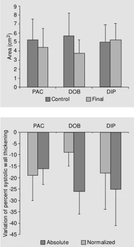
Baseline left ventricular end-diastolic area was not significantly different for the three modalities of stress. Before starting pacing, dobutamine and dipyridamole, left ventricular end-diastolic area was 5.24 ± 2.31, 5.64 ± 2.55 and 4.98 ± 1.94 cm2, respectively. Pacing did
not change end-diastolic area (4.42 ± 2.09 cm2; no significant difference from baseline),
dobutamine induced a significant reduction in area (3.38 ± 1.44 cm2, P = 0.03), while
dipy-ridamole infusion resulted in a small, nonsig-nificant increase in left ventricular diastolic dimension (5.21 ± 1.84 cm2) (Figure 2).
Se nsitivity o f stre ss te st m o dalitie s
In this investigation, the stress test was
H
e
a
rt
r
a
te
(
b
p
m
)
S
y
s
to
lic
b
lo
o
d
p
re
s
s
u
re
(
m
m
H
g
) 160
120
80
40
0
R
a
te
p
ro
d
u
c
t
(m
m
H
g
.b
p
m
) 3000025000
20000 15000 10000 5000 0 250 200 150 100 50 0
PAC DOB DIP
PAC DOB DIP
PAC DOB DIP
Control Final
considered to present an ischemic response (positive test) in the presence of a significant (P<0.05) reduction of or failure to increase absolute systolic wall thickening and per-cent systolic wall thickening in the circum-flex artery compared to the left anterior de-scending artery-related regions. Using this quantitative criterion, sensitivity was 36, 57 and 57%, for pacing, dobutamine and dipy-ridamole, respectively. The differences were not statistically significant.
Magnitude o f se gme ntal wall mo tio n
abno rm alitie s
Mean baseline values of absolute sys-tolic wall thickening and percent wall thick-ening were similar for all stress modalities, both in control and risk regions. Absolute systolic wall thickening in the left circum-flex and anterior descending regions was 0.40 ± 0.12 and 0.40 ± 0.11 cm before pac-ing, 0.45 ± 0.16 and 0.40 ± 0.12 cm before dobutamine and 0.46 ± 0.20 and 0.39 ± 0.09 cm before dipyridamole, respectively. Per-cent systolic wall thickening was 35 ± 10 and 37 ± 10%, 38 ± 12 and 36 ± 9%, 39 ± 14 and 35 ± 6%, for pacing, dobutamine and dipy-ridamole, respectively.
In dogs that presented an ischemic re-sponse to the stress test, a significant reduc-tion in percent systolic wall thickening was documented during pacing in the risk region (baseline: 40 ± 15%, pacing: 22 ± 11%; P = 0.01), while in the left anterior descending region no significant change occurred (base-line: 34 ± 8%, pacing: 32 ± 9%). Dobuta-mine also reduced percent systolic wall thick-ening in the risk region in dogs with a posi-tive stress test, but this change did not reach statistical significance (baseline: 37 ± 10%, dobutamine: 28 ± 12%). On the other hand, in the control region a significant increase of percent systolic wall thickening occurred (baseline: 41 ± 13%, dobutamine: 59 ± 15%; P = 0.02). A significant reduction of wall thickening was also documented during
dypiridamole in the left circumflex region (baseline: 36 ± 6%, dipyridamole: 19 ± 20%; P = 0.03), while in the control region a small, nonsignificant increase in percent wall thick-ening (baseline: 38 ± 10%, dipyridamole: 45 ± 8%) was observed.
Comparing the absolute variation of per-cent systolic wall thickening in the risk and control regions induced by the three stress modalities, more pronounced reduction oc-curred in systolic thickening during pacing (-19 ± 11%) and dipyridamole (-18 ± 16%) compared to dobutamine (-9 ± 6%). How-ever, when the variation of systolic thicken-ing was normalized to the control region, the magnitude of induced wall motion abnor-mality was not significantly different for the three modalities of stress (pacing: -16 ± 7%, dobutamine: -26 ± 10%, dipyridamole: -25 ± 16%) (Figure 3).
Figure 2. End-diastolic left ven-tricular area at baseline and at the end of each stress test. PAC, atrial pacing; DOB, dobutamine infusion; DIP, dipyridamole infu-sion.
A
re
a
(
c
m
2)
9 8 7
6 5
4
3
2
1 0
PAC DOB DIP
Control Final
V
a
ri
a
ti
o
n
o
f
p
e
rc
e
n
t
s
y
s
to
lic
w
a
ll
th
ic
k
e
n
in
g 0
-5
-10
-15
-20
-25
-30
-35
-40
-45
PAC DOB DIP
Absolute Normalized
Re latio nship be twe e n le ft ve ntricular
dysfunctio n induce d by ische mia and
ste no sis se ve rity
When positive tests for ischemia were considered, the correlation between the change of the relative systolic thickening in the risk region normalized to the control region and the severity of coronary stenosis, as evaluated by luminal diameter reduction, was statistically significant for dobutamine (r = 0.72, P = 0.046) but not for dipyridamole (r = -0.45, P = 0.26). However, a marked improvement in correlation was observed for dobutamine (r = 0.84, P = 0.036) and dipy-ridamole (r = 0.83, P = 0.043) when two dogs which had important left ventricular hypertro-phy and coronary stenosis severity in the lower range were excluded from the analysis.
D iscussio n
Characte ristics o f the e xpe rim e ntal m o de l
The experimental model used in this in-vestigation remained quite stable throughout the different phases of the study protocol as documented by the similar mean values for the hemodynamic variables measured (heart rate, systolic arterial pressure and rate-pressure product) and by the mean diastolic area of the left ventricle before each test. These observations indicate that, although dipyridamole was always the last stress test applied, this sequence probably had no sig-nificant effect, by itself, on the results ob-tained.
Using an animal model with one-vessel coronary stenosis ranging from 50 to 89% of luminal diameter reduction we were able to simulate coronary heart disease in a range of stenosis considered significant in the clini-cal setting. The variable degree of coronary stenosis used in this animal model appears to be adequate for sensitivity and specificity studies, considering that a 50% reduction of the luminal diameter represents the best
cut-off value for the functional significance of a stenosis (22). Moreover, conditions for com-paring sensitivities of different stress tests appear to be more adequate when a moder-ate degree of coronary stenosis such as that used in this investigation (mean: 69 ± 12% of luminal diameter reduction) is present.
Se nsitivity o f the stre ss te sts
In this study, the stress test was consid-ered to be positive for ischemia when it elicited a significant (P<0.05) reduction of or failure to increase systolic wall thickening and percent systolic wall thickening in the circumflex (stenosed) as compared to the left anterior descending artery (control)-re-lated regions. This quantitative approach for positivity of the test was important to avoid subjective evaluation of wall motion during stress. In addition, the comparative analysis of wall motion in risk and control regions has been shown to be valuable for the detec-tion of ischemia during stress echocardiog-raphy, considering that hyperkinesia may occur in the left ventricular region supplied by the non-stenosed coronary artery (23,24). The three tests did not differ significantly in sensitivity despite the higher value ob-served for dobutamine (57%) and dipy-ridamole (57%) as compared to the pacing stress test (36%). Probably, the tendency to a lower sensitivity during pacing was related to the relatively large number of tests prema-turely interrupted due to the occurrence of atrioventricular conduction disturbances.
pointed out. In their model, only critical coronary stenosis was produced as opposed to the large range of coronary stenosis used in the present study. In addition, dipyridamole was infused up to a maximal dose of 0.56 mg/kg body weight, while we used up to 0.84 mg/kg body weight in 12 out of 14 dogs. In the clinical setting, increased sensitivity has been documented when this larger dose of dipyridamole was used in stress echocar-diography (26). Segar et al. (27) reported sensitivity of 100% for dobutamine and 75% for dipyridamole in a swine model of ante-rior descending coronary artery stenosis, but they also used only critical coronary steno-sis. Therefore, under the conditions of the current investigation, dobutamine and dipy-ridamole seemed to be equally sensitive in producing systolic dysfunction secondary to myocardial ischemia, while atrial pacing showed a slightly lower, not significantly different, sensitivity.
There are no clinical investigations which directly compare these three stress tests in patients with one-vessel coronary disease. Some studies performed in groups of pa-tients with one-vessel disease have shown slightly higher sensitivity for dobutamine (range: 50-76%, mean: 66%) than for dipy-ridamole (range: 31-82%, mean: 52%) (28-32), while other studies have shown equiva-lent sensitivity for these stress tests (33,34). Although a comparison of the current exper-imental study with available clinical data cannot be directly performed considering the large number of factors interfering with the response to stress in patients, it should be mentioned that the reported mean values of sensitivity are very close to those obtained in the present experimental study. On the other hand, the sensitivity we obtained for pacing (36%) was much lower than that reported for patients with one-vessel disease (85%) (17), probably as a consequence of the large num-ber of pacing tests that were interrupted at an early stage due to atrioventricular conduc-tion disturbances.
Magnitude o f se gme ntal wall mo tio n
abno rm alitie s
Quantitative analysis of the magnitude of the segmental wall motion abnormalities in-duced by the stress tests showed that during pacing and dipyridamole the changes in per-cent systolic thickening in the risk region were comparable, but larger than those in-duced by dobutamine. However, when the variations in wall thickening were normal-ized for the control region supplied by the non-stenosed coronary artery, the three tests induced similar magnitudes of wall motion dysfunction. Our results are similar to those reported by Paulsen et al. (35) comparing exercise, pacing, dobutamine and dipy-ridamole in an experimental model. They demonstrated that pacing and exercise in-duced a more important wall motion abnor-mality, although the differences between all tests were reduced when the results were normalized to the non-ischemic region.
Re latio nship be twe e n le ft ve ntricular
dysfunctio n induce d by ische mia and
ste no sis se ve rity
hypertrophy, were excluded from analysis. This observation suggests that in these two animals ventricular hypertrophy may have contributed to the occurrence of wall motion dysfunction during stress.
In this one-vessel disease animal model, the dobutamine and dipyridamole stress tests showed identical sensitivities for the
detec-tion of myocardial ischemia in a wide range of left circumflex artery stenosis, while the pacing stress test tended to be less sensitive. On the other hand, the magnitude of segmen-tal left ventricular dysfunction induced by ischemia was similar for all stress tests evalu-ated.
Re fe re nce s
1. Picano E & Lattanzi F (1991). Dipyridamole echocardiography. A new diagnostic w in-dow on coronary artery disease. Circula-tion, 83 (Suppl III): III.19-III.26.
2. Picano E, Lattanzi F & M asini M (1987). Comparison of high-dose dipyridamole-echocardiography test and exercise tw o-dimensional echocardiography for diagno-sis of coronary artery disease. American
Journal of Cardiology, 59: 539-542.
3. M argonato A, Chierchia S & Cianflone D (1987). Limitations of dipyridamole-echo-cardiography in effort angina pectoris.
American Journal of Cardiology, 59:
225-230.
4. Picano E, Distante A, M asini M , M orales M A, Lattanzi F & L’Abbate A (1985). Dipy-ridamole-echocardiography test in effort angina pectoris. American Journal of
Car-diology, 56: 452-456.
5. Previtali M , Lanzarini L, Ferrario M , M us-sini A & M ontemartini C (1991). Dobuta-mine versus dipyridamole echocardiogra-phy in coronary artery disease. Circula-tion, 83 (Suppl III): III.27-III.31.
6. M azeika P, Nihoyannopoulos P, Joshi J & Oakley CM (1992). Uses and limitations of high dose dipyridamole stress echocdiography for evaluation of coronary ar-tery disease. British Heart Journal, 67: 144-149.
7. Berthe C, Pierard LA, Hiernaux M , Trot-teur G, Lempereur P, Carlier J & Kulbertus HE (1986). Predicting the extent and loca-tion of coronary artery disease in acute myocardial infarction by echocardiography during dobutamine infusion. Journal of
the American College of Cardiology, 58:
1167-1172.
8. Saw ada SG, Segar DS, Ryan T, Brow n SE, Dohan AM , Williams R, Fineberg NS, Armstrong WF & Feigenbaum H (1991). Echocardiography detection of coronary artery disease during dobutamine infu-sion. Circulation, 83: 1605-1614. 9. Saw ada SG, Segar DS, Ryan T, Dohan
AM , Williams R & Feigenbaum H (1992). Catecholamine stress echocardiography.
Echocardiography, 9: 177-187.
10. Segar DS, Brow n SE, Sada SG, Ryan T & Feigenbaum H (1992). Dobutamine stress echocardiography: Correlation w ith coro-nary lesion severity as determined by quantitative angiography. Journal of the
American College of Cardiology, 19:
1197-1202.
11. M arw ick T, D’ Hondt A, Baudhuin T, Willemart B, Wijns W, Detry JM & M elin J (1993). Optimal use of dobutamine stress for the detection and evaluation of coronary artery disease: Combination w ith echocardiography or scint igraphy, or both? Journal of the American College of
Cardiology, 22: 159-167.
12. Chapman PD, Doyle TP & Troup PJ (1984). St ress echocardiography w it h t rans-esophageal atrial pacing: Preliminary re-port of a new method for detection of ischemic w all motion abnormalities.
Cir-culation, 70: 445-450.
13. Iliceto S, D’Ambrosio G, Sorino M ,Papa A, Amico A, Ricci A & Rizzon P (1985). Comparison of post exercise and trans-esophageal atrial pacing tw o-dimensional echocardiography for detection of coro-nary artery disease. Feasibility, specificity and sensitivity. American Journal of
Car-diology, 57: 547-553.
14. Iliceto S, Sorino M & D’Am brosio G (1985). Detection of coronary artery dis-ease by tw o-dimensional echocardiogra-phy and transesophageal atrial pacing.
Journal of the American College of
Cardi-ology, 5: 1188-1197.
15. Iliceto S, Sorino M , D’Ambrosio G, Loprio-re V, Ricci A, Papa A, Amico A, Chiddo A & Rizzon P (1986). Atrial pacing in the detection and evaluation of coronary ar-tery disease. European Heart Journal, 7: 59-67.
16. L’Abbate A (1991). Pathophysiological ba-sis for noninvasive functional evaluation
of coronary st enosis. Circulat ion, 83 (Suppl III): III.2-III.7.
17. Lambertz H, Kreis A, Trumper H & Han-rath P (1990). Simultaneous transesopha-geal atrial pacing and transesophatransesopha-geal t w o-dim ensional echocardiography: a new method of stress echocardiography.
Journal of the American College of
Cardi-ology, 16: 1143-1153.
18. M ichael TA, Antonescu A, Bhambi B & Balasingam S (1996). Accuracy and use-fulness of atrial pacing in conjunction w ith transthoracic echocardiography in the de-tection of cardiac ischemia. American
Journal of Cardiology, 77: 187-190.
19. Kamp O, De Cock CC, Kupper SJ, Roos JP & Visser CA (1992). Simultaneous transesophageal tw o-dimensional echo-cardiography and atrial pacing for detect-ing coronary artery disease. American
Journal of Cardiology, 69: 1412-1416.
20. Popp R, Agaston A, Armstrong W, Nanda N, Pearlman A, Rakow ski H, Sew ard J, Silverman N, Smith M , Stew art W, Taylor R, Thys D & Davis C (1998). American Society of Echocardiography position pa-pers. Journal of the American Society of
Echocardiography, 11: 95-98.
21. American Heart Association (1985). Posi-tion of the American Heart AssociaPosi-tion on research animal use: a statement for health professionals by a task force ap-pointed by the board of directors of the American Heart Association. Circulation, 71: 849-850.
22. Babic R, Ostojic M , Stankovic G, Belesin B, Stojkovic S, Nedeljkovic M , Pantelic-Babic J & Djordjevic-Dikic A (1994). Quan-titative coronary arteriography versus stress echocardiography: Which is the “ best” angiographic cut-off? Journal of
the American College of Cardiology, 38A
(Abstract).
stress echocardiography? Circulation, 88 (Suppl I): I-403 (Abstract).
24. Ofili EO, St Vrain JA & Standeven JA (1993). Dobutamine stress echocardiog-raphy: Hem odynam ic correlat es and physiologic basis of w all thickening ab-normality. Journal of the American
Col-lege of Cardiology, 21: 175A (Abstract).
25. Fung AY, Gallagher KP & Buda AJ (1987). The physiologic basis of dobutamine as compared w ith dipyridamole stress inter-ventions in the assessment of critical cor-onary stenosis. Circulation, 76: 943-951. 26. Picano E, Lattanzi F, M asini M , Distante A
& L’Abbate A (1986). High dose dipy-ridamole echocardiography test in effort angina pectoris. Journal of the American
College of Cardiology, 8: 846-854.
27. Segar DS, Ryan T, Saw ada SG, Johnson M & Feigenbaum H (1995). Pharmacologi-cally induced myocardial ischemia: a com-parison of dobutamine and dipyridamole.
Journal of the American Society of
Echo-cardiography, 8: 9-14.
28. Previt ali M , Lanzarini L, Ferrario M , Tortorici M , M ussini A & M ontemartini C (1991). Dobutamine versus dipyridamole echocardiography in coronary artery dis-ease. Circulation, 83 (Suppl III): III.27-III.31.
29. Dagianti A, Penco M , Agati L, Sciomer S, Dagianti A, Rosanio S & Fedele F (1995). Stress echocardiography: comparison of exercise, dipyridamole and dobutamine in detecting the extent of coronary artery disease. Journal of the American College
of Cardiology, 26: 18-25.
30. Wilkenshoff UM , Schröder K, Völler H, Dissman R, Dingerkus H, Linderer T & Schröder R (1994). Comparison of dobu-tamine and dipyridamole stress echocar-diography in patients w ith single and multivessel disease undergoing PTCA.
Journal of the American College of
Cardi-ology, 144A (Abstract).
31. Lanzarini L, Fetiveau R, Poli A, M ussini A & Previtali M (1993). Com parison of dobutamine and dipyridamole stress-echo w ith exercise test for coronary artery dis-ease diagnosis. Journal of the American
College of Cardiology, 21: 89A (Abstract).
32. M artin TW, Seaw orth JF, Johns JP, Pupa LE & Condos WR (1992). Comparison of adenosine, dipyridamole and dobutamine in stress echocardiography. Annals of
In-ternal M edicine, 116: 190-196.
33. Belesin BD, Ostojic M , Stepanovic J, Djordjevic-Dikic A, Stojkovic S, Nedelj-kovic M , StanNedelj-kovic G, Petrasinovic Z, Gojkovic L & Vasiljevic-Pokrajcic Z (1994).
Stress echocardiography in the detection of myocardial ischemia. Head-to-head comparison of exercise, dobutamine and dipyridamole tests. Circulation, 90: 1168-1176.
34. Salustri A, Fioretti PM , M cNeill AJ, Pozzoli M M & Roetlandt JR (1992). Pharmacolo-gical stress echocardiography in the diag-nosis of coronary artery disease and myo-cardial ischaemia: a comparison betw een dobutamine and dipyridamole. European
Heart Journal, 13: 1356-1362.
35. Paulsen PR, Pavek T, Crampton M , Bache RJ, Boudreau R & Homans DC (1993). Which stress is best? Exercise, dobuta-mine, dipyridamole and pacing in an ani-mal model. Journal of the American
Col-lege of Cardiology, 21: 90A (Abstract).
36. White CW, Wright CB & Doty DB (1984). Does visual interpretation of the coronary arteriogram predict the physiologic impor-tance of a coronary stenosis? New
Eng-land Journal of M edicine, 310: 819-824.
37. Gould KL & Lipscomb K (1974). Effects of coronary stenoses on coronary flow re-serve and resistance. American Journal of
Cardiology, 34: 48-54.