Short-term evaluation of non-absorbable
m icro granular hydro xyapatite
infiltratio n in the guine a pig
sube pide rm al abdo m inal re gio n
1Departamento de O ftalmologia, O torrinolaringologia e
Cirurgia de Cabeça e Pescoço, Faculdade de Medicina de Botucatu, Universidade Estadual Paulista, Botucatu, SP, Brasil
2Departamento de Bioestatística, Instituto de Biociências, Universidade Estadual Paulista, Botucatu, SP, Brasil
3Departamento de Bioquímica, Faculdade de O dontologia de Bauru, Universidade de São Paulo, Bauru, SP, Brasil
A.P. Xavier1, S.A. Schellini1, F.F. Aragon2, C.R. Padovani2 and E.M. Taga3
Abstract
Non-absorbable microgranular hydroxyapatite was infiltrated into the subepidermal abdominal region of guinea pigs in order to assess the possibility of using this material to correct deficiencies in orbital volume. Microgranular hydroxyapatite (2.0 ml) was subepidermally infiltrated into the abdominal region of 20 guinea pigs. The animals were divided into four experimental groups of 5 animals each, which were killed 7 (G1), 15 (G2), 30 (G3) and 60 (G4) days after infiltration. The area and the largest and smallest diameters of the nodules formed by infiltration were evaluated at the site of infiltration and histological examination was performed. The mean granuloma area was similar in all groups. Histopathological examination showed that the material remained isolated from surrounding tissues by a pseudocapsule that became denser throughout the experiment. A host reaction started with young fibroblastic tissue that evolved to dense tissue until cartilaginous tissue was formed in G4, progressively advancing to-wards the center of the granuloma from G1 to G4. Non-absorbable microgranular hydroxyapatite is an inert material that was well toler-ated by the animals studied, with maintenance of the infiltrtoler-ated volume, and may perhaps be useful to fill anophthalmic cavities. Co rre spo nde nce
S.A. Schellini
Departamento de O ftalmologia, O torrinolaringologia e Cirurgia de Cabeça e Pescoço
Faculdade de Medicina de Botucatu UNESP
18618-000 Botucatu, SP Brasil
E-mail: sartioli@ fmb.unesp.br Research supported by PIBIC-CNPq. Publication supported by FAPESP.
Received November 27, 2000 Accepted August 27, 2001
Ke y wo rds
·Infiltration
·Microgranular hydroxyapatite
·Guinea pigs
·Histopathology
Intro ductio n
The loss of the ocular globe provokes undesirable aesthetic and psychological ef-fects. In order to minimize these effects it is necessary to reestablish the appearance by restoring the orbital volume, which is not always a simple procedure. Volume replace-ment can be made by the insertion of spheres
made of many materials and of different sizes using different surgical techniques.
synthetic HA and polyethylene.
Regardless of the type of material of which they are made, the spheres should not be very large, since this would facilitate their extrusion(2). The choice of sphere size is difficult and a deficit of orbital vol-ume persists in many individuals with anophthalmic cavities, causing a certain de-gree of enophthalmos.
In order to improve the adequacy of or-bital volume, one attempt was made by infil-trating a substance into the anophthalmic cavity. Thus, the objective of the present study was to demonstrate experimentally the effects of the infiltration of non-absorbable microgranular HA by infiltrating it subepi-dermally in guinea pigs.
Mate rial and Me tho ds
Microgranular semi-solid HA was sub-epidermally infiltrated into the abdominal region of 20 guinea pigs. The animals were supplied by the Central Animal House, Bo-tucatu Campus, UNESP.
The synthetic microgranular HA em-ployed (Ca10 (PO4)6 (OH)2) was available in semi-solid form and was developed by one of the authors (3). For infiltration it was necessary to dilute 2.0 ml of the material in 5.0 ml of distilled water.
We analyzed the tecidual reaction after subepidermal infiltration. The animals were divided into four experimental groups of 5 animals each, which received 2.0 ml of HA, using a 30 x 7-caliper needle and a 3.0-ml syringe. The animals were killed 7 (G1), 15 (G2), 30 (G3) and 60 (G4) days after infiltra-tion.
After killing the animals, a square (2.0 x 2.0 cm) piece of the abdominal region of guinea pigs where HA was infiltrated (skin and subepidermal tissues) was cut. The pieces were placed on white paper for 3 to 5 min and then placed in 10% formaldehyde solution.
After fixation, the pieces were removed from the formaldehyde solution and off the
paper, and measurements of the granulomas were performed using a computer image analyzer (Luzex-F-NIRECO). The smallest (d) and the largest diameters (D) were meas-ured in order to calculate the d/D ratio by reconstructing an ellipsoid shape. These pa-rameters were evaluated using one-way anal-ysis of variance (ANOVA) and comparisons between groups were made by the Tukey test (4), with the level of significance set at 0.05. The granuloma was cut out of each piece at its largest diameter using a blade over a silicon plate. Fragments were immersed in 70% alcohol solution for 48 h and embedded in wax, and sections were cut and stained with hematoxylin-eosin (HE).
Histopathological examination was per-formed with the experimenter blinded to the animals groups. Granulomas and surround-ing tissues were evaluated, as well as the host reaction.
Re sults
Clinical e valuatio n
The animals were in good general condi-tion throughout the experiment, presenting good physical activity and eating normally. Two animals (one from G1 and the other from G3) developed an infectious process that resulted in the extrusion of the injected material and were replaced. The remaining animals showed no alterations at the site of infiltration.
Granulo m a e valuatio n
Histo patho lo gical e valuatio n
The events that occurred during the study are summarized in Table 2.
Group 1. The injected material was
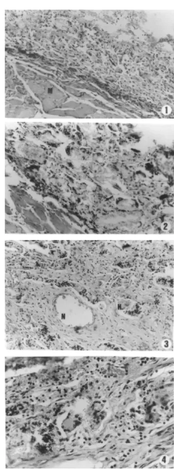
ob-served in the subepidermal region above the muscle layer. There were areas of hemor-rhage under the dermis and in the muscle layer closest to the HA. Between HA and host tissues there was a pseudocapsule formed by fibroblasts and inflammatory cells, which surrounded the injected material. HA was completely surrounded by a pseudocap-sule, and separated from neighboring tis-sues, forming an elongated nodule. In G1, the nodule was formed by inflammatory cells - neutrophils and monocytes - surrounding the HA, involving about 1/10 to 2/10 of its diameter. There were also young fibroblasts, and newly formed vessels, some of them with red blood cells (Figure 1).
Group 2. HA is seen as small birefringent granules surrounded by the pseudocapsule. Giant cells were observed around the gran-ules. There were also many subepidermal areas of hemorrhage. The pseudocapsule became thicker and denser after fibroblast proliferation. A fibrovascular and inflam-matory reaction occupied 2 to 3/10 of the diameter of the nodule. There were darkened areas, most likely the result of calcium crys-tal deposit, as well as pink homogeneous areas corresponding to collagen. There was a small number of inflammatory cells in the neighboring tissues (Figure 2).
Group 3. Fibrosis became denser (pseudo-capsule) around the nodule. HA was sur-rounded by a fibrovascular response at about the midpoint of the nodule, forming true septa with HA blocks. A rich inflamma-tory reaction consisting of macrophages, sometimes changing to giant cells was pres-ent. There were many newly formed vessels around the nodule which was full of red blood cells, and tissue inside the nodule became denser, with areas of collagen. The inflammatory reaction was almost absent in
Table 1. Granuloma measurements of groups.
Group Area d D d/D ratio Volume
G1 3.27 ± 1.43 1.62 ± 0.52 2.45 ± 0.54 0.65 ± 0.14 3.93 ± 2.24
G2 4.59 ± 1.36 1.98 ± 0.32 2.93 ± 0.46 0.68 ± 0.11 6.25 ± 2.97
G3 2.80 ± 0.93 1.43 ± 0.22 2.46 ± 0.53 0.60 ± 0.14 2.76 ± 1.33
G4 4.29 ± 2.05 2.02 ± 0.50 2.59 ± 0.61 0.78 ± 0.07 6.32 ± 4.63
Data are reported as means ± SD. D = largest diameter; d = smallest diameter. There w ere no significant differences among groups (ANOVA and Tukey test). the neighboring tissues (Figure 3).
Group 4. Nodules well separated from
neighboring tissues by a dense pseudocap-sule were observed. An inflammatory re-sponse filled the entire nodule and fibrosis surrounded all the HA. There were also many macrophages, giant cells, calcium deposits, as well as areas of collagen; however, this reaction was absent in the neighboring tis-sues (Figure 4).
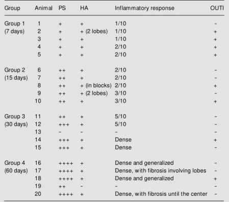
Table 2. Characteristics of histopathological evaluation of the granulomas in the experimental groups.
Group Animal PS HA Inflammatory response OUTI
Group 1 1 + + 1/10
-(7 days) 2 + + (2 lobes) 1/10 +
3 + + 1/10 +
4 + + 2/10 +
5 + + 2/10 +
Group 2 6 ++ + 2/10
-(15 days) 7 ++ + 2/10
-8 ++ + (in blocks) 2/10 +
9 ++ + (2 lobes) 3/10
-10 ++ + 3/10 +
Group 3 11 ++ + 5/10
-(30 days) 12 +++ + 5/10
-13 - - -
-14 +++ + Dense +
15 +++ + Dense
-Group 4 16 ++++ + Dense and generalized
-(60 days) 17 ++++ + Dense, w ith fibrosis involving lobes
-18 ++++ + Dense and generalized +
19 ++ - -
-20 ++++ + Dense, w ith fibrosis until the center
Figure 2. Periphery of the hydroxyapatite nodule. Ob-serve the hydroxyapatite granules surrounded by giant cells (arrow ). Homogeneous areas corresponding to collagen (G2 - HE, 100X).
Figure 3. The hydroxyapatite granules are surrounded by a fibrovascular and inflammatory reaction that forms true “ septa” dividing the hydroxyapatite (H) into “ blocks” . Collagen (* ) w as commonly detected in this animal group. N = new ly formed vessel (G3 - HE, 100X).
Figure 4. Birefringent hydroxyapatite granules in a dense fibrovascular response. Giant cells and collagen (* ). New ly formed vessels containing red blood cells (G4 - HE, 100X).
D iscussio n
Many studies are being conducted in an attempt to restore orbital volume after the loss of the eye or of its content. Currently, one of the most utilized material for this purpose in the United States is HA in the form of spheres surgically implanted into the orbital cavity. Since it is difficult to fit the volume of the sphere to the cavity, enoph-thalmos may remain even when spheres are utilized. Several investigators have attempted to use liquid HA, which could be infiltrated into the cavity and would not require surgery for insertion.
HA has been used for the reconstruction of tissues in dentistry and acts by accelerat-ing new bone and tissue formation by releas-ing calcium and phosphate ions that stimu-late the action of fibroblasts and macro-phages. HA has high biological compatibility and, depending on the size of its microgran-ules, can be absorbed by phagocytosis (5).
Histopathologically, we observed the hosts response to the HA, which consisted of engulfing and phagocytizing the material used. The giant cell response observed had been previously observed with the use of natural HA (6). A pseudocapsule was formed during the initial phase of the process, which persisted throughout the study period and became denser and thicker, preventing the loss of the substance into surrounding tis-sues. This fact led to a reduced inflammatory reaction outside the site of inoculation.
The inflammatory reaction close to the infiltrated material occupied an increasingly
larger space and its fibroblastic aspect be-came progressively denser, with the forma-tion of amorphous basophilic tissue similar to cartilaginous tissue. We believe that a longer follow-up of the animals is needed in order to observe the probable new bone for-mation that occurs with the use of natural HA.
The granuloma format remained un-changed from the HA infiltration until the end of the experiment (60 days). However, this was not homogeneous among the ani-mals. The mean d/D ratio varied significant-ly from the 1.00 target (corresponding to the spherical shape), indicating that the granu-loma was of irregular rather than spherical shape.
Nevertheless, the mean measurements of the granulomas did not vary significantly during the study period. It was possible to observe a more increased volume secondary to the inflammation against the HA at the beginning than at the end of the experimen-tal period. Perhaps with an experimenexperimen-tal period longer than 60 days HA granuloma might be significantly reduced.
Our results lead us to conclude that the non-absorbable microgranular semi-solid HA was well tolerated by the animals studied with maintenance of the infiltrated volume and may be useful for filling the anophthalmic cavities. However, it is necessary to carry out a study involving a longer time of obser-vation of the granulomas formed and to de-velop more precise techniques of HA infil-tration before this substance can be indi-cated for clinical use.
Re fe re nce s
1. Leitha T (1995). Three-phase bone scin-tigraphy of hydroxyapatite ocular implants. European Journal of Nuclear M edicine, 22: 308-314.
2. Hashimoto M , Rodrigues AC, M oraes-Silva M RB & Schellini SA (1994). Cavidade anoftálmica - causas, reconstrução e com-plicações. Revista Brasileira de Oftalmo-logia, 53: 517-522.
3. Taga EM (1991). Hidroxiapatita M icrogra-nular Viscosa (uso em odontologia na Fa-culdade de Odontologia de Bauru, USP). Apostila do Departamento de Bioquímica, USP, Bauru, SP, Brazil.
4. M ontgomery DC (1991). Design and Anal-ysis of Experiments. 3rd edn. John Wiley, New York, NY, USA.
5. Rosner M , Edw ard DP & Tso M OM
(1992). Foreign-body giant-cell reaction to the hydroxyapatite orbital implant. Ar-chives of Ophthalmology, 110: 173-174. 6. Goldberg RA, Dresner SC, Brasloso RA,