High prevalence of methicillin resistance and
PVL genes among
Staphylococcus aureus
isolates from the nares and skin lesions of
pediatric patients with atopic dermatitis
F.S. Cavalcante
1*, E.D. Abad
2*, Y.C. Lyra
1, S.B. Saintive
2, M. Ribeiro
2,
D.C. Ferreira
3,4and K.R.N. dos Santos
11Departamento de Microbiologia Me´dica, Instituto de Microbiologia Paulo de Go´es, Universidade Federal do Rio de Janeiro, Rio de Janeiro, RJ, Brasil 2Instituto de Puericultura e Pediatria Martaga˜o Gesteira, Universidade Federal do Rio de Janeiro, Rio de Janeiro, RJ, Brasil 3Centre for Ecological and Evolutionary Studies (Microbial Ecology), Faculty of Mathematics and Natural Science, University of Groningen, Groningen, The Netherlands 4
Programa de Po´s Graduac¸a˜o em Odontologia, Universidade Esta´cio de Sa´, Rio de Janeiro, RJ, Brasil
Abstract
Staphylococcus aureusis highly prevalent among patients with atopic dermatitis (AD), and this pathogen may trigger and aggravate AD lesions. The aim of this study was to determine the prevalence ofS. aureusin the nares of pediatric subjects and verify the phenotypic and molecular characteristics of the isolates in pediatric patients with AD. Isolates were tested for antimicrobial susceptibility, SCCmec typing, and Panton-Valentine Leukocidin (PVL) genes. Lineages were determined by pulsed-field gel electrophoresis and multilocus sequence typing (MLST). AD severity was assessed with the Scoring Atopic Dermatitis (SCORAD) index. Among 106 patients, 90 (85%) presentedS. aureusisolates in their nares, and 8 also presented the pathogen in their skin infections. Two patients had two positive lesions, making a total of 10S. aureusisolates from skin infections. Methicillin-resistantS. aureus(MRSA) was detected in 24 (26.6%) patients, and PVL genes were identified in 21 (23.3%), including 6 (75%) of the 8 patients with skin lesions but mainly in patients with severe and moderate SCORAD values (P=0.0095). All 24 MRSA isolates were susceptible to trimethoprim/sulfamethoxazole, while 8 isolates had a minimum inhibitory concentration (MIC) to mupirocin .1024mg/mL. High lineage diversity was found among the isolates including USA1100/ST30, USA400/ST1, USA800/ST5, ST83, ST188, ST718, ST1635, and ST2791. There was a high prevalence of MRSA and PVL genes among the isolates recovered in this study. PVL genes were found mostly among patients with severe and moderate SCORAD values. These findings can help clinicians improve the therapies and strategies for the management of pediatric patients with AD.
Key words: Atopic dermatitis;Staphylococcus aureus; Nasal colonization; Skin lesions; SCORAD
Introduction
Atopic dermatitis (AD) is a chronic inflammatory skin disease (1) that affects 10-20% of children worldwide (2). Several indexes have been proposed to assess AD severity; however, SCORAD (scoring atopic dermatitis) is the most widely used index (3). It adds a point for each symptom such as extension of eczema, dryness, pruritus, sleep disturbance, etc. Patients who score.25, 25-50, or.50 are considered to have mild, moderate, and severe AD, respectively.
Genetic predisposition, skin barrier defects, and environ-mental exposure are considered to be associated with the development of AD (4). Skin colonization byS. aureusmay also contribute to the onset and/or aggravation of lesions (5) because staphylococcal toxins such as Panton-Valentine Leukocidin (PVL) and superantigens can aggravate the eczema (6). The prevalence ofS. aureusin AD patients is up to 80% in nasal colonization (7-9) and can vary from 75%
Correspondence: K.R.N. dos Santos:,[email protected].. *These authors contributed equally to this study.
(7-10,12,13).
The resistance to methicillin inS. aureusis encoded in aStaphylococcal chromosome cassettemec(SCCmec), of which there are 11 types. Among AD patients, MRSA isolates usually carrymec cassettes commonly found in healthy individuals from the community, such as types IV and V (11). The genetic profile of most MRSA recovered from AD children belongs to well-established community lineages of different geographical regions such as ST188 in Korea (11).
Although some studies have reported S. aureus
prevalence in AD patients, as well as the detection of the bacterial virulence factors and their relation with clonality (7,9,11), this has not yet been analyzed in Brazil. Therefore, this study aimed to verify the prevalence of S. aureus colonization, including MRSA, in pediatric outpatients with AD and characterize the SCCmectypes, PVL genes, and clonality of isolates from nares and AD skin lesions. In addition, we correlated the presence of PVL genes with disease severity and isolate characteristics.
Material and Methods
Setting and study populations
A cross-sectional study was conducted between September 2011 and September 2012 at the Univer-sidade Federal do Rio de Janeiro hospital pediatric der-matology outpatient clinic, which provides assistance to about 130 AD pediatric patients. The target population of the study included patients diagnosed with AD of both genders who were 16 years old or less. The population was predominantly low income. The study was approved by the Ethics Committee of Instituto de Pediatria e Pueri-cultura Martaga˜o Gesteira, Universidade Federal do Rio de Janeiro (#51/11).
Collection and bacterial isolates
Swabs from the anterior nares and infected skin lesions were obtained from 106 patients. All infected skin sites of skin with infection were analyzed. Specimens were cultured on mannitol salt agar (Oxoid, UK) and characterized with standardized tests (14). The following controls were used:S. aureusstrains ATCC 25923 and ATCC 29213 (for suscep-tibility tests), Mu50 (SCCmectype II) (15), and the clinical isolates described previously (SCCmectypes II, III and IV; PVL genes positive) (16).
Antimicrobial susceptibility tests
All isolates were submitted to diffusion testing (17) and minimum inhibitory concentrations (MICs) for oxacillin and vancomycin by microdilution broth (18). The MIC test for
Bacterial DNA was extracted (19), and all S. aureus
isolates were submitted to polymerase chain reaction (PCR) for PVL genes (20). SCCmectyping was performed on all MRSA isolates (21).
PFGE, RM test, and MLST
Eighteen S. aureus isolates from 8 patients who pre-sented at least two clinical sites positive for the pathogen were evaluated for genotypes by pulsed-field electro-phoresis (PFGE) (22). This technique is based on DNA fragmentation followed by electrophoresis. Each isolate submitted to PFGE shows a band pattern. The isolates were grouped in clones according to band patterns similarities (23), and the clonality was obtained by comparisons with previously published images (24). A restriction-modification (RM) test was carried out to determine the bacterial clonal complexes (25). Isolates that were not included in a clonal complex by the RM test were submitted to the multilocus sequence typing (MLST) method (26).
Statistical analysis
All data were analyzed using the SPSS 20.0 software program for Windows (SPSS Inc., USA). The exact Fisher’s test and chi-square test were used to compare data. Significance was established at 5% (P,0.05).
Results
Bacterial isolates
Nasal and skin lesion swabs were collected from 106 AD patients. Ninety (85%) patients presentedS. aureusisolates in nares, and 8 (7.5%) also presented the pathogen in their skin infections. Two patients had two infected lesions positive for S. aureus, for a total of 10S. aureus isolates from skin infections. Among 90 patients with S. aureus
isolates, 24 (26.6%) had MRSA. The majority (86.6%) of patients with S. aureus had moderate or mild SCORAD values. Only 12 (13.4%) patients had severe SCORAD values (Table 1).
Antimicrobial susceptibility and MIC
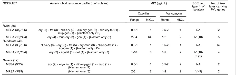
A total of 100S. aureusisolates (90 from nares and 10 from skin lesions) were evaluated, and 24 were positive for MRSA. All isolates were susceptible to ciprofloxacin, chloramphenicol, linezolid, rifampicin, teicoplanin, tigecy-cline, and trimethoprim/sulfamethoxazole. Antimicrobial resistance was detected for erythromycin (40%), clinda-mycin (15%), tetracycline (12%), mupirocin (8%), and gentamicin (7%) (Table 1).
For oxacillin, 76% of the isolates in both nasal and skin sites presented MIC values between 0.5 and 1mg/mL, but
Table 1. Characteristics of 100Staphylococcus aureusisolates from nares and skin lesions from 90 pediatric patients with atopic dermatitis and correlation with SCORAD index.
SCORADa Antimicrobial resistance profile (n of isolates) MIC (mg/mL) SCCmec type (n of isolates)
No. of iso-lates carrying
PVL genes Oxacillin Vancomycin
Range MIC90 Range MIC90
bMild (38)
MSSA (31/75.6) ery (5) tet (3) clinery (3) clinerygen (2) clinerytet (1) -mup-gen (1) -b-lactam only (16)
0.5-1 1 0.5-2 1 NA 2
MRSA (10/24.4) ery (4) - mup-ery (3) - gen (1) -b-lactam only (2) 2-64 64 1-2 2 IV (10) 5 cModerate (40)
MSSA (36/76.6) clinery (6) ery (5) tet (5) erymup (3) clinerytet (1) -ery-gen (1) -b-lactam only (15)
0.5-1 1 0.5-2 1 NA 14
MRSA (11/23.4) ery (2) - ery-tet (1) - tet (1) -b-lactam only (7) 1-16 8 1-2 2 IV (10) 4 nt (1)
Severe (12)
MSSA (9/75) ery (2) eryclin (1) clinerygen (1) mup (1) -b-lactam only (4)
0.5-1 1 0.5-2 2 NA 2
MRSA (3/25) b-lactam only (3) 2-8 2 1-2 2 IV (3) 2
aSCORAD is reported as (n of patients)/S. aureusstrain (n/% of isolates);bincludes 4 isolates from cutaneous lesion;cincludes 6 isolates from cutaneous lesion. SCORAD: scoring atopic dermatitis; MIC: minimum inhibitory concentration; SCCmec: Staphylococcal cassette chromosomemec; PVL: Panton-Valentine Leukocidin. MSSA: methicillin-susceptible Staphylococcus aureus; MRSA: methicillin-resistantS. aureus; ery: erythromycin; mup: mupirocin; tet: tetracycline; gen: gentamicin; clin: clindamycin; NA: not applicable; IV: SCCmectype IV; nt: nontypeable (mecA complex++ccr3 identified).
F.S.
Cavalcan
te
Biol
Res
48(7)
2015
www.bjournal.
>
SCCmectyping and PVL genes
Among the 24 MRSA isolates, 23 (95.8%) carried the SCCmecIV, and 1 isolate was nontypeable and presented the complex A for themecgene associated with theccr3 (Table 1).
Among 90 patients, 21 (23.3%) carried isolates with the PVL genes in the nares. Among the 8 patients with S. aureus isolates in their skin lesions, 6 (75%) possessed PVL genes. Among the children with moderate and severe SCORAD scores, 13 (32.5%) of 40 and 4 (33.3%) of 12 presented isolates positive for the PVL genes. Among patients with mild SCORAD, only 4 (10.5%) of the 38 presented this condition (P=0.0095). From the 100 S. aureus isolates evaluated, 18 (23.7%) of 76 methicillin-sensitiveS. aureus(MSSA) and 11 (46%) of 24 MRSA were positive for the PVL genes.
PFGE and MLST analysis
PFGE analysis showed that for 8 patients with at least two positive sites forS. aureus, 5 of them had isolates related to the USA1100/ST30 (3 patients) and USA800/ ST5 (2 patients) lineages (Table 2). Two patients had isolates related to the USA400/ST1 lineage on skin lesions. The same lineages ofS. aureuswere found in the nares and skin lesions of four patients. All USA1100/ST30 and USA400/ST1 isolates were PVL positive.
Eight samples did not have profiles related to any previously described lineage. Among them, two isolates recovered from skin lesions belonged to ST83 and ST1635. One nare isolate was included in ST718. Two isolates (nasal and skin lesion) from the same patient belonged to ST188, but one of them was MRSA and the other was MSSA. A new sequence type, ST2791 (allelic profile 3-1-1-8-12-1-1) that differs from ST188 with an alteration in theyqilallele, was found associated with three isolates recovered from the same patient.
Discussion
Various studies have characterizedS. aureusisolates from AD patients (8,9,11-13,27,28). In this investigation, we found a very high prevalence of S. aureus among the AD patients. Out of 106 patients, 90 (85%) exhibited nasal colonization by this pathogen. Other studies have reported a prevalence of nasal carriers of up to 80% among AD children around the world (7-9). Graber et al. (9) conducted a study in children suffering from different chronic skin diseases and demonstrated that patients with AD were the most densely colonized withS. aureus. This might be related to the supposed role of S. aureusin the pathogenesis of this disease and/or may be
Interestingly, Balma-Mena et al. (12) found that among 200 AD pediatric patients colonized by S. aureus who attended a dermatological outpatient clinic in Canada, 81% presented with the mild and moderate forms of AD, which is very similar to our findings. On the other hand, Pascolini et al. (13) verified that 77% of Italian children with high SCORAD values presented with S. aureus colonization, while only 15% of children had mild AD. Likewise, Rojo et al. (30) found a high prevalence rate ofS. aureusamong patients in Spain with moderate and severe AD. These conflicting results indicate that both the presence of the pathogen and its production of virulence factors may be relevant in AD aggravation.
In our study, MRSA isolates were detected in 26.6% of patients. However, studies in the literature have reported MRSA prevalence rates ranging from 0.5% to 16% in AD pediatric patients (7,9,12,13). The high level found in our study may be explained, in part, by the climatic character-istics of our country, the social aspects of the patients enrolled (largely low income), and the high prevalence of MRSA (7.5%) in the Brazilian healthy infant population (31). High methicillin resistance amongS. aureusisolates can be worrying because these patients require aggres-sive antibiotic therapy and theb-lactam drugs are the first choice in AD staphylococcal infections. Furthermore, the high resistance level to mupirocin that was observed in all mupirocin-resistant MRSA isolates in this study may have prevented decolonization of these patients.
Among the MRSA isolates, 95.8% carried SCCmecIV. Likewise, Chung et al. (11) showed the predominance of SCCmecIV among isolates from pediatric AD patients in South Korea. However, Lo et al. (32) conducted a study with AD pediatric patients in Taiwan and identified SCCmecV as the prevalent cassette. AsS. aureusbelonging to ST59/SCCmec
V is the most prevalent lineage in the Taiwan community (33), the lineage characteristics found in AD patients might be specific for each geographical region. This hypothesis could be supported by the PFGE and MLST analyses results of our isolates. Among 18 S. aureusisolates evaluated by these methods, 10 (55.5%) belonged to the USA1100/ST30, USA400/ST1, and USA800/ST5 lineages that are normally associated with MRSA isolates in Brazil (16,22). Also, USA300/ST8 and the isolates from clonal complexes 5, 45, and 80 are frequently found in both AD patients and healthy community populations in the USA and Canada (9,28).
Table 2. Characteristics of 18Staphylococcus aureusisolates present in at least two clinical sites in 8 pediatric patients with atopic dermatitis.
Patient No.
Social and behavioral aspects SCORAD index
Isolation site
S. aureustype/ SCCmectype
MIC (mg/mL)
Antimicrobial resistance
PVL genes
PFGE type
Clonality* ST
Attends school or kindergarten
No. of dwellers
Shared the bed with relatives
Oxa Van
1 N 3 Y Moderate Nares MSSA #0.5 1 cli-ery –– E1 ND 718
SL MSSA 1 1 tet ++ C1 USA 400 1
2 Y 5 N Mild Nares MRSA/IV 1 2 mup ++ A1 USA1100 30
SL A MRSA/IV 2 1 gen-mup ++ A1 USA1100 30
SL B MRSA/IV 4 1 mup ++ A1 USA1100 30
3 N 5 Y Mild Nares MSSA #0.5 #0.5 tet –– F ND 188
SL MRSA/IV 1 1 ery-tet ++ F ND 188
4 Y 8 N Mild Nares MSSA #0.5 1 tet ++ A3 USA1100 30
SL MRSA/IV 64 1 ery ++ E2 ND 83
5 N 4 Y Moderate Nares MSSA 1 2 –– + A2 USA1100 30
SL MSSA 1 1 –– + C2 USA 400 1
6 Y 3 N Moderate Nares MSSA #0.5 1 tet ++ D ND 2791
SL A MSSA #0.5 1 tet ++ D ND 2791
SL B MSSA #0.5 1 tet ++ D ND 2791
7 Y 3 Y Moderate Nares MSSA #0.5 1 clin-ery –– B2 USA 800 5
SL MSSA #0.5 1 ery-gen –– G ND 1635
8 Y 3 N Moderate Nares MSSA #0.5 1 clin-ery-tet –– B1 USA 800 5
SL MSSA #0.5 1 –– – B1 USA 800 5
* According to McDougal et al. (24). N: No; Y: Yes; SCORAD: scoring atopic dermatitis; SL: skin lesion; MIC: minimum inhibitory concentration; Oxa: oxacillin; Van: vancomycin; PVL: Panton-Valentine Leukocidin; PFGE: pulsed field gel electrophoresis; ST: obtained by multilocus sequence typing (MLST); MSSA: methicillin-susceptibleStaphylococcus aureus; MRSA: methicillin-resistantS. aureus; IV: SCCmectype IV; ery: erythromycin; mup: mupirocin; tet: tetracycline; gen: gentamicin; cli: clindamycin; (––): without resistance against the antimicrobials tested; SCCmec: Staphylococcal cassette chromosomemec; ND: not determined.
F.S.
Cavalcan
te
Biol
Res
48(7)
2015
www.bjournal.
individuals with AD, which is similar to our findings. Our molecular analysis of nares and skin lesion isolates recovered from the same patient showed that the isolates were identical in four of the eight (50%) cases. Other studies have also shown that the majority of S. aureus isolates recovered from the nares and skin lesions of the same pediatric AD patient exhibited the same genotypic profiles (9,13).
S. aureus is known to produce various potent toxins that can aggravate AD by triggering skin inflammation, and PVL is believed to play a key role in this recrudescence (6). In this study, PVL genes were detected in 29 isolates from 21 (23.3%) patients and were found in 75% of skin lesions. These findings differ from the majority of studies conducted in other countries that have found very low PVL rates ranging from 0% to 4.2% (11,13,28). However, Lo et al. (32) found that 71% of MRSA isolates recovered from skin lesions and nares of AD children in Taiwan were PVL positive. These authors detected isolates mainly belonging to ST59, a lineage strongly associated with PVL genes in that country, justifying its presence in AD patients (33). In the present study, all USA1100/ST30 isolates, a PVL-producing lineage prevalent in Rio de Janeiro (16), were positive for PVL genes. Furthermore, USA400/ST1 isolates were also positive for these genes, an unusual character-istic among isolates of this lineage in Brazil. Moreover, three isolates of a new lineage (ST2791) related to ST188 were also detected, and all of them were positive for PVL genes. This might be associated with the high occurrence of PVL-positive isolates recovered in this study.
Interestingly, we found that PVL genes were signifi-cantly more prevalent among children with moderate and severe SCORAD values (P=0.0095) compared to those with mild SCORAD values. Yeung et al. (28) evaluated 119
between these genes and increased disease severity. Although further studies are necessary to elucidate the role of PVL in AD recrudescence, our data suggest that PVL might contribute to the greater severity of this skin disease among the children in this study.
Our results increase the understanding of MRSA epidemiology in AD patients and can help clinicians to design improved therapies. Children colonized by MRSA underwent decolonization with topical mupirocin, except for those carrying mupirocin-resistant isolates, which were treated with trimethoprim/sulfamethoxazole (data not shown). Some authors have shown that community-acquired MRSA isolates are susceptible to trimethoprim/ sulfamethoxazole (34) and have suggested the use of this drug as an option in MRSA decolonization schemes (35,36). These data are in agreement with our study showing that all MRSA isolates were trimethoprim/sulfamethoxazole-susceptible. Thus, patients with cutaneous MSSA and MRSA infections were successfully treated with cefalexin and trimethoprim/sulfamethoxazole, respectively.
This study showed a high prevalence of S. aureus
and MRSA recovered from pediatric patients with AD in Brazil, including emergent lineages. We also found a high frequency of PVL genes among severe and moderate SCORAD patients. These factors may affect lesion severity and thus may contribute to improvements in the manage-ment policies of pediatric AD patients.
Acknowledgments
Research supported by FAPERJ, CNPq, CAPES, Fundac¸a˜o Universita´ria Jose´ Bonifa´cio (FUJB), and Programa de Nu´cleos de Exceleˆncia (PRONEX). D.C. Ferreira is a fellow of CAPES (#BEX9203).
References
1. Williams HC. Clinical practice. Atopic dermatitis.N Engl J Med2005; 352: 2314-2324, doi: 10.1056/NEJMcp042803. 2. Leung DY, Bieber T. Atopic dermatitis.Lancet2003; 361:
151-160, doi: 10.1016/S0140-6736(03)12193-9.
3. Hanifin JM, Thurston M, Omoto M, Cherill R, Tofte SJ, Graeber M. The eczema area and severity index (EASI): assessment of reliability in atopic dermatitis. EASI Evaluator Group.Exp Dermatol2001; 10: 11-18, doi: 10.1034/j.1600-0625.2001.100102.x.
4. Zheng T, Yu J, Oh MH, Zhu Z. The atopic march: progression from atopic dermatitis to allergic rhinitis and asthma.Allergy Asthma Immunol Res2011; 3: 67-73, doi: 10.4168/aair.2011. 3.2.67.
5. Leung DY, Boguniewicz M, Howell MD, Nomura I, Hamid QA. New insights into atopic dermatitis.J Clin Invest2004;
7. Suh L, Coffin S, Leckerman KH, Gelfand JM, Honig PJ, Yan AC. Methicillin-resistant Staphylococcus aureus coloniza-tion in children with atopic dermatitis. Pediatr Dermatol 2008; 25: 528-534, doi: 10.1111/j.1525-1470.2008.00768.x. 8. Chiu LS, Ho MS, Hsu LY, Tang MB. Prevalence and molecular characteristics ofStaphylococcus aureusisolates colonizing patients with atopic dermatitis and their close contacts in Singapore.Br J Dermatol2009; 160: 965-971, doi: 10.1111/j.1365-2133.2009.09038.x.
9. Graber CJ, Shane AL, Weintrub P, Chambers HF. Clonality of Staphylococcus aureuscolonization over time in attendees of a camp for children with chronic dermatoses.Pediatr Dermatol 2011; 28: 519-523, doi: 10.1111/j.1525-1470.2011.01508.x. 10. Farajzadeh S, Rahnama ZZ, Kamyabi Z, Ghavidel B.
aureusisolates from children with eczematous atopic derma-titis lesions.J Clin Microbiol2008; 46: 991-995, doi: 10.1128/ JCM.00698-07.
12. Balma-Mena A, Lara-Corrales I, Zeller J, Richardson S, McGavin MJ, Weinstein M, et al. Colonization with community-acquired methicillin-resistantStaphylococcus aureusin children with atopic dermatitis: a cross-sectional study.Int J Dermatol 2011; 50: 682-688, doi: 10.1111/j.1365-4632.2010.04751.x. 13. Pascolini C, Sinagra J, Pecetta S, Bordignon V, De Santis A,
Cilli L, et al. Molecular and immunological characterization of Staphylococcus aureus in pediatric atopic dermatitis: implications for prophylaxis and clinical management.Clin Dev Immunol 2011; 2011: 718708, doi: 10.1155/2011/ 718708.
14. Bannerman TL, Peacock SJ.Staphylococcus, Micrococcus and other catalase positive cocci. In: Murray PR, Baron EJ, Jorgensen JH, Landry ML, Pfaller MA (Editors),Manual of clinical microbiology. 9th edn. Washington: ASM Press; 2007. p 390-411.
15. Hiramatsu K, Hanaki H, Ino T, Yabuta K, Oguri T, Tenover FC. Methicillin-resistant Staphylococcus aureus clinical strain with reduced vancomycin susceptibility.J Antimicrob Chemother1997; 40: 135-136, doi: 10.1093/jac/40.1.135. 16. Caboclo RM, Cavalcante FS, Iorio NL, Schuenck RP, Olendzki
AN, Felix MJ, et al. Methicillin-resistantStaphylococcus aureus in Rio de Janeiro hospitals: dissemination of the USA400/ST1 and USA800/ST5 SCCmec type IV and USA100/ST5 SCCmec type II lineages in a public institution and polyclonal presence in a private one.Am J Infect Control2013; 41: e21-e26, doi: 10.1016/j.ajic.2012.08.008.
17. Anonymous. Performance standards for antimicrobial disk susceptibility tests. Approved standard. 9th-11th edn. Wayne: Clinical and Laboratory Standard Institute. M02-A11; 2012. 18. Anonymous.Methods for dilution antimicrobial susceptibility
tests for bacteria that grow aerobically. Approved standard. 8th edn. Wayne: Clinical and Laboratory Standard Institute. M07-A9; 2012.
19. Pitcher DG, Saunders NA, Owen RJ. Rapid extraction of bacterial genomic DNA withguanidiniumthiocyanate. Letters Appl Microbiol 1989; 8: 151-156, doi: 10.1111/j.1472-765X. 1989.tb00262.x.
20. Lina G, Piemont Y, Godail-Gamot F, Bes M, Peter MO, Gauduchon V, et al. Involvement of Panton-Valentine leukocidin-producingStaphylococcus aureusin primary skin infections and pneumonia.Clin Infect Dis1999; 29: 1128-1132, doi: 10.1086/313461.
21. Milheirico C, Oliveira DC, de Lencastre H. Update to the multiplex PCR strategy for assignment of mec element types in Staphylococcus aureus. Antimicrob Agents Chemother 2007; 51: 3374-3377, doi: 10.1128/AAC.00275-07. 22. Vivoni AM, Diep BA, de Gouveia Magalhaes AC, Santos KR,
Riley LW, Sensabaugh GF, et al. Clonal composition of Staphylococcus aureus isolates at a Brazilian university hospital: identification of international circulating lineages. J Clin Microbiol2006; 44: 1686-1691, doi: 10.1128/JCM.44.5. 1686-1691.2006.
23. van Belkum A, Tassios PT, Dijkshoorn L, Haeggman S, Cookson B, Fry NK, et al. Guidelines for the validation and application of typing methods for use in bacterial epidemiology. Clin Microbiol Infect2007; 13 (Suppl 3): 1-46, doi: 10.1111/ j.1469-0691.2007.01786.x.
24. McDougal LK, Steward CD, Killgore GE, Chaitram JM, McAllister SK, Tenover FC. Pulsed-field gel electrophoresis typing of oxacillin-resistant Staphylococcus aureus isolates from the United States: establishing a national database.J Clin Microbiol 2003; 41: 5113-5120, doi: 10.1128/JCM.41.11.5113-5120.2003. 25. Cockfield JD, Pathak S, Edgeworth JD, Lindsay JA. Rapid determination of hospital-acquired meticillin-resistant Staphylococcus aureus lineages. J Med Microbiol 2007; 56: 614-619, doi: 10.1099/jmm.0.47074-0.
26. Enright MC, Day NP, Davies CE, Peacock SJ, Spratt BG. Multilocus sequence typing for characterization of methicillin-resistant and methicillin-susceptible clones ofStaphylococcus aureus.J Clin Microbiol2000; 38: 1008-1015.
27. Kim DW, Park JY, Park KD, Kim TH, Lee WJ, Lee SJ, et al. Are there predominant strains and toxins ofStaphylococcus aureusin atopic dermatitis patients? Genotypic characteriza-tion and toxin determinacharacteriza-tion ofS. aureusisolated in adolescent and adult patients with atopic dermatitis.J Dermatol2009; 36: 75-81, doi: 10.1111/j.1346-8138.2009.00592.x.
28. Yeung M, Balma-Mena A, Shear N, Simor A, Pope E, Walsh S, et al. Identification of major clonal complexes and toxin
producing strains among Staphylococcus aureus
asso-ciated with atopic dermatitis. Microbes Infect 2011; 13: 189-197, doi: 10.1016/j.micinf.2010.10.023.
29. Boguniewicz M, Leung DY. Recent insights into atopic dermatitis and implications for management of infectious complications.J Allergy Clin Immunol2010; 125: 4-13, doi: 10.1016/j.jaci.2009.11.027.
30. Rojo A, Aguinaga A, Monecke S, Yuste JR, Gastaminza G,
Espana A. Staphylococcus aureus genomic pattern and
atopic dermatitis: may factors other than superantigens be involved?Eur J Clin Microbiol Infect Dis2014; 33: 651-658, doi: 10.1007/s10096-013-2000-z.
31. Lamaro-Cardoso J, Castanheira M, de Oliveira RM, Silva SA, Pignatari AC, Mendes RE, et al. Carriage of methicillin-resistantStaphylococcus aureusin children in Brazil.Diagn Microbiol Infect Dis 2007; 57: 467-470, doi: 10.1016/j. diagmicrobio.2006.10.008.
32. Lo WT, Wang SR, Tseng MH, Huang CF, Chen SJ, Wang CC. Comparative molecular analysis of meticillin-resistant Staphylococcus aureus isolates from children with atopic dermatitis and healthy subjects in Taiwan.Br J Dermatol2010; 162: 1110-1116, doi: 10.1111/j.1365-2133.2010.09679.x. 33. Chen FJ, Lauderdale TL, Huang IW, Lo HJ, Lai JF, Wang HY,
et al. Methicillin-resistant Staphylococcus aureus in Taiwan. Emerg Infect Dis2005; 11: 1760-1763, doi: 10.3201/eid1111. 050367.
34. Cavalcante FS, Schuenck RP, Caboclo RM, Ferreira DC, Nouer SA, Santos KR. Tetracycline and trimethoprim/ sulfamethoxazole at clinical laboratory: can they help to characterize Staphylococcus aureus carrying different SCCmec types?Rev Soc Bras Med Trop2013; 46: 100-102. 35. Horiuchi A, Nakayama Y, Kajiyama M, Fujii H, Tanaka N. Nasopharyngeal decolonization of methicillin-resistant Staphylococcus aureuscan reduce PEG peristomal wound infection. Am J Gastroenterol 2006; 101: 274-277, doi: 10.1111/j.1572-0241.2006.00366.x.