Effe ct o f co lle ctio n, transpo rt,
pro ce ssing and sto rage o f blo o d
spe cim e ns o n the activity o f lyso so m al
e nzym e s in plasm a and le uko cyte s
1Serviço de Genética Médica, Hospital de Clínicas de Porto Alegre and 2Departamento de Bioquímica, Universidade Federal do Rio Grande do Sul,
Porto Alegre, RS, Brasil
3Hospital Sarah Kubischeck, Brasília, DF, Brasil
4Serviço de Genética Médica, Universidade Federal da Paraíba,
João Pessoa, PB, Brasil
5Departamento de Genética, Universidade Federal do Pará, Belém, PA, Brasil
M. Burin1, C. Dutra-Filho2,
J. Brum3, T. Mauricio4,
M. Amorim5 and
R. Giugliani1
Abstract
This study was designed to evaluate the effect of different conditions of collection, transport and storage on the quality of blood samples from normal individuals in terms of the activity of the enzymes ß-glucuronidase, total hexosaminidase, hexosaminidase A, arylsulfatase A and ß-galactosidase. The enzyme activities were not affected by the different materials used for collection (plastic syringes or vacuum glass tubes). In the evaluation of different heparin concentrations (10% heparin, 5% heparin, and heparinized syringe) in the syringes, it was observed that higher doses resulted in an increase of at least 1-fold in the activities of ß-galactosidase, total hexosaminidase and hex-osaminidase A in leukocytes, and ß-glucuronidase in plasma. When the effects of time and means of transportation were studied, samples that had been kept at room temperature showed higher deterioration with time (72 and 96 h) before processing, and in this case it was impossible to isolate leukocytes from most samples. Comparison of heparin and acid citrate-dextrose (ACD) as anticoagulants revealed that ß-glucuronidase and hexosaminidase activities in plasma reached levels near the lower normal limits when ACD was used. In conclu-sion, we observed that heparin should be used as the preferable anticoagulant when measuring these lysosomal enzyme activities, and we recommend that, when transport time is more than 24 h, samples should be shipped by air in a styrofoam box containing wet ice.
Co rre spo nde nce
M. Burin
Serviço de Genética Médica Hospital de Clínicas de Porto Alegre Ramiro Barcelos, 2350
90035-003 Porto Alegre, RS Brasil
Fax: + 55-51-316-8010 E-mail: mburin@ hcpa.ufrgs.br Research supported by CNPq.
Received June 5, 1998 Accepted May 3, 2000
Ke y wo rds
·Lysosomal storage diseases ·Lysosomal enzymes ·Inborn errors of metabolism ·Reference laboratories ·Storage of blood samples ·Sample handling
Intro ductio n
Lysosomal storage diseases are inherited metabolic disorders caused by the deficiency of an enzyme activity, protein activator or transporting protein, leading to the accumu-lation of specific substrates in the lysosomes,
which secondarily causes clinical symptoms (1). In most cases, the enzyme assays in blood samples (plasma or leukocytes) pro-vide a definitive diagnosis (2).
these disorders and because of the complex-ity of the laboratory techniques involved. These centers usually receive samples from other laboratories and/or hospitals located far away. As a consequence, some problems may arise regarding the quality of the samples and the activity of some lysosomal enzymes due to their exposure to different conditions of transport and storage. Some authors sug-gest, for instance, that isolation of leuko-cytes and plasma for lysosomal enzyme as-says should be completed within 18 and 24 h after blood collection (3,4). This is espe-cially difficult in large countries like Brazil, where facilities for transport are poor.
When blood samples are not processed soon after collection, changes may occur in the levels of sodium, potassium, lactate, ammo-nia, glucose and pH, among other parameters. These changes occur due to the residual meta-bolic activity of blood cells, to partial degrada-tion of these cells, and to the imbalance of the processes which influence the half-life of many blood components (5-7). On this basis, it is advisable to identify the optimal time between blood collection at the original service and its processing at the reference laboratory, before alterations of biochemical results occur, as well as the best conditions for sample collec-tion and transport.
Since the data available in the literature on this issue are scarce, we designed this study to evaluate the influence of different conditions of collection, transport and stor-age of blood samples on the activity of ß-glucuronidase (EC 3.2.1.31), total hex-osaminidase (EC 3.2.1.50) and hexosamini-dase A, assayed in plasma, and arylsulfatase A (EC 3.1.6.1), ß-galactosidase (EC 3.2.1. 23), total hexosaminidase and hexosamini-dase A, assayed in leukocytes.
Mate rial and Me tho ds
Pro to co l and sample pre paratio n
Blood samples were obtained from 132
healthy individuals (males and females) aged 18 to 60 years. Informed consent was ob-tained from each individual before collec-tion of blood, and the procedures followed were in accordance with the ethical recom-mendations of our institution. We designed the following 5-stage protocol: a) stage 1 -evaluation of the influence of the material used for collection: 16 ml of heparinized blood was collected from five individuals, 8 ml into a vacuum glass tube and 8 ml into a plastic syringe, and plasma and leukocytes were isolated 30 min after collection; b)
stage 2 - evaluation of the effect of the
heparin concentration used for collection on the time until processing after collection: 24 ml of blood was collected from 15 individu-als, divided into three subgroups (10% hep-arin, 5% hephep-arin, and heparinized syringe); each sample was divided into three aliquots of 8 ml, kept at 4o
C and processed at differ-ent storage times after collection (6 to 8 h, 30 to 32 h, and 54 to 56 h); plasma and leuko-cyte pellets were isolated after storage; c)
stage 3 - evaluation of the impact of
trans-portation and shipping time to the labora-tory: samples from 60 subjects were col-lected in three different cities (Brasília, João Pessoa and Belém), with distances from our laboratory ranging from 2000 to 5000 km; in each city two 8-ml aliquots of heparinized blood were collected from 20 individuals into plastic syringes (4 remittances of 5 samples each). One syringe was shipped in a styrofoam box with ice by air, and the other in a cardboard box (without ice) by regular mail. The samples were divided into groups according to the time elapsed between col-lection and the initial processing operation at the laboratory; d) stage 4 - evaluation of the impact of time between collection and processing and of different temperatures during storage: 16 ml of blood was collected from 40 subjects and divided into two aliquots of 8 ml. One of them was stored at room temperature and the other was kept at 4o
of 10, according to time between collection and isolation of plasma and leukocytes which was 24, 48, 72 and 96 h; e) stage 5 - compari-son of the effect of two anticoagulants at different times between collection and pro-cessing: 32 ml of blood was collected from 10 subjects and divided into two aliquots of 16 ml. Heparin was used for one of them (heparinized syringe) and 2.8 ml of an acid citrate-dextrose (ACD) solution was added to the other at the following concentrations: 85 mM Na citrate, 42 mM citric acid, and 136 mM dextrose.
The samples were kept at 4oC and divided
into two groups (8 ml each), according to time between collection and isolation of plasma and leukocytes which was 24 and 72 h.
Leukocytes were isolated by the method of Skoog and Beck (8) using ACD-dextran-dextrose for sedimentation. Plasma and leu-kocytes were kept frozen at -40o
C until the time for enzyme assays. Lysosomal enzymes were released by 3 cycles of freezing and thawing. Protein was measured by the method of Lowry et al. (9).
Enzym e assays
All reagents and substrates were pur-chased from Sigma. ß-Galactosidase was assayed by the method of Suzuki (10). Leu-kocytes were incubated for 1 h at 37o
C using 1.33 mM 4-methylumbelliferyl-ß-D-galac-toside diluted in 0.1 M citrate-phosphate buffer, pH 4.0, containing 0.2 M NaCl. The reaction was stopped with 0.5 M glycine-NaOH buffer, pH 10.3, and fluorescence was measured.
Hexosaminidases were assayed by the method of Singer et al. (11). Because hex-osaminidase A is labile at 50o
C, 50 µl of leukocytes (diluted 1:60) and plasma (di-luted 1:30) were incubated for 3 h at 50o
C in 1 M citrate/0.2 M phosphate buffer, pH 4.45, containing 0.75% bovine serum albumin. These samples and duplicates kept at 0o
C were then incubated at 37oC for 1 h with 1
mM 4-methylumbelliferyl-N-acetyl-ß-D-glu-copyranoside. The reaction was stopped with 0.5 M glycine-NaOH buffer, pH 10.3, and fluorescence was measured.
ß-Glucuronidase was assayed by the method of Beaudet et al. (12). Plasma was incubated for 1 h at 37o
C with 2 mM 4-methylumbelliferyl-ß-D-glucuronide in 0.5 M sodium acetate buffer, pH 3.0. The reaction was stopped with 0.5 M glycine-NaOH buffer, pH 10.3, and fluorescence was measured.
Arylsulfatase A was assayed by the method of Lee-Vaupel and Conzelmann (13). Leukocytes were incubated at 0oC with 10
mM p-nitrocatechol sulfate in 0.5 M sodium acetate buffer, pH 5.0, containing 0.5 mM sodium pyrophosphate and 10% NaOH. The reaction was stopped with 1 M NaOH and absorbance was measured at 515 nm.
The enzyme activities are reported as nmol substrate hydrolyzed h-1
mg protein-1
(leukocytes) and nmol substrate hydrolyzed h-1
ml plasma-1
. Hexosaminidase A is re-ported as percentage of total hexosamini-dase activity. All assays were performed in duplicate.
The normal reference ranges for each enzyme are 5-20 nmol h-1
mg protein-1
for arylsulfatase A, 70-280 nmol h-1 mg protein-1
for ß-galactosidase, 552-1662 nmol h-1
mg protein-1
for total hexosaminidase, 48-89% for hexosaminidase A in leukocytes, 30-300 nmol h-1
ml-1
for ß-glucuronidase, 1000-2860 nmol h-1
ml-1
for total hexosaminidase, and 45-72% for hexosaminidase A in plasma.
Statistical analysis
300 250
240
180
120
60
0
200
150
100
50 0 asa B-gal hex (x 0.1) % hex A
Leukocytes
E
n
zy
m
e
a
c
ti
v
it
ie
s
B-glu hex (x 0.1) % hex A Plasma
syr vac
A B
Re sults
Figure 1 shows the influence of the mate-rial used for collection on the enzyme activi-ties studied. It can be seen that no significant difference was observed between blood samples collected into a vacuum glass tube and into a plastic syringe for any of the parameters evaluated.
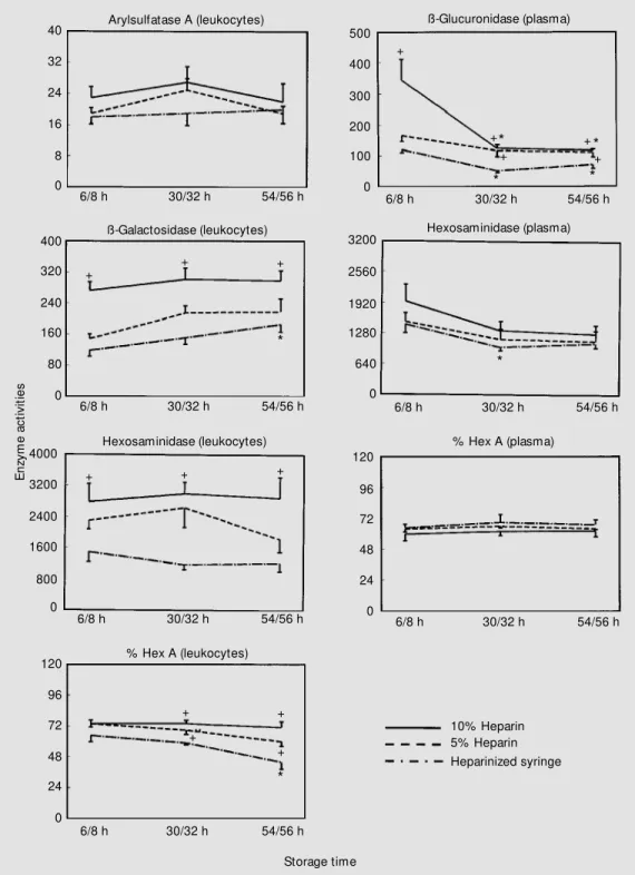
Next, we evaluated the influence of hep-arin concentration (10% hephep-arin, 5% hepa-rin, and heparinized syringe) at different times (6 to 8 h, 30 to 32 h, and 54 to 56 h) between collection and processing (Figure 2). The activities of arylsulfatase A in
leuko-cytes and hexosaminidase A in plasma were not affected by different heparin concentra-tions at any time of storage. The effect of heparin concentration was observed mainly when blood was collected with 10% heparin, compared to a heparinized syringe. The ß-galactosidase, total hexosaminidase, hex-osaminidase A (leukocytes), and ß-glucu-ronidase (plasma) enzymes exhibited higher activities with this concentration. In addi-tion, various enzyme activities (ß-galactosi-dase, hexosaminidase A in leukocytes, and ß-glucuronidase and total hexosaminidase in plasma) exhibited significant differences according to the period of processing. In plasma, ß-glucuronidase and total hex-osaminidase activities diminished, compared to 6/8 h of storage. In leukocytes the same effect occurred for hexosaminidase A, whereas the opposite effect was seen for ß-galactosidase.
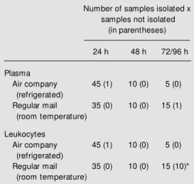
When analyzing the impact of type of transportation to the laboratory and shipping time (the time between collection in the dif-ferent cities and initial processing at our laboratory), we decided first to divide the 60 samples into 3 groups, i.e., samples pro-cessed within 24, 48 and 72/96 h after col-lection. One blood sample was lost during transportation, and one plasma during pro-cessing (Table 1). As observed in this table, leukocytes could not be isolated from 10 of 15 samples sent by regular mail (room tem-perature) arriving after 72 to 96 h. When analyzing the number of leukocyte samples Figure 1 - Comparison of
leuko-cyte (A) and plasma (B) activities of different lysosomal enzymes w hen blood collection w as per-formed w ith a plastic syringe (syr) or w ith a vacuum glass tube (vac). Values are the mean ± SEM and are reported as nmol h-1 mg protein-1 and nmol h-1
ml-1 for leukocytes and plasma,
respectively. asa, Arylsulfatase A; B-gal, ß-galactosidase; hex (x 0.1), hexosaminidase; % hex A, hexosaminidase A percentage; B-glu, ß-glucuronidase.
Table 1 - Distribution of plasma and leukocytes from 60 individuals divided into tw o aliquots according to mode of transportation and time of processing.
* P<0.05 compared to 24 h (chi-square test).
Number of samples isolated x samples not isolated
(in parentheses)
24 h 48 h 72/96 h
Plasma
Air company 45 (1) 10 (0) 5 (0) (refrigerated)
Regular mail 35 (0) 10 (0) 15 (1) (room temperature)
Leukocytes
Air company 45 (1) 10 (0) 5 (0) (refrigerated)
E
n
zy
m
e
a
c
ti
v
it
ie
s
40
32
24
16
8
0
6/8 h 30/32 h 54/56 h Arylsulfatase A (leukocytes)
10% Heparin 5% Heparin Heparinized syringe
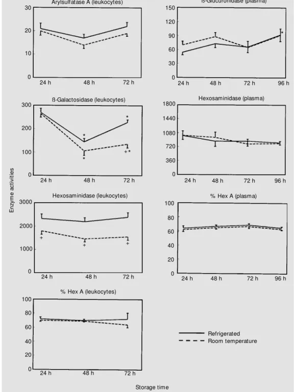
Storage time which could be isolated adequately after arriving by air company or regular mail, a significant failure in obtaining leukocytes was observed for samples shipped by regular mail after 72/96 h (P<0.05, chi-square test). Figure 3 shows that there was no
signifi-cant effect of type of transport or time of processing on the activities of arylsulfatase A in leukocytes, and of ß-glucuronidase, total hexosaminidase and hexosaminidase A in plasma. However, the impact of transport (temperature of storage) was observed on
Figure 2 - Effect of different heparin concentrations and stor-age times on the activity of ly-sosomal enzymes. Values are the mean ± SEM and are re-ported as nmol h-1 mg protein-1
and nmol h-1 ml-1 for leukocytes
and plasma, respectively. Hex-osaminidase A (Hex A) is re-ported as % hexosaminidase. * P<0.05 compared to 6/8 h at the same heparin concentra-tion. +P<0.05 compared to the
heparinized syringe group at the same storage time.
4000
3200
2400
1600
800
0
6/8 h 30/32 h 54/56 h
+ + +
Hexosaminidase (leukocytes)
500
400
300
200
100
0
6/8 h 30/32 h 54/56 h +
+ +
* +
ß-Glucuronidase (plasma)
*
*
120
96
72
48
24
0
6/8 h 30/32 h 54/56 h % Hex A (plasma)
400
320
240
160
80
0
6/8 h 30/32 h 54/56 h ß-Galactosidase (leukocytes)
+
+ +
*
120
96
72
48
24
0
6/8 h 30/32 h 54/56 h % Hex A (leukocytes)
+ +
+
* +
3200
2560
1920
1280
640
0
6/8 h 30/32 h 54/56 h Hexosaminidase (plasma)
*
the activities of ß-galactosidase, total hex-osaminidase, and hexosaminidase A in leu-kocytes. Lower total hexosaminidase activ-ity and hexosaminidase A percentage were observed at 24 h for samples shipped by regular mail without refrigeration, when
com-pared to samples shipped by air. In contrast, we observed that ß-galactosidase exhibited a higher activity when samples arrived by regu-lar mail. In addition, total hexosaminidase analyzed within 48 and 72/96 h and hosaminidase A analyzed within 72/96 h
ex-E
n
zy
m
e
a
c
ti
v
it
ie
s
20
10
0
24 h 48 h 72/96 h
Arylsulfatase A (leukocytes)
Refrigerated (air company) Room temperature (mail)
M ailing time Figure 3 - Effect of different
m eans of t ransport at ion and shipping times on the activity of lysosomal enzymes. Values are the mean ± SEM and are re-ported as nmol h-1 mg protein-1
and nmol h-1 ml-1 for leukocytes
and plasma, respectively. Hex-osaminidase A (Hex A) is re-ported as % hexosaminidase. * P<0.05 compared to 24 h con-sidering the sam e m eans of transportation. +P< 0.05 com
-pared to samples shipped by air considering the same shipping time.
3000
2000
1000
0
24 h 48 h 72/96 h
Hexosaminidase (leukocytes)
+
+
*
+
*
* *
120
96
72
48
24
0
24 h 48 h 72/96 h
ß-Glucuronidase (plasma)
120
96
72
48
24
0
24 h 48 h 72/96 h
% Hex A (plasma) 300
200
100
0
24 h 48 h 72/96 h
ß-Galactosidase (leukocytes)
90 80 70 60
0
24 h 48 h 72/96 h
% Hex A (leukocytes)
+
* *
+
50 40 30 20 10
*
2000
1600
1200
800
400
0
24 h 48 h 72/96 h
Hexosaminidase (plasma)
hibited higher activities in samples sent by regular mail. On the other hand, when study-ing time of processstudy-ing, total hexosaminidase and hexosaminidase A activities in leuko-cytes were lower at 48 and 72/96 h, and at 72/96 h, respectively, in samples shipped by
air with refrigeration when compared to samples tested at 24 h.
In another set of experiments, the impact of time between sample collection and sample processing in the laboratory was evaluated (Figure 4). In this stage it was not possible to
E
n
zy
m
e
a
c
ti
v
it
ie
s
30
20
0
24 h 48 h 72 h
Arylsulfatase A (leukocytes)
Refrigerated Room temperature
Storage time 10
Figure 4 - Effect of different tem-peratures and times of storage. Values are the mean ± SEM and are reported as nmol h-1 mg
pro-tein-1 and nmol h-1 ml-1 for
leu-kocytes and plasm a, respec-tively. Hexosaminidase A (Hex A) is reported as % hexosamini-dase. * P<0.05 compared to 24 h. +P<0.05 compared to the
re-frigerated group at the same storage time.
0
24 h 48 h 72 h
3000
1000
Hexosaminidase (leukocytes)
+ +
2000
+
120
90
60
30
0
24 h 48 h 72 h
150
ß-Glucuronidase (plasma)
96 h *
100
80
60
40
20
0
24 h 48 h 72 h
% Hex A (plasma)
96 h 300
200
100
0
24 h 48 h 72 h
ß-Galactosidase (leukocytes)
*
*
+ *
*
24 h 48 h 72 h
% Hex A (leukocytes)
0 100
80
60
40
20
1440
1080
720
360
0
24 h 48 h 72 h Hexosaminidase (plasma) 1800
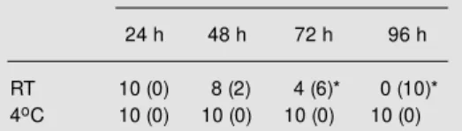
separate leukocytes from any of the 10 samples kept at room temperature for 96 h before processing (Table 2). When we ana-lyzed these data by the chi-square test we found a significant difference (P<0.01) be-tween room temperature and 4o
C after 72 h
and 96 h. These results confirm those ob-served when samples were sent by regular mail at 72/96 h (Table 1). It can be seen in Figure 4 that total hexosaminidase activities in leukocytes were lower at all storage times studied for samples kept at room
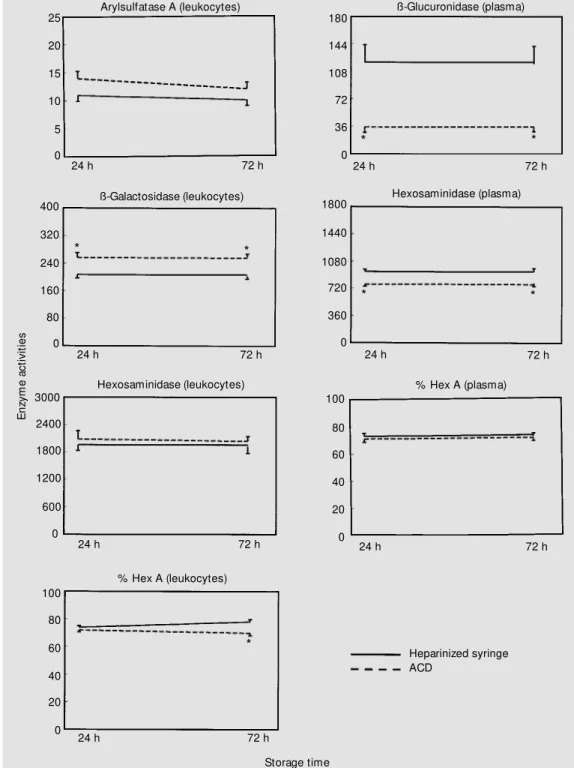
tempera-Figure 5 - Effect of different an-ticoagulants for samples pro-cessed at different times after collection. Values are means ± SEM and are reported as nmol h-1 mg protein-1 and nmol h-1
ml-1 for leukocytes and plasma,
respectively. Hexosaminidase A (Hex A) is reported as % hexosam inidase. * P< 0.05 com -pared to the heparinized group at the same storage time. ACD, Acid citrate-dextrose.
E
n
zy
m
e
a
c
ti
v
it
ie
s
25
20
0
24 h 72 h
Arylsulfatase A (leukocytes)
Heparinized syringe ACD
Storage time 15
10
5
0
24 h 72 h
3000
1800
Hexosaminidase (leukocytes)
2400
1200
600
144
108
72
36
0
24 h 72 h
180
ß-Glucuronidase (plasma)
* *
100
80
60
40
20
0
24 h 72 h
% Hex A (plasma) 400
240
160
0
24 h 72 h
ß-Galactosidase (leukocytes)
* 320
80
*
24 h 72 h
% Hex A (leukocytes)
0 100
80
60
40
20
*
1440
1080
720
360
0
24 h 72 h
Hexosaminidase (plasma) 1800
ture compared to samples kept at 4o
C. The same occurred for ß-galactosidase activity after 72 h of storage. For this enzyme, leuko-cyte activities decreased when compared to samples stored for 24 h. Moreover ß-glucu-ronidase showed higher activity in samples stored for 96 h at 4o
C.
Finally, the comparison between ACD and heparin as anticoagulants at different times (24 and 72 h) of sample processing is shown in Figure 5. First, it can be observed that there was no effect of different times of processing on the enzyme activities for ei-ther anticoagulant. However, when analysis was performed for ACD compared to hepa-rin, higher activities of ß-galactosidase in leukocytes were observed for ACD at both processing times. In contrast, plasma ß-glu-curonidase and total hexosaminidase activi-ties were lower when ACD was used at both processing times, while the same occurred at 72 h of sample processing for hexosamini-dase A activity in leukocytes.
D iscussio n
In the present study, no difference was observed between blood collected with a vacuum glass tube and blood collected with a plastic syringe. This finding is in contrast to the data reported by Sasakawa and Tokunaga (6), who demonstrated that blood stored in plastic bags showed smaller varia-tions due to the air permeability of the mate-rial, which preserved better the pH and blood viscosity when compared to blood stored in glass bottles.
The choice of anticoagulant for biochemi-cal analysis is very important and depends directly on the assay to be performed. EDTA, for instance, has a chelating effect which causes loss of activity of some lysosomal enzymes (14). Some authors refer to heparin as a powerful inhibitor of leukocytic lysoso-mal enzymes (15-18) when added directly to the incubation medium. Although a very low concentration of heparin may be present
dur-ing assays measurdur-ing plasma enzyme activi-ties, only traces of heparin (if any) are ex-pected after the leukocyte preparation pro-cedure. However, there are many biochemi-cal genetic laboratories which use heparin as anticoagulant (3,4,19-21), and some refer-ence centers recommend the collection of blood with heparin concentrations as high as 10% (Shin Y, personal communication). In the present study, higher lysosomal enzyme activities were observed when blood was collected with 10% heparin. In addition, the activities seemed to be better preserved dur-ing different periods of processdur-ing when blood was collected with the higher heparin concentration. ß-Glucuronidase activity was affected by heparin and time of processing, with the highest activity being observed within 6/8 h with 10% heparin, in agreement with Triebling et al. (22), who reported that heparin raised the total activity of ß-glucu-ronidase.
Furthermore, there was a clear effect of time of transportation. In many cases samples are processed more than 48 h after collection due to the large number of steps involved between collection and processing at the reference laboratory, where large distances are an important factor. Firstly, we observed a clear deterioration of samples sent by regu-lar mail. The failure to obtain leukocytes occurred in samples processed 72 and 96 h after collection and shipped at room temper-ature. Leukocyte pellets could not be iso-Table 2 - Distribution of leukocyte samples ac-cording to time betw een collection and process-ing and storage temperature.
* P<0.05 compared to 24 h (chi-square test). RT, Room temperature.
Storage Number of samples isolated x condition samples not isolated (in parentheses)
24 h 48 h 72 h 96 h
lated from 10 of 15 blood specimens arriving after 72-96 h. This observation agrees with the findings of Branum et al. (23), who stud-ied the effect of heparin and ACD on leuko-cyte yield and function, and on lysosomal enzyme activity. According to these authors, ACD is the best choice of anticoagulant for those specimens submitted to leukocyte iso-lation after a time longer than 48 h. ACD is a good anticoagulant for blood preservation (24,25), while heparin has been cited only as an anticoagulant and not as a preservative (26). Samples which came by regular mail exhibited higher enzyme activities in most cases. However, a higher number of unsuc-cessful leukocyte preparations was observed under these conditions. Bailey and Bove (5) demonstrated that routine transportation, pro-cessing and handling of blood may lead to alterations in the biochemical characteristics of the sample.
The results obtained when the effect of transport was eliminated emphasize the dif-ficulties for the isolation of leukocytes from samples kept at room temperature for long periods of time. When the samples were collected at our laboratory (stage 4), we also observed smaller variations in the activity of the enzymes studied in comparison to the samples which traveled long distances, indi-cating that transport itself influences the be-havior of the enzymes.
Arylsulfatase A activity was highly stable under all conditions analyzed here, in agree-ment with Draper et al. (27). The activities of ß-galactosidase, total hexosaminidase and hexosaminidase A in leukocytes varied in-dependently of the processing conditions. Lombardo et al. (28) consider ß-galactosi-dase to be the most labile enzyme. They observed that ß-galactosidase and ß-glucu-ronidase are released to a great extent from leukocytes and platelets, leading to an
in-crease in plasma activities during refriger-ated storage of blood (7). This seems to be the case for the increase in ß-glucuronidase activity observed in the present study.
Since the literature (23) indicates the ex-istence of other anticoagulants such as ACD which preserves leukocytes better than hep-arin, we compared the effects of ACD and heparin. The results indicate that the activi-ties of ß-glucuronidase and total hexosamini-dase in plasma reached levels near the lower normal limits when ACD was used. This kind of information is important if one de-sires to maintain consistent normal-range values for all samples tested, particularly for the detection of carriers and affected indi-viduals. On the other hand, our findings are in agreement with Branum et al. (23) in terms of arylsulfatase A activity in leuko-cytes, which did not differ with the use of one anticoagulant or the other. According to these results, the use of a heparinized syringe seems to be preferable to ACD. Heparin, which is an inexpensive anticoagulant used worldwide, has been used in our routine for a long time, and all normal ranges of lysoso-mal enzyme activities have been well estab-lished in the laboratory with this anticoagu-lant.
Re fe re nce s
1. Tager JM , Jonsson LM , Aerts JG, Oude EJ, Schram AW, Erickson AH & Barranger JA (1984). M etabolic consequences of genetic defects in lysosomes. Biochemi-cal Society Transactions, 12: 902-905. 2. Low den JA (1982). Enzymological
diagno-sis of lysosomal storage diseases. In: Willey AM , Carter TP, Kelly S & Porter IH (Editors), Clinical Genetics - Problems in Diagnosis and Counseling. Academ ic Press, New York.
3. Wenger DA & Williams C (1991). Screen-ing for lysosomal disorders. In: Hommes FA (Editor), Techniques in Diagnostic Hu-man Biochemical Genetics - A Laboratory M anual. Wiley-Liss Inc., New York. 4. Landels EC, Ellis IH, Bobrow M & Fensom
AH (1991). Tay-Sachs disease heterozy-gote detection: use of centrifugal analyser for automation of hexosaminidase assays w ith tw o different artificial substrates.
Journal of M edical Genetics, 28: 101-109. 5. Bailey DN & Bove JR (1975). Chemical and hematological changes in stored CPD blood. Transfusion, 15: 244-249. 6. Sasakaw a S & Tokunaga E (1976).
Physi-cal and chemiPhysi-cal changes of ACD pre-served blood: a comparison of blood in glass bottles and plastic bags. Vox San-guinis, 31: 199-210.
7. Lombardo A, Goi G, Guagnellini E, Fabi A, Sciorelli G, Burlina AB & Tettamanti G (1984). Behaviour of several enzymes of lysosomal origin in human plasma during w hole blood storage. Clinica Chimica Acta, 143: 343-353.
8. Skoog WA & Beck WS (1956). Studies on the fibrogen, dextran and phytohemag-glutinin methods of isolating leukocytes.
Blood, 11: 436-454.
9. Low ry OH, Rosebrough NJ, Farr AL & Randall RJ (1951). Protein measurement w ith the Folin phenol reagent. Journal of Biological Chemistry, 193: 265-275. 10. Suzuki K (1977). Globoid cell
leukodystro-phy (Krabbe disease) and GM 1-gangliosi-dosis. In: Glew RH & Peters SP (Editors),
Practical Enzymology of Sphingolipidoses. A.R. Liss, New York.
11. Singer JD, Cotlier E & Krimmer R (1973). Hexosaminidase A in tears and saliva for rapid identification of Tay-Sachs diseases and its carriers. Lancet, 2: 116-119. 12. Beaudet AL, Diferrante NM , Ferry GD,
Nichols BL & M ullins CE (1975). Variation in the phenotypic expression of ß-glucu-ronidase deficiency. Pediatrics, 86: 388-394.
13. Lee-Vaupel M & Conzelmann E (1987). A simple chromogenic assay for arylsulfa-tase A. Clinica Chimica Acta, 164: 171-180.
14. Kolodny EH & M umford RA (1976). Hu-man leukocyte acid hydrolases: character-ization of eleven lysosomal enzymes and study of reaction conditions for their auto-mated analysis. Clinica Chimica Acta, 70: 247-257.
15. Avila JL & Convit J (1975). Inhibition of leucocytic lysosomal enzymes by gly-cosaminoglycans in vitro. Biochemical Journal, 152: 57-64.
16. Avila JL & Convit J (1976). Physicochemi-cal characteristics of the glycosaminogly-can lysosomal enzyme interaction in vitro.
Biochemical Journal, 160: 129-136. 17. M ikulíková D & Trnavský K (1982).
Influ-ence of a glycosaminoglycan polysulfate (arteparon) on lysosomal enzyme release from human polymorphonuclear leuko-cytes. Zeitschrift für Rheumatologie, 41: 50-53.
18. Lund-Hansen T, Hoyer PE & Andersen H (1984). A quantitative cytochemical assay of ß-galactosidase in single cultured hu-man skin fibroblasts. Histochemistry, 81: 321-330.
19. Singer HS, Nankervis GA & Schafer IA (1972). Leukocyte beta-galactosidase ac-tivity in the diagnosis of generalized GM 1 gangliosidosis. Pediatrics, 49: 352-361. 20. Raghavan S, Gajew ski A & Kolodny EH
(1977). GM 1-Ganglioside ß-galactosidase in leukocytes and cultured fibroblasts.
Clinica Chimica Acta, 81: 47-56. 21. M utoh T, Kiuchi K, Sobue I & Naoi M
(1984). Application of a GM 1 ganglioside ß-galactosidase microassay method to di-agnosis of GM 1 gangliosidosis. Clinica Chimica Acta, 140: 223-230.
22. Triebling A, Dlugosz J, Brzozow ski J, Lukaszew icz W & Gabryelew icz A (1979). Heparin effect on the activity of certain lysosomal hydrolases in dog pancreas (in vivo investigations). Acta Physiologica Polonica, 30: 527-532.
23. Branum E, Cummins L, Bartilson M , Hop-per M , Pruett S & O’Brien JF (1988). Ef-fect of tw o anticoagulants on leukocyte yield and function, and on lysosomal en-zyme activity. Clinical Chemistry, 34: 110-113.
24. Schechter DC & Sw an H (1962). Bio-chemical alterations of preserved blood.
Archives of Surgery, 84: 269-276. 25. M oore SB, Beckala H, DeGoey S &
Leavelle D (1981). A report on the use of ACD (solution B) as w hole blood transport medium for recovery of lymphocytes for HLA typing. In: Zachary AA & Braun WE (Editors), Laboratory M anual, American Association for Clinical Histocompatibility Testing. American Association for Clinical Histocompatibility Testing, New York. 26. Walker RH (1990). Blood and blood
com-ponents: preparation, storage, and ship-ment. In: Walker RH (Editor), Technical M anual. American Association of Blood Banks, Arlington.
27. Draper RK, Fiskum GM & Edmond J (1976). Purification, molecular w eight, amino acid and subunit composition of arylsulfatase A from human liver. Archives of Biochemistry and Biophysics, 177: 525-538.