ISSN 0100-879X
BIOMEDICAL SCIENCES
AND
CLINICAL INVESTIGATION
www.bjournal.com.br
www.bjournal.com.br
Volume 44 (7) 606-728 July 2011
Braz J Med Biol Res, July 2011, Volume 44(7) 647-651
doi: 10.1590/S0100-879X2011007500061
Effect of methylprednisolone on perivascular pulmonary edema,
inflammatory infiltrate, VEGF and TGF-beta immunoexpression in the
remaining lungs of rats after left pneumonectomy
F. Guimarães-Fernandes, M.N. Samano, R.P. Vieira, C.R. Carvalho, R. Pazetti, L.F.P. Moreira,
P.M. Pêgo-Fernandes and F.B. Jatene
Faculdade de Medicina de Ribeirão Preto Campus
Ribeirão Preto
Institutional Sponsors
The Brazilian Journal of Medical and Biological Research is partially financed by
analiticaweb.com.br S C I E N T I F I C
Effect of methylprednisolone on perivascular
pulmonary edema, inflammatory infiltrate, VEGF
and TGF-beta immunoexpression in the remaining
lungs of rats after left pneumonectomy
F. Guimarães-Fernandes
2, M.N. Samano
2, R.P. Vieira
1, C.R. Carvalho
1,
R. Pazetti
2, L.F.P. Moreira
2, P.M. Pêgo-Fernandes
2and F.B. Jatene
21Departamento de Clínica Médica, Faculdade de Medicina, Universidade de São Paulo, São Paulo, SP, Brasil 2Departamento de Cardiopneumologia, Instituto do Coração, Hospital das Clínicas,
Faculdade de Medicina, Universidade de São Paulo, São Paulo, SP, Brasil
Abstract
Pneumonectomy is associated with high rates of morbimortality, with postpneumonectomy pulmonary edema being one of the leading causes. An intrinsic inflammatory process following the operation has been considered in its physiopathology. The use of corticosteroids is related to prevention of this edema, but no experimental data are available to support this hypothesis. We evaluated the effect of methylprednisolone on the remaining lungs of rats submitted to left pneumonectomy concerning edema and inflammatory markers. Forty male Wistar rats weighing 300 g underwent left pneumonectomy and were randomized to re-ceive corticosteroids or not. Methylprednisolone at a dose of 10 mg/kg was given before the surgery. After recovery, the animals were sacrificed at 48 and 72 h, when the pO2/FiO2 ratio was determined. Right lung perivascular edema was measured by the index between perivascular and vascular area and neutrophil density by manual count. Tissue expressionof vascular endothelial growth factor (VEGF) and transforming growth factor-beta (TGF-β) were evaluated by immunohistochemistry light microscopy. There was perivascular edema formation after 72 h in both groups (P = 0.0031). No difference was observed between oper-ated animals that received corticosteroids and those that did not concerning the pO2/FiO2 ratio, neutrophil density or TGF-β expression. The tissue expression of VEGF was elevated in the animals that received methylprednisolone both 48 and 72 h after surgery (P = 0.0243). Methylprednisolone was unable to enhance gas exchange and avoid an inflammatory infiltrate and TGF-β expression also showed that the inflammatory process was not correlated with pulmonary edema formation. However, the overexpression of VEGF in this group showed that methylprednisolone is related to this elevation.
Key words: Pneumonectomy; Glucocorticoids; Pulmonary edema; Vascular endothelial growth factor; Animal model; Transforming growth factor beta.
Introduction
Correspondence: M.N. Samano, Av. Dr. Enéas C. Aguiar, 44, 2º andar, Bloco II, Sala 9, 05403-000 São Paulo, SP, Brasil. Fax: +55-11-3069-5351. E-mail: marcos.samano@incor.usp.br
Received September 24, 2010. Accepted April 18, 2011. Available online May 13, 2011. Published July 25, 2011. The mortality of pneumonectomy is very high in
com-parison to other pulmonary surgical procedures, reaching
up to 9.4% in some studies (1,2). When compared to
lobectomy, the mortality increases almost 10-fold. Although pulmonary lobectomy is the standard surgical treatment for lung cancer, pneumonectomy is still required in some
cases, such as those involving central or bulky tumors. Some other inflammatory diseases such as tuberculosis,
bronchiectasis and other non-malignant diseases may also
require extirpation of one lung.
Even though there have been improvements in surgical management with reduction of mortality, the incidence of complications is about 60%. Respiratory complications are the most frequent, corresponding to 15%. In this group of
patients the mortality can reach 30%. Among the most rel -evant complications are acute respiratory failure, pneumo-nia, pulmonary thromboembolism, and pulmonary edema.
When pulmonary edema occurs after pneumonectomy with no association with pre-existing heart disease, acute
648 F. Guimarães-Fernandes et al.
called postpneumonectomy pulmonary edema (PPPE) (3). The physiopathology of PPPE is not completely known
and multifactorial causes seem to be involved in the develop-ment of edema. Endothelial damage and increased capillary permeability seem to be the most accepted hypothesis (4).
Thus, the use of anti-inflammatory agents may prevent or
alter the development of pulmonary edema. The treatment of acute lung injury/acute respiratory distress syndrome (ALI/ ARDS) with corticosteroids is well established and due to the similarity of clinical and radiologic features of these diseases, it may also play a major role in PPPE. Although some clinical reports of the use of corticosteroids to prevent PPPE have
been published, there are no experimental studies that cor
-relate corticosteroids and PPPE and explain the mechanism
of this protective effect (5).
The association of transforming growth factor-beta (TGF-β) with the acute inflammatory process is well established as well
its effect on the increase of vascular permeability in ARDS
and ALI. TGF-β belongs to the dimeric polypeptide family of
growth factors and all human body cells produce and have
receptors for this factor. TGF-β is present in the initial phases of the inflammatory process in animal models and in human
studies, and probably causes the development of edema due to a decrease in the number of gap junctions between epithelial
cells. Thus, it is an important mediator of lung inflammation (6). The vascular endothelial growth factor (VEGF) also plays
an important role in the regulation of tonus and permeability
of the alveolo-capillary membrane, participating in the inflam
-matory process (7). Its regulation is a very complex process and depends on hypoxia, cytokines, endotoxins, and many
other intracellular signs. In the lungs it is important during embryogenesis and development and is also
important in the pathogenesis of emphysema and ALI.
In a previous study, we observed the forma-tion of pulmonary perivascular edema in rats after left pneumonectomy without evidence of
oxidative stress related to nitric oxide synthase
or neutrophil migration (8). In the present study, we analyzed the effect of methylprednisolone
on the partial O2 pressure (pO2)/inspired O2
fraction (FiO2) ratio, pulmonary perivascular
edema, neutrophil infiltrate, and the expression of TGF-β and VEGF in the remaining lung of
rats after left pneumonectomy.
Subjects and Methods
The study was approved by the Scientific
and Ethics Committee of the Instituto do
Cora-ção (InCor, protocol #SDC 3125/08/041). Forty male Wistar rats weighing on average 300 g
were included in the study. The animals were randomized into two groups: 20 underwent left pneumonectomy (control group) and 20
underwent left pneumonectomy after a single
intraperito-neal injection of 10 mg/kg methylprednisolone (corticosteroid group). In the control group, 10 animals were sacrificed 48 h after pneumonectomy and 10 were sacrificed 72 h after the
procedure. Similarly, the corticosteroid group was divided into
two groups according to the time of sacrifice.
All animals were sedated and anesthetized with isoflurane
(Cristália, Brazil) in an anesthetic chamber and a tracheal tube was inserted under light laryngoscopy. They were randomized to receive or not corticosteroid administered as an
intraperi-toneal injection of 10 mg/kg methylprednisolone sodium suc
-cinate (União Química, Brazil). The animals were ventilated in a Harvard Apparatus model 683 with a gas mixture of oxygen and isoflurane at a tidal volume of 10 mL/kg. The respiratory
rate was 80 cycles per minute. Thoracotomy was performed
in the fifth left intercostal space and pneumonectomy was
performed with en bloc ligation of pulmonary hilum. After resection of left lung a chest tube was introduced through the incision and removed after chest closure. The animals were sent to the animal house and maintained for 48 or 72 h with food and water ad libitum.
At the time of sacrifice, the animals were once again se -dated and intubated. After sternolaparotomy the abdominal aorta was accessed to collect an arterial blood sample for gas
analysis and the animals were sacrificed by exsanguination via resection of the aorta. The lower lobe was maintained inflated with trapped air by ligation of the corresponding bronchus, fixed
in buffered formaldehyde, embedded for 24 h and submitted
to histological examination.
The pO2/FiO2 ratio was determined by gas analysis of
an arterial blood sample collected just before sacrifice for
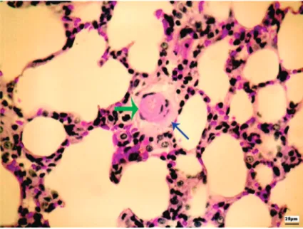
Figure 1. Digitized small-caliber vessel (green arrow) with sorrounding
the assessment of pulmonary function at 48 and 72 h.
Histological sections stained with hema
-toxylin/eosin (HE) were analyzed in order to
quantify pulmonary edema. Digitized images of parenchymal small caliber vessels were
analyzed with the UTHSCSA-Image-Tool®
software, version 3.0 (9). An index for
semi-quantitative quantification of perivascular
pulmonary edema (perivascular edema index)
was calculated on the basis of the relationship between the perivascular area and the vascular
area (Figure 1), as described in Ref. 8.
Neutrophil density was used to quantify the
inflammatory infiltrate in histological sections stained with HE. A manual count of neutrophils
present in the alveolar septa was performed by an observer blind to the study. Twenty
non-coin-cident fields, enlarged to 400X, were quantified
by the point-counting technique (8,10).
TGF-β and VEGF were quantified on the
basis of the immunohistochemistry in bronchial epithelial cells. The polyclonal antibodies
anti-TGF-β and anti-VEGF (Santa Cruz Biotechonology Inc., USA)
were used together by the biotin-streptavidin technique on lung sections. Digital quantitative analysis was performed using the Image Pro Plus 4.0 software (Media Cybernetics, USA), and
expression was determined as the ratio between the positive area and the entire corresponding epithelial area in five com
-plete airway sections from each animal (Figure 2).
Statistical analysis was performed using the GraphPad software for Windows. Data were analyzed by two-way ANOVA
and complemented by the Bonferroni t-test. The results obtained were parametric, and the values were reported as
averages with a 95% confidence interval. The level of signifi -cance was set at 5%.
Results
The average pO2/FiO2 values for the pulmonary function
of animals submitted to pneumonectomy were not significantly
different from those of the animals submitted to
pneumonec-tomy and receiving methylprednisolone at 48 or 72 h. However,
there was an increase in perivascular pulmonary edema, as
shown in Figure 3, in the animals of both groups sacrificed after 72 h compared to 48 h (P = 0.0031).
Neutrophil density did not differ significantly between groups. However, a decrease was observed in the corticos -teroid group 48 and 72 h after pneumonectomy. The average neutrophil density was 501.6 neutrophils/105 µm2 after 48 h
and 734.7 neutrophils/105 µm2 after 72 h in the control group
and 424.8 and 479.3 neutrophils/105 µm2, respectively, in the
corticosteroid group. The expression of TGF-β also did not
differ between the control and corticosteroid groups (P =
0.5525). However, the graph in Figure 4 shows that the im
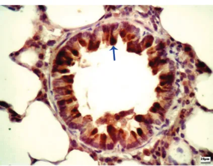
-Figure 2. Photomicrograph showing tumor growth factor-β immunoreactivity in
the bronchial wall (blue arrow). Magnification = 400X.
Figure 3. Pulmonary edema index graph showing edema forma
-tion 72 h after pneumonectomy (*P = 0.0031), without distinc-tion between the control and corticosteroid groups. Data are reported as means ± SEM for 10 animals in each group (two-way ANOVA, complemented by the Bonferroni t-test).
Figure 4. Vascular endothelial growth factor showing a
650 F. Guimarães-Fernandes et al.
munohistochemical expression of VEGF was significantly
increased in the corticosteroid group 72 h after pneumo-nectomy (P < 0.001).
Discussion
The hemodynamic and pulmonary physiological changes occurring after pneumonectomy have not been fully determined. The contribution of the right lung to the
total pulmonary capacity is about 53 to 55%, and that of
the left lung is 45 to 47%. In rats, the left lung corresponds
to only 35% of total pulmonary mass (11). Pulmonary func -tion declines in all patients who undergo pneumonectomy and depends on the side of the procedure, patient age and the remaining lung function. The forced vital capacity
(FVC) and the forced expiratory volume in the first second (FEV1) decline to less than 50% of the initial values. Usually there is no alteration in oxygen saturation, pO2 or pCO2,
but oxygen consumption is reduced from 17 to 28% after pulmonary resection (2). In the present experimental model, as described before, the pO2/FiO2 ratio showed absence
of functional loss after pneumonectomy. Similarly, the ad-ministration of corticosteroid before the operation did not
improve the levels of pO2 after pneumonectomy (8).
The perivascular space of rodents is formed by
capillar-ies and connective tissue. During inflammatory processes or allergic reactions this space can be infiltrated by leukocytes.
The importance of this space has been long neglected due to the studies focused on other pulmonary compartments such as the interstice, bronchoalveolar space and basement
membrane of the bronchi. However, it is well known now that the perivascular space is related to the extravascular
lung water accumulation that occurs in pulmonary edema
(12-15). In an experimental model of asthma, there is rapid
migration of eosinophils to this space (16). In the present study, pneumonectomy caused perivascular pulmonary
edema in the group sacrificed after 72 h, which was not
affected by corticosteroid administration. This is evidence that pulmonary edema occurs as a result of pneumonectomy
and that the administration of an anti-inflammatory agent
has no protective effect on edema formation.
The association of pulmonary edema and acute inflam -mation is evident in ALI/ARDS, where neutrophils play an important role in endothelial injury and vascular permeability.
Although a surgical procedure can trigger an inflammatory
process, the neutrophil infiltrate is reduced 48 and 72 h after pneumonectomy (8). Despite the well-known effect of corti -costeroid on neutrophil migration, we observed only a trend
to a reduction of the neutrophil infiltrate after corticosteroid administration, which was not statistically significant.
TGF-β has been shown to be related to edema forma -tion in animal and human studies. There is an increase of
its expression after the induction of ARDS with tracheal
instillation of Escherichia coli toxin. The importance of
TGF-β in pulmonary edema formation has been observed in another study in which TGF-β knock-out animals showed
lower pulmonary edema formation compared to normal
animals (17). There is a relationship between TGF-β expression and ARDS in humans, where its expression
is increased in the initial phases of ARDS and its higher
concentration is related to a worse pO2/FiO2 ratio. There is
a lack of studies concerning analyses of TGF-β expression
after surgical procedures. It is reasonable to assume that
surgery may act as a trauma and elevate the expression of inflammatory markers. However, we did not observe an effect of corticosteroid on the immunoexpression of TGF-β
after pneumonectomy.
VEGF is a protein that regulates endothelial cell dif -ferentiation and angiogenesis, which was discovered as a vascular permeability factor (18). In humans, most studies on lung injuries have shown a reduction of intrapulmonary
VEGF levels in ALI/ARDS, especially in the early stages (19). However, several in vitro studies revealed that VEGF
increases the endothelial permeability (20,21). In fact,
there are few studies concerning VEGF and its effect on
pulmonary edema. In the present study, we observed that
corticosteroid caused increased expression of VEGF but
without a correlation with pulmonary edema formation. Although there is no consensus about the real effect of
VEGF, an anti-inflammatory action was observed in the
present study.
Pneumonectomy caused perivascular pulmonary edema 72 h after the procedure and corticosteroid did not
prevent this edema. We did not observe an anti-inflammatory action of corticosteroid concerning the neutrophil infiltrate or TGF-β expression. This finding supports the idea that
perivascular edema formation occurs after pneumonectomy
and is not related to inflammatory involvement. Methylpred
-nisolone was responsible for an increase in VEGF levels
after pneumonectomy.
References
1. Bernard A, Deschamps C, Allen MS, Miller DL, Trastek VF, Jenkins GD, et al. Pneumonectomy for malignant disease: factors affecting early morbidity and mortality. J Thorac Cardiovasc Surg 2001; 121: 1076-1082.
2. Kopec SE, Irwin RS, Umali-Torres CB, Balikian JP, Conlan AA. The postpneumonectomy state. Chest 1998; 114: 1158-1184.
3. Parquin F, Marchal M, Mehiri S, Herve P, Lescot B. Post-pneumonectomy pulmonary edema: analysis and risk fac-tors. Eur J Cardiothorac Surg 1996; 10: 929-932.
4. Deslauriers J, Aucoin A, Gregoire J. Postpneumonectomy pul-monary edema. Chest Surg Clin N Am 1998; 8: 611-31, ix.
Bartolucci AA. Intraoperative solumedrol helps prevent postpneumonectomy pulmonary edema. Ann Thorac Surg
2003; 76: 1029-1033.
6. Dhainaut JF, Charpentier J, Chiche JD. Transforming growth factor-beta: a mediator of cell regulation in acute respiratory distress syndrome. Crit Care Med 2003; 31: S258-S264.
7. Lahm T, Crisostomo PR, Markel TA, Wang M, Lillemoe KD, Meldrum DR. The critical role of vascular endothelial growth factor in pulmonary vascular remodeling after lung injury.
Shock 2007; 28: 4-14.
8. Samano MN, Pazetti R, Prado CM, Tiberio IC, Saldiva PH, Moreira LF, et al. Effects of pneumonectomy on nitric oxide synthase expression and perivascular edema in the remain-ing lung of rats. Braz J Med Biol Res 2009; 42: 1113-1118.
9. Monteiro RJ, Jatene FB, Pazetti R, Correia AT, Manoel LA, Bernardo WM, et al. Evaluation of the cardiac morphological alterations secondary to the pulmonary emphysema: ex-perimental study in rats. Braz J Cardiovasc Surg 2004; 19: 341-347.
10. Finkelstein R, Fraser RS, Ghezzo H, Cosio MG. Alveolar inflammation and its relation to emphysema in smokers. Am J Respir Crit Care Med 1995; 152: 1666-1672.
11. Brown LM, Rannels SR, Rannels DE. Implications of post-pneumonectomy compensatory lung growth in pulmonary physiology and disease. Respir Res 2001; 2: 340-347.
12. Drake RE, Laine GA, Allen SJ, Katz J, Gabel JC. A model of the lung interstitial-lymphatic system. Microvasc Res 1987; 34: 96-107.
13. Nagai H, Kira S, Mimoto T, Inatomi K, Yoneda R. Sequential changes of perivascular edema cuffs in models of
perme-ability and hemodynamic pulmonary edema. Respiration
1991; 58: 57-61.
14. Pabst R, Tschernig T. Perivascular capillaries in the lung: an important but neglected vascular bed in immune reactions?
J Allergy Clin Immunol 2002; 110: 209-214.
15. Schmiedl A, Tschernig T, Luhrmann A, Pabst R. Leukocyte infiltration of the periarterial space of the lung after allergen provocation in a rat asthma model. Pathobiology 2005; 72: 308-315.
16. Pabst R. The periarterial space in the lung: its important role in lung edema, transplantation, and microbial or allergic inflammation. Pathobiology 2004; 71: 287-294.
17. Pittet JF, Griffiths MJ, Geiser T, Kaminski N, Dalton SL, Huang X, et al. TGF-beta is a critical mediator of acute lung injury. J Clin Invest 2001; 107: 1537-1544.
18. Lahm T, Crisostomo PR, Markel TA, Wang M, Lillemoe KD, Meldrum DR. The critical role of vascular endothelial growth factor in pulmonary vascular remodeling after lung injury.
Shock 2007; 28: 4-14.
19. Kosmidou I, Karmpaliotis D, Kirtane AJ, Barron HV, Gibson CM. Vascular endothelial growth factors in pulmonary ede-ma: an update. J Thromb Thrombolysis 2008; 25: 259-264. 20. Koh H, Tasaka S, Hasegawa N, Yamada W, Shimizu M,
Nakamura M, et al. Protective role of vascular endothelial growth factor in endotoxin-induced acute lung injury in mice.
Respir Res 2007; 8: 60.
21. Koh H, Tasaka S, Hasegawa N, Asano K, Kotani T, Morisaki H, et al. Vascular endothelial growth factor in epithelial lining fluid of patients with acute respiratory distress syndrome.