Received: January 10, 2012 Accepted: August 13, 2012
Conflict of Interests: The authors state that there are no financial and personal conflicts of interest that could have inappropriately influenced their work.
Copyright: © 2012 Coutinho Filho et al.; licensee EDIPUCRS. This is an Open Access article distributed under the terms of the Creative Commons Attribution-Noncommercial-No Derivative Works 3.0 Unported License.
Behavior of subcutaneous tissue of rats in
response to infected dentine associated with
different endodontic irrigants
Comportamento do tecido subcutâneo de ratos frente à dentina
infectada e associada a diferentes irrigantes endodônticos
Tauby S. Coutinho-Filho a
Cláudio M. A. Ferreira b
Emmanuel J. N. L. da Silva b
Francisco J. Souza-Filho c
a Department of Restorative Dentistry, Endodontic
Division, State University of Rio de Janeiro (UERJ), Rio de Janeiro, RJ, Brazil
b Specialty Training Course in Endodontics, State
University of Rio de Janeiro (UERJ), Rio de Janeiro, RJ, Brazil
c Department of Restorative Dentistry – Endodontic
Division, Piracicaba Dental School, State University of Campinas (UNICAMP), Piracicaba, SP, Brazil
Correspondence:
Emmanuel João Nogueira Leal da Silva State University of Rio de Janeiro Department of Restorative Dentistry Endodontic Division
Bl. 28 de setembro, 157 Rio de Janeiro, RJ – Brazil 20550-900
E-mail: [email protected] Abstract
Purpose: To compare rat subcutaneous connective tissue reaction to dentine contaminated with
Enterococcus faecalis associated with 0.9% sterile saline, 5.25% sodium hypochlorite (NaOCl) and 2% chlorhexidine gel (CHX).
Methods: Dentine was crushed into powder and inoculated with E. faecalis. Tested substances were mixed with contaminated dentine and placed in polyethylene tubes. Ten male Wistar rats had their backs divided into four quadrants that received an implant containing one of the tested substances. An empty tube was used as a control. Five rats were randomly distributed for evaluation at time intervals of 24 hours and 72 hours. Tissue samples were histologically processed. Tissue reactions to experimental groups were evaluated under optical microscopes. Results: Groups of 5.25% NaOCl induced greater inflammatory response after 24 hours and 72 hours. Compared to groups of 2% CHX, the groups of 0.9% sterile saline showed milder inflammatory reactions after 24 hours and more severe after 72 hours.
Conclusion: The results indicate that 5.25% NaOCl group showed a higher inflammatory reaction to rat subcutaneous connective tissue and the 2% chlorhexidine group showed the least reaction.
Key words: Chlorhexidine; root canal irrigants; sodium hypochlorite; subcutaneous tissue
Resumo
Objetivo: Comparar a resposta do tecido subcutâneo de ratos frente a dentina contaminada com Enterococcus faecalis associado ao soro fisiológico 0.9% , hipoclorito de sódio 5.25% (NaOCl) ou clorexidina gel 2% (CHX).
Metodologia: Foi realizada a contaminação de dentina em pó com E. faecalis. As substâncias testadas foram misturadas com a dentina contaminada e inseridas em tubos de polietileno. Dez ratos Wistar tiveram os dorsos divididos em quatro quadrantes e cada quadrante recebeu um tubo com cada uma das misturas testadas. Um tubo vazio foi utilizado como controle. Os ratos foram distribuídos em dois grupos para avaliação no período de 24 e 72 horas. Os tecidos foram processados histologicamente e as reações teciduais foram avaliadas sobre microscopia de luz.
Resultados: Os grupos de NaOCl 5.25% promoveram maiores reações inflamatória após 24 e 72 horas. Quando comparado com os grupos de CHX 2%, os grupos de soro fisiológico 0.9% mostraram inflamação moderada após 24 horas e severa após 72 horas.
Conclusão: Os resultados indicaram que o grupo de NaOCl 5.25% apresentou maior reação inflamatória aos tecidos subcutâneos de rato e que o grupo de CHX 2% apresentou menor reação inflamatória.
Introduction
One of the goals of endodontic therapy is the removal of all vital or necrotic tissue, microorganisms, and microbial by-products from root canal system (1,2). The mechanical action of endodontic instruments may remove most bacteria found in the canal space; however, in several situations the complete debridement of the root canal system is complicated by the presence of a complex anatomy (1). This complex anatomy can provide ideal locations for organic residues and bacteria lodged deep inside isthmuses, accessory canals, deltas, and dentinal tubules that cannot be reached even after careful mechanical instrumentation (3). To aid in the removal of debris and the disinfection of these areas, the use of various intracanal irrigants has been advocated (4).
Sodium hypochlorite (NaOCl) is the most used irrigant in endodontic therapy, and its antimicrobial property has been widely reported, acting also as a lubriicant for instrumentation and lushing loose debris from root canals (5,6). Although NaOCl has these great qualities, it is known to be extremely irritating to the periapical tissues, especially at higher concentrations (3). Spilling of NaOCl beyond the foramen may cause inlammatory reaction in the periapical tissues and severe pain (7).
Among the alternatives, chlorhexidine gluconate has been recommended as an auxiliary chemical substance (3,8); in addition to being relatively non-toxic when compared to NaOCl (9) it has excellent antimicrobial power and prolonged time of action within the canal, called substantivity (3). These properties may offer a clinical advantage of chlorhexidine over NaOCl in infected teeth postoperative against resistant microorganisms (3,8).
During canal instrumentation and irrigation, dentine chips, pulp tissue fragments, necrotic tissue, microorganisms, and intracanal irrigants may be extruded from the apical foramen (10). Nevertheless, little data is available about tissue reaction to endodontic irrigants associated with dentine chips and microorganisms. Thus, the aim of the present study was to compare the reaction of rat subcutaneous connective tissue in response to the implant of dentine infected with Enterococcus faecalis associated with 0.9% sterile saline, 5.25% sodium hypochlorite and 2% chlorhexidine gluconate gel.
Methodology
Thirty teeth from the Teeth Bank of the Rio de Janeiro State University (UERJ) had the crown portion removed and the roots sanded to cementum removal. Dentine was broken into smaller particles, which were then ground through a Ball Mill (SPEX CertiPrep, Metuchen, NJ, USA). Particles were obtained with size between 0.2 and 20 μm. Bacterial strains of Enterococcus faecalis (ATCC 29212, Rockville, MD, USA) were cultured in a TSB medium (Difco, MI, USA) for 24 hours to obtain the microbial pellet. After this growth, the strains were centrifuged and washed three times with buffered saline solution (PBS, Phosphate buffered saline)
and the pH was adjusted to 7.2. The strains were then diluted in saline solution to obtain 0.5 turbidity on the McFarland scale, corresponding to 1.5×10⁸ colony-forming units per
milliliter.
The samples were divided into four groups according to the material to be tested:
• Group 1: empty tube (control group); • Group 2: 0.9% sterile saline;
• Group 3: 5.25% sodium hypochlorite (B’Herzog – Rio de Janeiro, RJ, Brazil);
• Group 4: 2% chlorhexidine gluconate gel (Endogel®
Essencial Pharma – Itapetininga, SP, Brazil).
For each group, 150 μL aliquots of bacterial suspension were mixed with 2g of dentin powder until obtaining a homogenous pellet. Then, the contaminated dentin was mixed with 0.5 mL of the corresponding substance to the experimental group and placed inside polyethylene tubes.
Ten Wistar rats weighing 250-270g were used for experiments. The animals were housed in a temperature-controlled environment with water and food ad libitum. All experiments were conducted in accordance with the National guidelines on the welfare of experimental animals and after approval by the Ethics in Research Committee of the Dental School of Rio de Janeiro State University. Under general anesthesia with xylazine (10 mg/kg body weight) and 5% ketamine hydrochloride (25 mg/kg body weight), the dorsal of each animal was divided into four quadrants and a tube containing a different sample was inoculated in each quadrant. After the implantation of the cylinders, the incisions were closed by simple suture.
Evaluations were made 24 and 72 hours after the inoculation. In each examination period, ive animals were euthanized with an overdose of anesthesia. The surgical parts containing tubes wrapped in conjunctive tissue were excised, stored in 10% formalin solution for 48 hours, washed in running water and thereafter embedded in parafin blocks using standard procedures. Six-micrometer-thick sections were obtained from the parafin-embedded specimens and stained with hematoxylin and eosin for histomorphological analysis under optical microscopy with a Zeiss – Axioplan 2 optical microscope (Carl Zeiss AG, Germany).
The microscopic analysis of the inlammatory process after the tube implantation consisted of a description or notiication of the inlammatory iniltrate presence or absence. The type of iniltrate, acute or chronic, their intensity and extent were also evaluated. Exudate characteristics could be assessed by edema, abscess, or micro abscess presence. The presence or absence of tissue necrosis was also described.
Results
Fig. 1. Reaction of rat subcutaneous connective tissue to the empty tube
(control group). (A) After 24 hours
the subcutaneous tissue showed mild inflammatory infiltrate, moderate edema areas (double-headed arrow) and
presence of blood vessels (arrowhead) in small number. (B) After 72 hours is possible to observe an edema reduction and chronic
inflammatory infiltrate (H&E, original magnification X100).
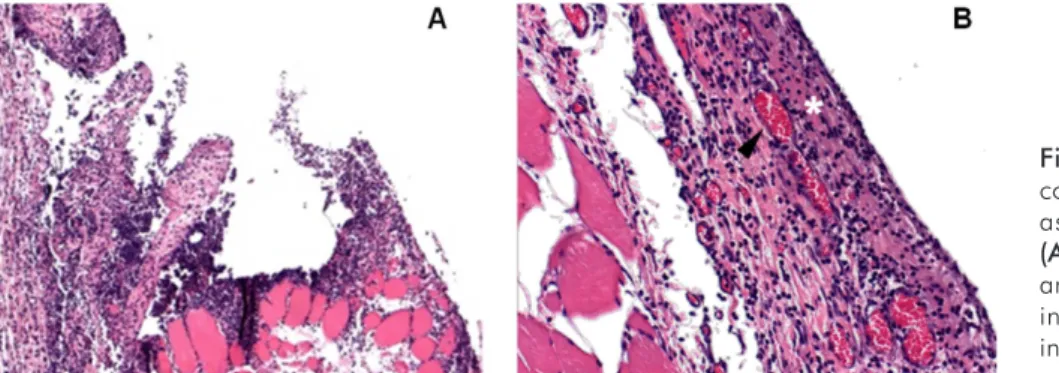
Fig. 2. Reaction of rat subcutaneous connective tissue to 0.9% sterile saline associated with infected dentine.
(A) After 24 hours severe mononuclear
and polymorph nuclear inflammatory infiltrate rich in neutrophils and intense edema areas (H&E, original
magnification X100). (B) After 72 hours
the assessed area presented an acute inflammation rich in neutrophils, mononuclear infiltrate severe edema, hyperemic blood vessels (arrowhead), small areas of tissue necrosis and micro abscesses (white star) (H&E, original magnification X200).
Fig. 3. Reaction of rat subcutaneous connective tissue to 5.25% NaOCl associated with infected dentine. (A) After 24 hours subcutaneous tissue presented large areas of intense inflammatory infiltrate rich in neutrophils, severe edema, some areas of tissue destruction and many dilated and hyperemic blood vessels (H&E, original magnification
X100). (B) After 72 hours the assessed
area presented intense inflammatory infiltrate rich in neutrophils, large areas of edema, larger areas of tissue necrosis than in the 24 hours period, presence of abscesses and a large number of dilated and hyperemic blood vessels (H&E, original magnification X200).
Fig. 4. Reaction of rat subcutaneous connective tissue to 2.0% Chlorhexidine gel associated with infected dentine.
(A) After 24 hours it is possible to observe moderate to intense inflammatory infiltrate (white star), with presence of polymorphonuclear and mononuclear cells, severe edema, some areas of tissue necrosis and small number of hyperemic blood vessels (H&E, original
magnification X100). (B) After 72 hours
Empty tube (control)
As shown in Fig. 1, the histological indings in this group were similar in 24 hours and 72 hours. In the 24-hour period, the subcutaneous tissue showed mild inlammatory iniltrate, moderate edema areas, and presence of blood vessels in small number. In the 72-hour period an edema reduction and chronic inlammatory iniltrate presence were observed.
Saline solution
In the 24-hour period, the histological section showed severe mononuclear and polymorph nuclear inlammatory iniltrate rich in neutrophils, and intense edema areas. In the 72-hour period, the assessed area presented an acute inlammation rich in neutrophils, mononuclear iniltrate, severe edema, hyperemic blood vessels, small areas of tissue necrosis, and micro abscesses (Fig. 2).
Sodium hypochlorite
In the 24-hour period, the subcutaneous tissue presented large areas of intense inlammatory iniltrate rich in neutrophils, severe edema, some areas of tissue destruction, and many dilated and hyperemic blood vessels. In the 72-hour period, the assessed area presented intense inlammatory iniltrate rich in neutrophils, large areas of edema, larger areas of tissue necrosis than in the 24h period, a large number of dilated and hyperemic blood vessels, and presence of abscess (Fig. 3).
Chlorhexidine
In the 24-hour period, moderate to intense inlamma-tory iniltrate was observed, with the presence of poly-morphonuclear and mononuclear cells, severe edema, some areas of tissue necrosis, and a small number of hyperemic blood vessels. In the 72-hour period, the area assessed showed intense inlammatory iniltrate rich in neutrophils and mononuclear cells, intense edema, larger areas of necrosis, low number of hyperemic blood vessels, and no abscess (Fig. 4).
Discussion
During endodontic treatment, debris, auxiliary chemical substances, and bacteria are often pushed out of the apex, coming into direct contact with the periapical vital tissues (9). Infection is considered the most signiicant factor in the lare-up pathogenesis (11,12). The organic tissue associated with microorganisms that spill into the apical region during the canal preparation may cause acute inlammation reaction and severe pain. Enterococcus faecalis was the microorganism selected for this study for several reasons: it is the key species in several persistent endodontic infections; it is more resistant to some auxiliary chemical substances than other microorganisms; it is able to be active in these substances; and it is easy to grow and to identify (13-15).
Good tolerance of subcutaneous connective tissue to polyethylene tubes was shown in the present study. Little
inlammatory response was observed in the control group. This inlammation could be caused by the aggression during tubes surgical implantation (9,16). In the saline solution group intense inlammatory response was observed within the irst 24 hours. This response was more intense after 72 hours. The results suggest that despite the low inlammatory reaction of the saline solution, the association with pathogenic microorganisms can lead to intense inlammatory response, as this solution does not have antimicrobial action (17).
The substances 5.25% NaOCl and 2% chlorhexidine gel presented similar results at 24 hours. However, when compared to the 72-hour period, a more acute response was characterized in the hypochlorite group, with features not found in the chlorhexidine group, such as abscess formation, larger areas of necrosis, and more severe vascular changes. These results suggest the possibility of better antibacterial activity against E. faecalis of chlorhexidine compared to NaOCl. Previous studies reported that 2% chlorhexidine has a more effective antimicrobial action after several days of the chemomechanical preparation (10,18,19). This residual antimicrobial effect of chlorhexidine, called substantivity may be related to less tissue reaction caused after a 72-hour period. Another factor that may be attributed to this difference in inlammatory response between the two groups is the inability of chlorhexidine to dissolve vital and necrotic tissue (11,18,20), which makes it less aggressive than NaOCl. Tissue dissolution may cause severe effects, such as hemolysis, ulceration, inhibition of neutrophil migration, damage to endothelial, damage to ibroblast cells, facial nerve weakness, and necrosis if the solution is extruded during endodontic treatment (21-24).
When compared to the saline solution group, the chlorhexidine group results in the 72-hour period had differences similar to those found in the hypochlorite group. Again, the antimicrobial power and the chlorhexidine substantivity may be related to these results. However, in the 24-hour period, the chlorhexidine group showed greater irritation to tissues, with necrosis areas and inlammatory iniltrate more intense than the saline solution group. These results may be attributed to an initial cytotoxicity of chlorhexidine (21). Gomes-Filho et al. (9) suggest that the chlorhexidine gel may promote a greater inlammatory response in the conjunctive tissue because the tissues would take longer to absorb it than when it’s in its liquid form.
1. Burleson A, Nusstein J, Reader A, et al. The in vivo evaluation of hand/rotary/ultrasound instrumentation in necrotic, human mandibular molars. J Endod 2007;33:782-7. 2. Ricucci D, Siqueira JF Jr. Fate of the tissue in lateral canals and apical ramifications in
response to pathologic conditions and treatment procedures. J Endod 2010;36:1-15. 3. Ferraz CC, Gomes BP, Zaia AA, Teixeira FB, Souza FJ Filho. In vitro assessment of the
antimicrobial action and the mechanical ability of chlorhexidine gel as an endodontic irrigant. J Endod 2001;27:452-5.
4. Card SJ, Sigurdsson A, Orstavik D, Trope M. The effectiveness of increased apical enlargement in reducing intracanal bacteria. J Endod 2002;28:779-83.
5. Christensen CE, McNeal SF, Eleazer P. Effect of lowering the pH of sodium hypochlorite on dissolving tissue in vitro. J Endod 2008;34:449-52.
6. Arias-Moliz MT, Ferrer-Luque CM, Espigares-García M, Baca P. Enterococcus faecalis biofilms eradication by root canal irrigants. J Endod 2009;35:711-4.
7. Gernhardt CR, Eppendorf K, Kozlowski A, Brandt M. Toxicity of concentrated sodium hypochlorite used as an endodontic irrigant. Int Endod J 2004;37:272-80.
8. Dametto FR, Ferraz CC, Gomes BP, Zaia AA, Teixeira FB, de Souza-Filho FJ. In vitro
assessment of the immediate and prolonged antimicrobial action of chlorhexidine gel as an endodontic irrigant against Enterococcus faecalis. O Surg O Med O Pathol O Radiol Endod 2005;99:768-72.
9. Gomes-Filho JE, Aurélio KG, Costa MM, Bernabé PF. Comparison of the biocompatibility of different root canal irrigants. J Appl Oral Sci 2008;16:137-44.
10. Ferraz CC, Gomes NV, Gomes BP, Zaia AA, Teixeira FB, Souza-Filho FJ. Apical extrusion of debris and irrigants using two hand and three engine-driven instrumentation techniques. Int Endod J 2001;34:354-8.
11. Tsesis I, Faivishevsky V, Fuss Z, Zukerman O. Flare-ups after endodontic treatment: a meta-analysis of literature. J Endod 2008;10:1177-81.
12. Alves Vde O. Endodontic flare-ups: a prospective study. Oral Surg Oral Med Oral Pathol Oral Radiol Endod 2010;110:68-72.
13. Haapasalo HK, Sirén EK, Waltimo TM, Ørstavik D, Haapasalo MP. Inactivation of local root canal medicaments by dentine: an in vitro study. Int Endod J 2000;33:126-31. 14. Rôças IN, Siqueira JF Jr. Characterization of the microbiota of root canal-treated teeth with
post-treatment disease. J Clin Microbiol 2012. [Epub ahead of print]
15. Dagna A, Arciola CR, Florindi F, Scribante A, Saino E, Visai L, Poggio C. In vitro evaluation of antimicrobial efficacy of endodontic irrigants. Int J Artif Organs 2011;34:914-9. 16. Semenoff TA, Semenoff Segundo A, de Figueiredo JA. Biocompatibility of different
intracanal medications in rat bucal submucosa tissue. J Appl Oral Sci 2008;16:12-7. 17. Vianna ME, Gomes BP, Berber VB, Zaia AA, Ferraz CC, de Souza-Filho FJ. In vitro evaluation
of the antimicrobial activity of chlorhexidine and sodium hypochlorite. O Surg O Med O Pathol O Rad Endod 2004;97:79-84.
18. Onçağ O, Hoşgör M, Hilmioğlu S, Zekioğlu O, Eronat C, Burhanoğlu D Comparison of antibacterial and toxic effects of various root canal irrigants. I Endod J 2003;36:423-32. 19. Withe RR, Hays GL, Janer LR. Residual antimicrobial activity after canal irrigation with
chlorhexidine. J Endod 1997;23:229-31.
20. Okino LA, Siqueira EL, Santos M, Bombana AC, Figueiredo JA. Dissolution of pulp tissue by aqueous solution of chlorhexidine digluconate and chlorhexidine digluconate gel. Int Endod J 2004;37:38-41.
21. Spångberg LS, Engström B, Langeland K. Biologic effects of dental materials. Toxicity and antimicrobial effect of endodontic antiseptics in vitro. O Surg O Med Oral Pathol 1973;36:856-71.
22. Pelka M, Petschelt A. Permanent mimic musculature and nerve damage caused by sodium hypochlorite: a case report. Oral Surg Oral Med Oral Pathol Oral Radiol Endod 2008;106:80-3. 23. De Sermeño RF, da Silva LA, Herrera H, Herrera H, Silva RA, Leonardo MR. Tissue damage
after sodium hypochlorite extrusion during root canal treatment. Oral Surg Oral Med Oral Pathol Oral Radiol Endod 2009;108:46-9.
24. Kerbl FM, DeVilliers P, Litaker M, Eleazer PD. Physical effects of sodium hypochlorite on bone: an ex vivo study. J Endod 2012;38:357-9.
25. Zehnder M, Kosicki D, Luder H, Sener B, Waltimo T. Tissue-dissolving capacity and antibacterial effect of buffered and unbuffered hypochlorite solutions. O Surg O Med O Pathol O Rad Endod 2002;94:756-62.
Conclusions
In conclusion, the results of the present study indicate that 5.25% NaOCl group showed the highest inlammatory reaction in rat subcutaneous connective tissue and the 2% chlorhexidine group showed the lowest.