MFG-E8 Released by Apoptotic Endothelial Cells Triggers
Anti-Inflammatory Macrophage Reprogramming
Marie-Joe¨lle Brissette1,2, Ste´phanie Lepage1,2, Anne-Sophie Lamonde1,2, Isabelle Sirois1, Jessika Groleau1, Louis-Philippe Laurin1,3, Jean-Franc¸ois Cailhier1,2,3*
1Centre de Recherche du Centre hospitalier de l’Universite´ de Montre´al (CRCHUM), Universite´ de Montre´al, Montreal, Quebec, Canada,2Institut du Cancer de Montre´al, Montreal, Quebec, Canada,3Nephrology Division, Centre hospitalier de l’Universite´ de Montre´al (CHUM), Department of Medicine, Universite´ de Montre´al, Montreal, Quebec, Canada
Abstract
Apoptotic endothelial cells are an important component of the ‘‘response to injury’’ process. Several atherosclerosis risk factors such as hyperglycemia and oxidized low-density lipoproteins, and immune injuries, such as antibodies and complement, induce endothelial cell apoptosis. While endothelial cell apoptosis is known to affect neighboring vascular wall cell biology, its consequences on macrophage reprogramming are ill defined. In this study, we report that apoptosis of human and mouse endothelial cells triggers the release of milk fat globule-epidermal growth factor 8 (MFG-E8) and reprograms macrophages into an anti-inflammatory cells. We demonstrated that MFG-E8 is released by apoptotic endothelial cells in a caspase-3-dependent manner. When macrophages were exposed to conditioned media from serum-starved apoptotic endothelial cells, they adopt a high anti-inflammatory, low pro-inflammatory cytokine/chemokine secreting phenotype that is lost if MFG-E8 is absent from the media. Macrophage treatment with recombinant MFG-E8 recapitulates the effect of conditioned media. Finally, we showed that MFG-E8-mediated reprogramming of macrophages occurs through increased phosphorylation of signal transducer and activator of transcription-3 (STAT-3). Taken together, our study suggests a key role of MFG-E8 release from apoptotic endothelial cells in macrophage reprogramming and demonstrates the importance of the apoptotic microenvironment in anti-inflammatory macrophage responses.
Citation:Brissette M-J, Lepage S, Lamonde A-S, Sirois I, Groleau J, et al. (2012) MFG-E8 Released by Apoptotic Endothelial Cells Triggers Anti-Inflammatory Macrophage Reprogramming. PLoS ONE 7(4): e36368. doi:10.1371/journal.pone.0036368
Editor:Jo¨rg Hermann Fritz, McGill University, Canada
ReceivedFebruary 7, 2012;AcceptedApril 5, 2012;PublishedApril 30, 2012
Copyright:ß2012 Brissette et al. This is an open-access article distributed under the terms of the Creative Commons Attribution License, which permits unrestricted use, distribution, and reproduction in any medium, provided the original author and source are credited.
Funding:This work was funded in part by research grants from the Kidney Foundation of Canada, the Fonds de la recherche en sante´ du Que´bec (FRSQ), the Foundation from the Centre hospitalier de l’Universite´ de Montre´al (CHUM), and from l’Institut du Cancer de Montre´al. JFC was the recipient of a Kidney Foundation of Canada scholarship and is now a FRSQ clinician-scientist scholar. The authors thank the J.-L. Le´vesque Foundation and the CHUM Renal Division for their support. The funders had no role in study design, data collection and analysis, decision to publish, or preparation of the manuscript.
Competing Interests:The authors have declared that no competing interests exist. * E-mail: [email protected]
Introduction
Apoptosis, an ubiquitous form of cell death, occurs during embryogenesis in normal tissues and during inflammation. It has been classically associated with a silent form of cell dismissal [1]. However, recent evidence suggests that apoptotic cells can modulate their microenvironment and neighboring cell biology. Apoptotic cells are known to release ‘‘eat-me’’ and ‘‘find-me’’ signals aimed at coordinating the non-phlogistic recruitment of professional phagocytes to allow swift clearance of apoptotic cells and inhibition of neutrophil influx [2,3,4,5,6,7]. Mounting evidence indicates that the paracrine component of the apoptotic program is not limited to the regulation of leukocyte trafficking, but also prepares the cellular microenvironment for remodeling after apoptotic cell deletion. Apoptotic endothelial cells (EC) are a key component of the ‘‘response to injury’’ process. It is recognized that most atherosclerosis risk factors (such as hypertension [8], hyperglycemia [9] and oxidized low-density lipoproteins [10]) and antibody-complement-mediated immune injuries [11] induce EC apoptosis, which can generate a local microenvironment that will affect cell survival [12,13,14,15], activation [16,17] and differen-tiation [13,18] of neighboring vascular wall cells. Apoptotic cells can modify their local environment through classical and
non-classical secretion of various biological agents [15]. However, the reprogramming consequences of this apoptotic microenvironment on macrophages have not yet been fully characterized.
Macrophages are essential for initiating both inflammation and the repair of injured tissues [19]. In inflammation, they respond destructively to the damage identified; they also promote resolution of inflammation and contribute to tissue repair [20]. Initiation of inflammation, tissue damage and fibrosis are promoted by macrophages through reprogramming induced by the local microenvironment [11,20,21,22,23]. Indeed, the macro-phage phenotype is affected by various signaling cues that vary according to the inflammation phase when they are recruited [20,23]. The injury-inducing and repair-promoting role of macrophages has been well described: macrophage depletion during the fibrosis phase reduces scarring, but depletion during recovery inhibits matrix degradation [24]. Furthermore, there is evidence to suggest that macrophages have a crucial role in conditions where endothelial apoptosis is present [25,26,27,28].
anti-inflammatory (M2). Pro-anti-inflammatory M1 macrophages stem from classical and innate activation. They produce pro-inflamma-tory cytokines such as tumor necrosis factor, monocyte chemo-tactic protein-1 (MCP-1) and macrophage inflammatory protein-2 (MIP-2). Anti-inflammatory, pro-repair M2 macrophages derive from alternative activation or reprogramming induced by the phagocytosis of apoptotic cells. This phenotype is characterized by the production of anti-inflammatory molecules such as interleukin-10 (IL-interleukin-10), transforming growth factor (TGF)-b1, and vascular endothelial growth factor (VEGF) [30,31,32]. However, consider-ing phenotype as described is a simplistic view, as these macrophage phenotypes represent a continuum where transitional states are possible [33].
Our previous work, via a proteomic approach, suggested that milk fat globule-epidermal growth factor 8 (MFG-E8) could be secreted by apoptotic EC [15]. It could be produced by various cell types and, essentially, by activated macrophages. MFG-E8 is crucial for the phagocytosis of apoptotic cells [34] and in macrophage activation [35,36]. We have further investigated MFG-E8 release by apoptotic endothelial cells due to its importance in inflammation and especially in macrophage function.
Here, we show MFG-E8 release by apoptotic EC. We propose that apoptotic EC may be the initial cellular source of MFG-E8, before its production by activated macrophages [34]. Our study suggests that apoptotic cell-conditioned media contribute to macrophage reprogramming into anti-inflammatory, pro-repair macrophages, via MFG-E8 release in a phagocytosis-independent manner.
Results
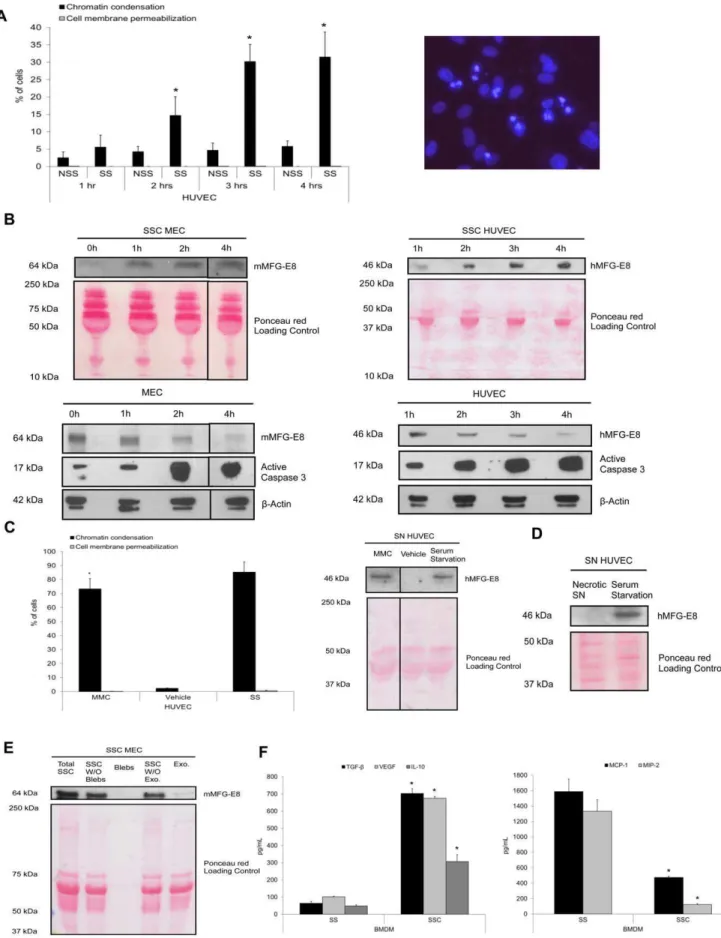
Apoptotic endothelial cell-conditioned medium contains MFG-E8 and could program macrophages
We first assessed whether apoptotic EC could release MFG-E8. Apoptosis was inducedin vitroby serum starvation (SS) for 4 h as reported previously [12,13,15,37,38]. This model is relevant to situations where EC apoptosis is found: chronic transplant vasculopathy and ischemia-reperfusion [11,39]. In our study, serum-starved human umbilical vein endothelial cells (HUVEC) (Figure 1a), evaluated with Hoechst 33342 (HO) and propidium iodide (PI) staining, showed a progressive time-dependent increase of chromatin condensation in the absence of cell membrane permeabilization, indicative of apoptosis, as described elsewhere [12,13,15,37,38]. Necrosis, indicated by cell membrane permea-bilization (inclusion of PI), was not significantly induced by SS. Chromatin condensation was present after 2 h of SS (Figure 1a), concomitantly to caspase-3 activation (Figure 1b, lower panels). Murine EC (MEC) and HUVEC apoptosis was associated with MFG-E8 release in the serum starved-conditioned media (SSC) (Figure 1b, upper panels), whereas the intracellular MFG-E8 content declined in EC, whilst simultaneously exhibiting height-ened expression of active caspase-3 fragments (Figure 1b, lower panels). No significant differences were found in MFG-E8 release between BALB/c and C57BL/6 serum-starved EC (data not shown). We used mitomycin C (MMC) as another pro-apoptotic stimulus [15,37]. MMC treatment of EC augmented the percentage of cells with chromatin condensation and promoted MFG-E8 release (Figure 1c). Furthermore, MFG-E8 was absent from media conditioned by necrotic EC suggesting that this protein is not released passively as a consequence of cell membrane permeabilization (Figure 1d). Since MFG-E8 can be secreted as a soluble or as a small membrane vesicle protein (like exosomes), we then investigated which form serum-starved EC
released MFG-E8. Equal volumes of total unfractionated SSC were centrifuged at 50 000 g to remove apoptotic bodies and apoptotic cells. Supernatants and bleb pellets were collected. Obtained supernatants were then ultracentrifuged at 200 000 g to sediment small membrane vesicles, the resulting supernatants and vesicle pellets were harvested. MFG-E8 levels were detected in total unfractionated SSC, in supernatants from the centrifugation at 50 000 g and in supernatants from the 200 000 g ultracentri-fugation (Figure 1e). The small membrane vesicle fraction contained MFG-E8, but at lower levels than in the supernatants. MFG-E8 was absent in the apoptotic blebs fraction (Figure 1e). These results suggest that MFG-E8 is mostly released by apoptotic EC as a soluble molecule rather than associated to small membrane vesicles.
To evaluate the phenotypic consequences of SSC on macro-phage reprogramming, murine bone marrow-derived macrophag-es (BMDM) were stimulated with SSC for 24 h. Experiments performed with BMDM from C57BL/6 and BALB/c mice showed no differences in cytokine production between strains (data not shown). They produced more TGF-b1, VEGF and IL-10 in response to SSC from apoptotic EC than control serum-starved macrophages (Figure 1f, left panel, values in Table 1). Further-more, the production of pro-inflammatory chemokines MCP-1 and MIP-2 was significantly lower with SSC than with SS exposure (Figure 1f, right panel, values in Table 1). Similar results were obtained with human monocytes-derived macrophages (HMDM). TGF-b1 production by HMDM was increased 2.5 times in response to SSC from apoptotic EC compared to serum starvation alone whereas production of pro-inflammatory cyto-kines IL-8, MCP-1 and IL-6 were 90%, 93% and 97% reduced respectively with SSC exposure compared to SS (p,0.001, n = 3). Therefore, macrophages exposed to conditioned media from serum-starved apoptotic endothelial cells, adopt a high anti-inflammatory, low pro-inflammatory cytokine/chemokine secret-ing phenotype.
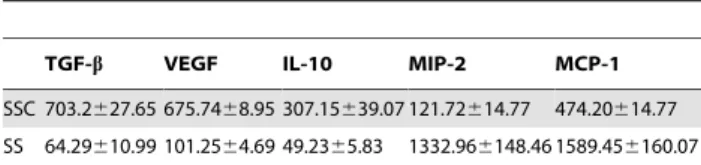
Caspase-3 activation is necessary for MFG-E8 release and subsequent macrophage programming
MFG-E8 production from apoptotic EC programs macrophages into an anti-inflammatory phenotype.
MFG-E8 plays an important and sufficient role in macrophage programming
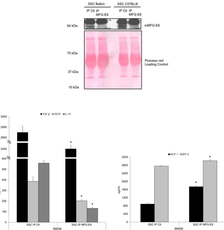
To clearly establish that macrophage programming in response to apoptotic MEC is dependent on MFG-E8 release from apoptotic MEC, we immunoprecipitated MFG-E8 from apoptotic MEC-conditioned media. Figure 3 demonstrates that the absence of MFG-E8 significantly inhibited macrophage reprogramming by apoptotic EC. Indeed, MFG-E8-immunodepleted SSC (Figure 3, upper panel) attenuated the production of the anti-inflammatory cytokines TGF-b1, VEGF and IL-10 (Figure 3, lower left panel, values in Table 3) and increased the production of the pro-inflammatory chemokines MCP-1 and MIP-2 (Figure 3, lower right panel, values in Table 3) compared to control.
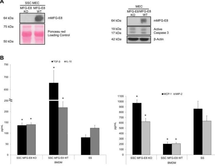
To further highlight the importance of EC-derived MFG-E8 in macrophage programming, we studied MEC derived from MFG-E8 KO mice. Immunoblotting for MFG-MFG-E8 content in superna-tants and cell extracts from serum-starved KO and WT EC confirmed the absence of MFG-E8 in the KO (Figure 4a). In addition, immunoblotting revealed similar active caspase-3 levels in both KO and WT MEC (Figure 4a, right panel). SSC from MFG-E8 KO mice attenuated the production of anti-inflamma-tory cytokines TGF-b1 and IL-10 and increased the pro-inflammatory chemokines, MCP-1 and MIP-2, compared to SSC from MFG-E8 WT mice (Figure 4b, values in Table 4). SSC MFG-E8 KO-induced cytokine/chemokine production by mac-rophages was similar to that observed with the SS control (Figure 4b, values in Table 4). This suggests that the absence of MFG-E8 significantly altered macrophage programming by inducing comparable cytokine/chemokine production as seen in SS-stimulated macrophages.
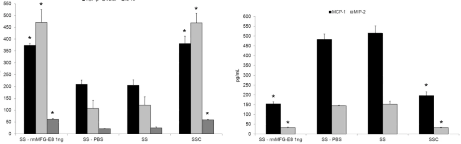
To demonstrate the essential role of MFG-E8 in reprogram-ming macrophage phenotype, we performed studies with recom-binant murine (rm)MFG-E8 at the same concentration as found in SSC, 1 ng/ml (data not reported). Macrophages were stimulated with rmMFG-E8 (1 ng/mL), phosphate buffered saline (PBS) (vehicle for rmMFG-E8), SS or SSC for 48 h and the supernatants were harvested. Stimulation of macrophages with rmMFG-E8 increased production of the anti-inflammatory cytokines TGF-b1, VEGF and IL-10 and decreased the production of pro-inflammatory chemokines MCP-1 and MIP-2 compared to control PBS-treated macrophages (Figure 5, values in Table 5). Chemokine/cytokine production by macrophages treated with rmMFG-E8 was similar to that seen with SSC-treated macro-phages. Similar results were obtained with HMDM. TGF-b1 production by HMDM was increased by 46% in response to rhMFG-E8, whereas production of pro-inflammatory cytokines IL-8 and MCP-1 were reduced by 73% and 70% respectively with rhMFG-E8 treatment compared to control PBS-treated macro-phages (p,0.001, n = 2). Taken together, these results demon-strated the importance of MFG-E8 in the induction of an anti-inflammatory macrophage phenotype.
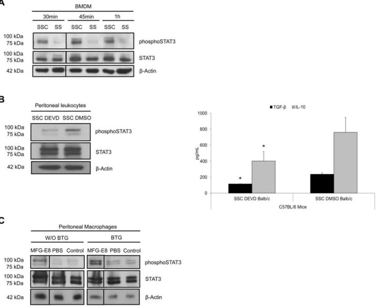
Apoptotic endothelial cell-conditioned media activated the signal transducer and activator of transcription-3 (STAT-3) pathway in macrophages
STAT family of transcription factors are involved in repro-gramming of macrophages. STAT-1 activation is classically associated with the pro-inflammatory cytotoxic macrophage phenotype, whereas STAT-3 activation characterizes the pro-repair macrophages [20]. We, therefore, studied STAT-3 activation in macrophage reprogramming by apoptosis-condi-tioned media. Phosphorylated STAT-3 levels were higher after SSC stimulation compared to SS in BMDM (Figure 6a). We tested an experimental peritonitis model to assess thevivoreprogramming of macrophages by SSC. Pre-conditioning of peritoneal leukocytes with SSC-DMSO for 3 h increased STAT-3 phosphorylation in the cellular extracts 2 h after the induction of Brewer thioglycol-late (BTG) peritonitis in mice compared to SSC-DEVD (Figure 6b, left panel). This, in turn, resulted in increased production of
TGF-b1and IL-10 (Figure 6b, right panel, see Table 6 for values). In additional studies, we determined the role of rmMFG-E8 (0.6mg)
in resident peritoneal macrophage pre-conditioning in STAT-3 activation. The administration of rmMFG-E8 increased the levels of STAT-3 phosphorylation in immunomagnetically-isolated peritoneal macrophages compared to PBS and unmanipulated control, prior to BTG injection. This STAT-3 activation persisted and increased further after BTG-induced peritonitis in isolated peritoneal macrophages compared to both controls (Figure 6c),
(upper panels) and cells (lower panels) were harvested. Immunoblotting of MEC protein extracts showed that MFG-E8 levels decreased over time in parallel with increased active caspase-3 levels (lower left panel). HUVEC also exhibited reduced intracellular MFG-E8 levels over time (lower right panel). MFG-E8 levels increased over time in serum-starved conditioned medium (SSC) from EC (upper panels).b-Actin and Ponceau red staining were loading controls. Representative of 3 experiments.CPercentage of cells with increased chromatin condensation and cell membrane permeabilization (as evaluated with HO and PI staining) in HUVEC exposed to MMC 0.01 mg/mL or vehicle in normal medium and serum starvation (as positive control) for 15 h (left panel), *p,0.0001 versus vehicle,n= 3. Immunoblot for hMFG-E8 in supernatant of EC treated with MMC (right panel). Ponceau red staining is shown as loading control. Representative of 2 experiments.DImmunoblot for hMFG-E8 in supernatants conditioned by necrotic HUVEC (3 freeze-thaw cycles) and serum-starved HUVEC as positive control. Ponceau red staining included as loading control. Representative of 2 experiments.
E Immunoblot for mMFG-E8 from total medium conditioned by apoptotic EC (Total SSC), supernatant after removal of apoptotic blebs by centrifugation at 50 000 g (SSC without (W/O) blebs) and apoptotic blebs (Blebs) purified from total SSC by centrifugation, supernatant obtained from the supernatant after 50 000 g and 200 000 g centrifugation (SSC W/O exo.) and exosome-like nanovesicle fraction pelleted after the 200 000 g centrifugation (Exo.). Proteins from equal initial volumes were precipitated by TCA. Ponceau red staining is shown as loading control of samples. Representative of 2 experiments.F MEC were serum-starved for 4 h, the SSC were harvested, centrifuged to remove apoptotic cells. Murine macrophages were exposed to SSC or serum starvation (SS) for 24 h. ELISA were performed for TGF-b1, VEGF, IL-10, (left panel) MCP-1 and MIP-2
(right panel), *p,0.05, representative ofn= 14, 12, 4, 7 and 9 separate experiments respectively. doi:10.1371/journal.pone.0036368.g001
Table 1.Phenotypic analysis of murine bone-marrow-derived macrophages exposed to SSC vs SS.
TGF-b VEGF IL-10 MIP-2 MCP-1
SSC 703.2627.65 675.7468.95 307.15639.07 121.72614.77 474.20614.77 SS 64.29610.99 101.2564.69 49.2365.83 1332.966148.46 1589.456160.07
Figure 2. Caspase-3 activation is necessary for MFG-E8 release and subsequent macrophage reprogramming.MEC were pre-treated with an irreversible caspase-3 inhibitor, DEVD-FMK (SSC-DEVD, 100mM) to prevent apoptosis, and then serum-starved for 4 h. Control MEC were
pre-treated with vehicle (DMSO) for 2 h, washed and serum-starved for 4 hAMurine MFG-E8 was immunoblotted in SSC and cell extracts. DEVD-FMK-treated murine EC released less MFG-E8 compared to the vehicle (DMSO) (left panel), whereas their intra-cellular content remained higher than DMSO-treated EC (right panel). Ponceau red andb-Actin were loading controls. Representative of 3 experiments.BImmunoblot for murine MFG-E8 of SSC from caspase-3 KO EC compared to EC from WT mice. Representative of 2 experiments.CMurine macrophages produced more TGF-b1, VEGF,
IL-10 (left panel) and less pro-inflammatory chemokines MCP-1 and MIP-2 (right panel) when exposed to media where apoptosis was not inhibited. *p,0.05, representative ofn= 11, 9, 3, 5 and 8 separate experiments respectively.
doi:10.1371/journal.pone.0036368.g002
indicating that MFG-E8 activated the STAT-3 pathway. Alto-gether, these results suggest that STAT-3 activation is present in the observed anti-inflammatory reprogramming of macrophages.
Discussion
Apoptotic cells release various elements that modify their microenvironment. This includes numerous chemokines or chemokine-like compounds, such as lysophosphatidylcholine [3], fractalkine [40] and nucleotides [41]. Existing evidence indicates that apoptotic EC induces resistance to apoptosis and contributes to changes in the phenotype of neighboring vascular wall cells [12,13]. EC apoptosis, through cathepsin L release, degrades perlecan and generates the pro-fibrotic fragment LG3 [37]. Recently, other reports have suggested that the apoptotic milieu could also promote survival [14] and increase the phagocytosis of apoptotic cells by macrophages [42].
Apoptotic cells could activate classical and non-classical secretion pathways involving the exosomal release of proteins. Using proteomic analysis of media conditioned by apoptotic cells, we have previously suggested that MFG-E8 could be secreted, perhaps from the exosomal compartment [15]. However, this observation warranted further evaluation as presented here. Dendritic cells can secrete MFG-E8 through the release of exosomes [43]. Macrophages produce MFG-E8 upon activation whereas resident macrophages do not [34].
Our data highlights caspase-3-dependent MFG-E8 release by apoptotic EC as the primary source of an important protein introducing a novel mechanism of macrophage programming by the microenvironment. This apoptosis-conditioned microenviron-ment induces a phenotypic switch in macrophages, promoting anti-inflammatory and pro-repair macrophages, independent of apoptotic cell phagocytosis-induced reprogramming of macro-phages. It suggests that, in addition to the anti-inflammatory function of apoptotic cells per se through their engulfment by macrophages and subsequent reprograming [30,31], the apoptotic microenvironment could similarly reprogram the neighboring resident and recruited macrophages as a consequence of MFG-E8 secretion. Considering the pro-inflammatory mediators that can be produced by apoptotic EC, such as extra-cellular matrix fragments [37], a local dampening molecule could be essential to attenuate the local inflammatory response due to tissue injury. Apoptotic cells could constitute the initial source of MFG-E8 in the early inflammatory response, before production by activated macrophages [34]. The new role we are postulating for MFG-E8 could be important to maintain local tissue homeostasis by cellular death itself, to promote the pro-repair programming of macro-phages and ensure the early presence of a potent apoptotic cell-opsonizing molecule [34]. This would endow MFG-E8 with another potentially crucial function. Indeed, MFG-E8 is critical for apoptotic cell phagocytosis [34] and in macrophage biology [35,36]. During apoptotic cell engulfment, MFG-E8 opsonizes phosphatidylserine, allowing its recognition by macrophages
through avb3 and avb5 integrins [34]. This process seems to occur in activated macrophages through a granulocyte-macro-phage-colony-stimulating-factor induced mechanism of MFG-E8 expression [35]. In response to bacterial lipopolysaccharides (LPS) stimulation, MFG-E8 has been shown to reduce macrophage activation by modulating integrin signaling [36]. Data from an ischemia-reperfusion injury model indicates that MFG-E8 admin-istration protects mice by promoting apoptotic cell engulfment [44]. MFG-E8 may bind lung collagen to facilitate its clearance in pulmonary fibrosis [45]. The role of MFG-E8 in inflammation extends beyond phagocytosis. Effectively, local release of TGF-b1 and CCL22 through MFG-E8 expression may foster the recruitment and maintenance of FoxP3+ Tregs, promoting allograft tolerance [35]. MFG-E8 can modify macrophage behavior by increasing IL-10 production [36]. Significantly, most of these studies have implicated macrophages as the main source of MFG-E8 production and studied its role as an inhibitor of LPS stimulation. We suggest here that apoptotic cell-conditioned media and MFG-E8 reprogram macrophages through increased STAT-3 phosphorylation. However, the signaling pathways involved in STAT-3 activation by MFG-E8 during SS are still unknown. After LPS treatment, MFG-E8 could induce STAT-3 and suppressor of cytokine signaling-3 (SOCS3) activation to attenuate the pro-inflammatory stimulation of macrophages [46]. STAT-3 has recently been implicated in MFG-E8 stimulation of cancer stem cells produced by tumor-associated macrophages [47].
In clinical situations, such as transplant vasculopathy and highly proliferative cancers, where EC apoptosis is important, the constant presence of apoptotic EC could promote an unregulated repair response by macrophages with the constant production of pro-fibrotic and immunosuppressive mediators. This could lead to tissue fibrosis and/or impaired immune response. Therefore, better understanding of MFG-E8’s role in macrophage repro-gramming and associated signaling pathway activated by the apoptotic cell microenvironment, is central to the development of new therapeutic approaches in transplantation and cancer biology.
Materials and Methods
Cell culture and generation of conditioned media
Human umbilical vein endothelial cells (HUVECs) (Clonetics, San Diego, CA, USA) were cultured as described elsewhere [37] and used at passages 4–5. Serum-free media conditioned by apoptotic or caspase-inhibited EC, were obtained as described (42). Equal EC numbers (2.56104cells/cm2) were preincubated for 2 h in normal medium containing either DMSO (vehicle) or DEVD-FMK (100mM) (R&D Systems, Minneapolis, MN, USA)
for caspase-3 inhibition, washed and the culture medium was changed for serum-free RPMI medium (Wisent, St-Bruno, Que´bec, Canada) and then EC were serum-starved for 4 h to obtain SSC-DMSO and SSC-DEVD respectively. To induce apoptosis in another way, EC were treated with MMC (0.01 mg/ ml, Sigma, Oakville, Ontario, Canada) for 15 h. Conditioned
Table 2.Caspase-3 dependent production of MFG-E8 in EC reprograms bone-marrow-derived macrophages.
TGF-b VEGF IL-10 MIP-2 MCP-1
SSC-DEVD 151.2864.17 111.4967.89 61.6462.69 417.33642.48 966.94667.87 SSC-DMSO 499.45613.11 160.0668.60 203.25624.90 61.6165.86 431.43637.06
Data are presented as value mean6SD in pg/mL; SSC: apoptotic serum-starved conditioned medium; TGF: transforming growth factor; VEGF: vascular endothelial growth factor; IL: interleukin; MIP: macrophage inflammatory protein; MCP: monocyte chemotactic protein.
Figure 3. MFG-E8 immunoprecipitation from SSC alters macrophage reprogramming.Serum-Starved Conditioned medium (SSC) from MEC were treated with an anti-MFG-E8 antibody (or isotype control) to deplete the MFG-E8 content. Immunoblotting of MFG-E8 protein in SSC is shown for MEC (top panel). BMDM treated with SSC depleted of MFG-E8 produced less TGF-b1, VEGF, IL-10 (lower left panel), and more MCP-1 and
MIP-2 (lower right panel). *p,0.05, representative ofn= 2, 2, 3, 5 and 3 separate experiments respectively. doi:10.1371/journal.pone.0036368.g003
Table 3.Immunoprecipitation of MFG-E8 in SSC reduces the anti-inflammatory reprogramming of macrophages by SSC.
TGF-b VEGF IL-10 MIP-2 MCP-1
SSC IP-MFGE8 2401.33634.79 202.78613.12 131.91610.40 3021.53638.68 1743.41641.15 SSC IP-ctl 2849.18660.47 385.23643.70 558.26625.46 2757.97623.73 887.54638.36
Data are presented as value mean6SD in pg/mL; SSC: apoptotic serum-starved conditioned medium; TGF: transforming growth factor; VEGF: vascular endothelial growth factor; IL: interleukin; MIP: macrophage inflammatory protein; MCP: monocyte chemotactic protein.
doi:10.1371/journal.pone.0036368.t003
media were collected and stored at220uC. Conditioned media were centrifuged at 50 kG to eliminate apoptotic bodies. Necrotic conditioned media were produced by submitting EC to 3 freeze-thaw cycles. Sequential centrifugation protocol was done with 30 mL of SSC with proteases inhibitors (PMSF, Pepstatine A 2 mM, Leupeptine 2 mM), 10 mL of total SSC unfractioned was
kept. 20 mL of this total SSC was then centrifuged at 50 000 g at 4uC for 15 min, blebs pellets was then resuspend in 20 mL of SS supplemented of proteases inhibitors and 10 mL were kept aside. The residual 10 mL were then ultracentrifuged at 200 000 g at 4uC for 18 h. Supernatants were kept and small membrane vesicle pellets were resuspended in 10 mL of SS supplemented with proteases inhibitors.
Murine endothelial cell (MEC) isolation
Thoracic aortae were removed surgically from anaesthetized mice, the endothelial side was placed on Matrigel (BD Bioscience, Mississauga, Ontario, Canada) for approximately a week, or until endothelial spreading was sufficient, in Dulbecco’s modified Eagle’s medium (DMEM) low glucose (Gibco, Burlington, Ontario, Canada) supplemented with fetal bovine serum (FBS)(Wisent, St-Bruno, Que´bec, Canada), calf serum (Gibco, Burlington, Ontario, Canada), endothelial cell growth supplement (VWR, Radnor, Pennsylvania, USA), heparine (Sigma, Oakville, Ontario, Canada), fugizon (Gibco, Burlington, Ontario, Canada) and penicillin/streptomycin (Wisent, St-Bruno, Que´bec, Canada).
Figure 4. SSC from MFG-E8 KO mice do not reprogram macrophages into anti-inflammatory macrophages. AMFG-E8 KO or WT MEC were serum-starved for 4 h. Supernatants and cell extracts were immunoblotted for mMFG-E8 confirming KO status. Caspase-3 activation was similar between the 2 groups. B Murine macrophages were stimulated with Serum-Starved Conditioned medium (SSC) from MFG-E8 KO or WT EC. Supernatant were analyzed by ELISA. The results indicate that MFG-E8 in SSC is necessary to induce an anti-inflammatory macrophage phenotype. *p,0.05, representative ofn= 5 separate experiments.
doi:10.1371/journal.pone.0036368.g004
Table 4.SSC from MFG-E8 KO mice reduces the anti-inflammatory reprogramming of macrophages.
TGF-b IL-10 MIP-2 MCP-1
SSC MFG-E8 KO 136.56611.06 140.1168.61 622.70662.00 972.82658.59 SSC MFG-E8 WT 676.93649.23 218.72627.32 209.42619.16 203.65633.51 SS 80.80612.49 124.01612.90 635.39697.45 863.236145.10
When the desired confluence was reached, MEC were harvested after dispase treatment and seeded on plastic flasks(BD Bioscience, Mississauga, Ontario, Canada). Cultured MEC were expanded between passages 4 and 6 and tested experimentally.
Isolation of blood monocyte-derived macrophages and preparation of bone marrow-derived macrophages
Peripheral monocytes were isolated from healthy donors by Ficoll gradient (Wisent, St-Bruno) followed by CD14+ immuno-magnetic selection (Stem Cell, Vancouver). Both CD14++/ CD162 and CD14low/CD16+ monocyte populations were collected. HMDM were matured in Iscove DMEM (Gibco, Burlington, Ontario, Canada) with penicillin/streptomycin (Wi-sent, St-Bruno, Que´bec, Canada), glutamine (Wi(Wi-sent, St-Bruno, Que´bec, Canada) and 10% decomplemented autologous human serum for 5–7 days before being exposed to experimental media for 24 h. Informed written consent was obtained from healthy donors according to the hospital ethics committee (comite´ d’e´thique de la recherche du CHUM). The data were not shown. Bone marrow-derived macrophages (BMDM) were prepared from C57BL/6 mice. Bone marrow was isolated from femurs by standard sterile techniques and matured for 7 days in culture plastic in Dulbecco’s modified Eagle’s medium (DMEM) (Wisent, St-Bruno, Que´bec, Canada) with 10% FBS (Wisent, St-Bruno, Que´bec, Canada), penicillin/streptomycin (100mg/ml) (Wisent,
St-Bruno, Que´bec, Canada), and 20% L929 cell-conditioned medium as a source of macrophage-colony stimulating factor. BMDM were more than 96% positive for the macrophage marker F4/80 by flow cytometry.
Immunoblotting and reagents
In all experiments, equal volumes of all conditioned media were concentrated by centrifugation in a 10-kD vivaspin concentrator, according to the manufacturer’s specifications (Sigma, Oakville, Ontario, Canada) as described previously [37] or by a trichlor-oacetic acid (TCA) precipitation protocol of supernatants 9:1, washed with cold acetone and solubilization in sample buffer [15]. A fixed volume in all conditions was loaded onto gel. Proteins were separated by SDS-PAGE electrophoresis and transferred to nitrocellulose membranes. Western blotting was performed [37] and the membranes probed with human MFG-E8 and anti-mouse MFG-E8 antibodies (R&D Systems, Minneapolis, MN, USA and Santa Cruz Biotechnology, Santa Cruz, CA, USA, respectively). Ponceau red staining of membranes served as protein-loading control. Proteins were extracted from cell pellets with protease or phosphatase inhibitor cocktail separated by electrophoresis, transferred to nitrocellulose or PVDF membranes, and probed. The membranes were probed with anti-active caspase-3 (Cell Signaling Technology, Pickering, Ontario, Cana-da), anti-b actin (Abcam, Cambridge, MA, USA), anti-phospho STAT3 and STAT3 total antibodies (Cell Signaling Technology, Pickering, Ontario, Canada).
Fluorescence microscopy for quantification of cells with chromatin condensation and cell membrane
permeabilization
For fluorescence microscopy, unfixed/unpermeabilized adher-ent EC were stained with Hoechst 33342 (29 -(4-ethoxyphenyl)-5-(4-methyl-1-piperazinyl)-2.59-bi-1H-benzimidazole) (HO) and PI. They were grown to confluence in 24-well culture plates (BD
Figure 5. Recombinant murine MFG-E8 recapitulates SSC-induced macrophage reprogramming.Murine macrophages were stimulated with rmMFG-E8 (1 ng/mL) resuspended in RPMI (SS), vehicle (PBS), SS or SSC for 48 h and supernatant were harvested. rmMFG-E8 induced an anti-inflammatory macrophage phenotype with an increased production of TGF-b1, VEGF and IL-10 and reduced MCP-1 and MIP-2 compared to the
vehicle control (PBS resuspended in SS). *p,0.05 vs respective controls, mean6SD , representative ofn= 3 separate experiments. doi:10.1371/journal.pone.0036368.g005
Table 5.rmMFG-E8 reproduces the anti-inflammatory macrophage phenotype.
TGF-b VEGF IL-10 MIP-2 MCP-1
SS+rmMFG-E8 373.16610.02 470.82653.29 61.1463.69 32.6563.02 154.69610.61 SS+PBS 209.27618.04 107.35634.55 21.9460.70 145.1362.75 483.50627.13
Data are presented as value mean6SD in pg/mL; TGF: transforming growth factor; VEGF: vascular endothelial growth factor; IL: interleukin; MIP: macrophage inflammatory protein; MCP: monocyte chemotactic protein.
doi:10.1371/journal.pone.0036368.t005
Bioscience, Mississauga, Ontario, Canada). HO (1mg/ml) was added to a final concentration of 5mg/ml immediately before fluorescence microscopy analysis (excitation filter I = 360– 425 nm). Apoptotic cells show increased HO fluorescence in the absence of PI positivity. Secondary and primary necrotic cells present PI positivity.
Enzyme-linked immunosorbent assay (ELISA)
Human IL-6, -8, active TGF-b1, MCP-1 protein levels, as well as murine IL-10, MCP-1, macrophage inflammatory protein-2 (MIP-2) and vascular endothelial growth factor (VEGF) were measured by ELISA) according to the supplier’s protocol (BD
Figure 6. STAT-3 activation in macrophage reprogramming by apoptosis-conditioned media. ABMDM were stimulated with Serum-Starved Conditioned medium (SSC) or serum starvation (SS) for 30 minutes to 1 h. Total protein extracts were harvested and immunoblotted for phospho-STAT-3, STAT-3 and b-actin as loading control. Phosphorylated STAT-3 levels are higher after SSC stimulation compared to SS. Representative of 4 experiments.B C57BL/6 mice were pre-conditioned with SSC-DEVD or SSC-DMSO intraperitoneally for 3 h. Experimental peritonitis was induced with thioglycollate for 2 h and followed by peritoneal lavage to harvest peritoneal cellular exudates and supernatants. Protein extracts from the cellular exudates showed increased STAT-3 phosphorylation in mice pre-conditioned with SSC-DMSO compared to SSC-DEVD (B, left panel). ELISA of the supernatants revealed that SSC-DMSO pre-treatment increased TGF-b1and IL-10 production compared to SSC-DEVD (B, right
panel). *p,0.05, representative ofn= 3 separate experiments.CMFG-E8 conditioning increased STAT-3 phosphorylation compared to PBS pre-conditioned or control immunomagnetically-isolated peritoneal macrophages prior to Brewer thioglycollate (BTG) administration (W/O BTG). STAT-3 activation persisted and increased further 2 h following the induction of BTG peritonitis (BTG) in pre-conditioned macrophages. Total STAT-3 levels are depicted.b-Actin were loading controls. Representative of 2 experiments.
doi:10.1371/journal.pone.0036368.g006
Table 6.Apoptosis-conditioned mediain vivo pre-conditioning reprogram peritoneal macrophages.
TGF-b IL-10
SSC-DEVD 116.5461.21 401.886108.47 SSC-DMSO 237.55617.64 760.506184.76
Bioscience, Mississauga, Ontario, Canada and R&D Systems, Minneapolis, MN, USA).
MFG-E8 immunoprecipitation from MEC and HUVEC SSC
SSC were incubated with specific antibodies against MFG-E8 or control antibody for 6 h. Protein A/G linked beads were incubated overnight at 4uC under agitation. SSC were then centrifuged and immunoblotted for MFG-E8 (Santa Cruz Biotechnology, Santa Cruz, CA, USA).
Recombinant murine MFG-E8
BMDM matured for 7 days were stimulated with rmMFG-E8 (R&D Systems, Minneapolis, MN, USA) 1 ng/mL in serum-free media (Wisent, St-Bruno, Que´bec, Canada) for 48 h. PBS added to serum-free media (Wisent, St-Bruno, Que´bec, Canada) served as negative control. Cytokines/chemokines from the supernatants were evaluated by ELISA.
Experimental animals and induction of experimental peritonitis
C57BL/6 and BALB/c mice were bought from Charles River (Canada). We acquired the MFG-E8 KO mice on the C57BL/6 background were generously donated to us by Professor S. Nagata. Caspase-3 KO mice were obtained from Jackson Laboratory. Mice were housed in CRCHUM animal facilities. They were injected intraperitoneally (IP) with 0.5 mL of conditioned media or rmMFG-E8 (0.6mg) for 3 h and then injected with 3% Brewer’s
thioglycollate (BTG) (Difco) and underwent peritoneal lavage with 5 mL of phosphate buffered saline (PBS) (Wisent, St-Bruno, Que´bec, Canada) at 2 h thereafter. Peritoneal lavage fluid was centrifuged and stored at 280uC until analyzed for cytokine/ chemokine production by ELISA. Macrophages were immuno-magnetically-isolated with magnetic beads (Miltenyi Biotech, Auburn, CA, USA) after the peritoneal lavages and kept for immunoblotting. Performed animal experiments were approved by our institutional Animal Care Committee (Comite´ institution-nel de protection des animaux, CRCHUM).
Statistical analysis
The results are expressed as mean 6 SD were analyzed by Student’sT-test (with Bonferroni correction when appropriate) or ANOVA, as appropriate.p,0.05 was deemed to be significant for all tests.
Acknowledgments
We would like to thank Mrs. M. Soulez for her help with the EC culture and Mr. O. Da Silva for editing this manuscript.
Author Contributions
Conceived and designed the experiments: MJB SL JG JFC. Performed the experiments: MJB SL ASL JG IS JFC. Analyzed the data: MJB SL JG IS JFC. Contributed reagents/materials/analysis tools: IS. Wrote the paper: MJB LPL JG IS JFC.
References
1. Savill J, Dransfield I, Gregory C, Haslett C (2002) A blast from the past: clearance of apoptotic cells regulates immune responses. Nat Rev Immunol 2: 965–975.
2. Schaub FJ, Han DK, Liles WC, Adams LD, Coats SA, et al. (2000) Fas/FADD-mediated activation of a specific program of inflammatory gene expression in vascular smooth muscle cells. Nat Med 6: 790–796.
3. Lauber K, Bohn E, Krober SM, Xiao YJ, Blumenthal SG, et al. (2003) Apoptotic cells induce migration of phagocytes via caspase-3-mediated release of a lipid attraction signal. Cell 113: 717–730.
4. Bournazou I, Pound JD, Duffin R, Bournazos S, Melville LA, et al. (2009) Apoptotic human cells inhibit migration of granulocytes via release of lactoferrin. J Clin Invest 119: 20–32.
5. Ravichandran KS (2010) Find-me and eat-me signals in apoptotic cell clearance: progress and conundrums. The Journal of experimental medicine 207: 1807–1817.
6. Ravichandran KS (2011) Beginnings of a good apoptotic meal: the find-me and eat-me signaling pathways. Immunity 35: 445–455.
7. Chekeni FB, Ravichandran KS (2011) The role of nucleotides in apoptotic cell clearance: implications for disease pathogenesis. Journal of molecular medicine 89: 13–22.
8. Hamet P, deBlois D (2001) Endothelial and myocyte apoptosis–role of angiotensin II. Can J Cardiol 17 Suppl A: 26A–28A.
9. Baumgartner-Parzer SM, Wagner L, Pettermann M, Grillari J, Gessl A, et al. (1995) High-glucose–triggered apoptosis in cultured endothelial cells. Diabetes 44: 1323–1327.
10. Sata M, Walsh K (1998) Oxidized LDL activates fas-mediated endothelial cell apoptosis. J Clin Invest 102: 1682–1689.
11. Cailhier JF, Laplante P, Hebert MJ (2006) Endothelial apoptosis and chronic transplant vasculopathy: recent results, novel mechanisms. Am J Transplant 6: 247–253.
12. Raymond M, De´sormeaux A, Laplante P, Vigneault N, Filep J, et al. (2004) Apoptosis of endothelial cells triggers a caspase-dependent anti-apoptotic paracrine loop active on vascular smooth muscle cells. FASEB J 18: 705–707. doi:710.1096/fj.1003-0573fje.
13. Laplante P, Raymond MA, Gagnon G, Vigneault N, Sasseville AMJ, et al. (2005) Novel fibrogenic pathways are activated in response to endothelial apoptosis: implications in the pathophysiology of systemic sclerosis. J Immunol 174: 5740–5749.
14. Weigert A, Johann AM, von Knethen A, Schmidt H, Geisslinger G, et al. (2006) Apoptotic cells promote macrophage survival by releasing the anti-apoptotic mediator sphingosine-1-phosphate. Blood 108(5): 1635–1642.
15. Sirois I, Raymond MA, Brassard N, Cailhier JF, Fedjaev M, et al. (2011) Caspase-3-dependent export of TCTP: a novel pathway for antiapoptotic intercellular communication. Cell Death Differ 18: 549–562.
16. Chen W, Frank ME, Jin W, Wahl SM (2001) TGF-beta released by apoptotic T cells contributes to an immunosuppressive milieu. Immunity 14: 715–725. 17. Lepage S, Cailhier JF (2009) Chronic transplant vasculopathy
microenviron-ment present in the renal allograft reprograms macrophage phenotype. Transplant Proc 41: 3311–3313.
18. Rovere P, Vallinoto C, Bondanza A, Crosti MC, Rescigno M, et al. (1998) Bystander apoptosis triggers dendritic cell maturation and antigen-presenting function. J Immunol 161: 4467–4471.
19. Sean Eardley K, Cockwell P (2005) Macrophages and progressive tubulointer-stitial disease. Kidney Int 68: 437–455.
20. Kluth DC, Erwig LP, Rees AJ (2004) Multiple facets of macrophages in renal injury. Kidney Int 66: 542–557.
21. Cailhier JF, Sawatzky DA, Kipari T, Houlberg K, Walbaum D, et al. (2006) Resident pleural macrophages are key orchestrators of neutrophil recruitment in pleural inflammation. Am J Respir Crit Care Med 173: 540–547.
22. Cailhier JF, Partolina M, Vuthoori S, Wu S, Ko K, et al. (2005) Conditional macrophage ablation demonstrates that resident macrophages initiate acute peritoneal inflammation. J Immunol 174: 2336–2342.
23. Lin SL, Castano AP, Nowlin BT, Lupher ML, Jr., Duffield JS (2009) Bone marrow Ly6Chigh monocytes are selectively recruited to injured kidney and differentiate into functionally distinct populations. J Immunol 183: 6733–6743. 24. Duffield JS, Forbes SJ, Constandinou CM, Clay S, Partolina M, et al. (2005) Selective depletion of macrophages reveals distinct, opposing roles during liver injury and repair. J Clin Invest 115: 56–65.
25. Martinet W, Verheye S, De Meyer GR (2007) Selective depletion of macrophages in atherosclerotic plaques via macrophage-specific initiation of cell death. Trends in cardiovascular medicine 17: 69–75.
26. De Meyer I, Martinet W, De Meyer GR (2012) Therapeutic strategies to deplete macrophages in atherosclerotic plaques. British journal of clinical pharmacology doi: 10.1111/j.1365-2125.2012.04211.x.
27. Libby P (2002) Inflammation in atherosclerosis. Nature 420: 868–874. 28. Kitchens WH, Chase CM, Uehara S, Cornell LD, Colvin RB, et al. (2007)
Macrophage depletion suppresses cardiac allograft vasculopathy in mice. Am J Transplant 7: 2675–2682.
29. Stout RD, Jiang C, Matta B, Tietzel I, Watkins SK, et al. (2005) Macrophages sequentially change their functional phenotype in response to changes in microenvironmental influences. J Immunol 175: 342–349.
30. Voll RE, Herrmann M, Roth EA, Stach C, Kalden JR, et al. (1997) Immunosuppressive effects of apoptotic cells [letter]. Nature 390: 350–351. 31. Fadok VA, Bratton DL, Konowal A, Freed PW, Westcott JY, et al. (1998)
Macrophages that have ingested apoptotic cells in vitro inhibit proinflammatory cytokine production through autocrine/paracrine mechanisms involving TGF-beta, PGE2, and PAF. J Clin Invest 101: 890–898.
32. Golpon HA, Fadok VA, Taraseviciene-Stewart L, Scerbavicius R, Sauer C, et al. (2004) Life after corpse engulfment: phagocytosis of apoptotic cells leads to VEGF secretion and cell growth. Faseb J 18: 1716–1718.
33. Mosser DM, Edwards JP (2008) Exploring the full spectrum of macrophage activation. Nat Rev Immunol 8: 958–969.
34. Hanayama R, Tanaka M, Miwa K, Shinohara A, Iwamatsu A, et al. (2002) Identification of a factor that links apoptotic cells to phagocytes. Nature 417: 182–187.
35. Jinushi M, Nakazaki Y, Dougan M, Carrasco DR, Mihm M, et al. (2007) MFG-E8-mediated uptake of apoptotic cells by APCs links the pro- and antiinflammatory activities of GM-CSF. J Clin Invest 117: 1902–1913. 36. Aziz MM, Ishihara S, Mishima Y, Oshima N, Moriyama I, et al. (2009)
MFG-E8 attenuates intestinal inflammation in murine experimental colitis by modulating osteopontin-dependent alphavbeta3 integrin signaling. J Immunol 182: 7222–7232.
37. Cailhier JF, Sirois I, Laplante P, Lepage S, Raymond MA, et al. (2008) Caspase-3 activation triggers extra-cellular cathepsin L release and endorepellin proteolysis. J Biol Chem 283(40): 27220–27229.
38. Laplante P, Sirois I, Raymond MA, Kokta V, Beliveau A, et al. (2010) Caspase-3-mediated secretion of connective tissue growth factor by apoptotic endothelial cells promotes fibrosis. Cell death and differentiation 17: 291–303.
39. Cornell LD, Smith RN, Colvin RB (2008) Kidney transplantation: mechanisms of rejection and acceptance. Annual review of pathology 3: 189–220. 40. Truman LA, Ford CA, Pasikowska M, Pound JD, Wilkinson SJ, et al. (2008)
CX3CL1/fractalkine is released from apoptotic lymphocytes to stimulate macrophage chemotaxis. Blood 112(13): 5026–5036.
41. Elliott MR, Chekeni FB, Trampont PC, Lazarowski ER, Kadl A, et al. (2009) Nucleotides released by apoptotic cells act as a find-me signal to promote phagocytic clearance. Nature 461: 282–286.
42. Scannell M, Flanagan MB, deStefani A, Wynne KJ, Cagney G, et al. (2007) Annexin-1 and peptide derivatives are released by apoptotic cells and stimulate phagocytosis of apoptotic neutrophils by macrophages. J Immunol 178: 4595–4605.
43. Thery C, Regnault A, Garin J, Wolfers J, Zitvogel L, et al. (1999) Molecular characterization of dendritic cell-derived exosomes. Selective accumulation of the heat shock protein hsc73. J Cell Biol 147: 599–610.
44. Cui T, Miksa M, Wu R, Komura H, Zhou M, et al. (2010) Milk fat globule epidermal growth factor 8 attenuates acute lung injury in mice after intestinal ischemia and reperfusion. Am J Respir Crit Care Med 181: 238–246. 45. Atabai K, Jame S, Azhar N, Kuo A, Lam M, et al. (2009) Mfge8 diminishes the
severity of tissue fibrosis in mice by binding and targeting collagen for uptake by macrophages. J Clin Invest 119: 3713–3722.
46. Aziz M, Jacob A, Matsuda A, Wu R, Zhou M, et al. (2011) Pre-treatment of recombinant mouse MFG-E8 downregulates LPS-induced TNF-alpha produc-tion in macrophages via STAT3-mediated SOCS3 activaproduc-tion. PLoS One 6: e27685.