Received: 6 November, 2012. Accepted: 8 July, 2013
ABSTRACT
Here, we describe the development of sporangial and gametangial conceptacles for Amphiroa beauvoisii and A. van-bosseae; sporangial conceptacles only for A. misakiensis; and gametangial conceptacles only for A. cryptarthrodia and
A. rigida. The descriptions are based on the observation of histological preparations obtained from 112 specimens collected from the Gulf of California, in Mexico, and the Azores archipelago of Portugal. Information on the deve-lopment of the sporangial conceptacle pore and conceptacle senescence is here described and illustrated for the first time. Four development patterns were observed: two for sporangial conceptacles; one for spermatangial conceptacles; and one for carposporangial conceptacles. The phases of development of the sporangial conceptacle were found to be useful in delimiting species within the genus. Based on the sporangium location on the cavity floor and the pore canal anatomy, the species A.beauvoisii, A. misakiensis and A. vanbosseae can be distinguished from each other.
Key words: carposporangial filament origin, sporangium location, sporangial pore canal anatomy, senescence, taxonomy
Development of conceptacles
in Amphiroa (Corallinales, Rhodophyta)
Edgar Francisco Rosas-Alquicira1,6, Rafael Riosmena-Rodríguez2, Gustavo Hernández-Carmona3 and Ana Isabel Neto4,5
1 Universidad del Mar, Oaxaca, Mexico
2 Universidad Autónoma de Baja California Sur, Programa de Investigación en Botánica Marina, Departamento de Biología Marina, Mexico 3 Centro Interdisciplinario de Ciencias Marinas, Instituto Politécnico Nacional, Mexico
4 Universidade dos Azores, Centro de Investigação de Recursos Naturais, Departamento de Biologia, Azores, Portugal 5 Centro Interdisciplinar de Investigação Marinha e Ambiental, Laboratório de Investigação Aquática Insular, Portugal 6 Author for correspondence: [email protected]
Introduction
Information concerning the reproduction of articulated coralline algae has been provided by several authors, thus expanding knowledge of the Corallinaceae family. Never-theless, the reproductive structures are still poorly described for many genera of articulated coralline algae. In general, all members of the Corallinaceae produce sporangia, carpos-porangia and spermatangia within uniporate conceptacles, whose development undergoes a number of stages (Johan-sen 1968). Although the transformation from vegetative to fertile tissue is not fully understood in the Corallinaceae, it is known to involve the local destruction of cells, which, when combined with continued accelerated growth of surrounding tissue, results in the depression of peripheral cells (Johansen 1972), a process that differs among genera (Johansen 1968). Historically, the morphology and anatomy of conceptacles, together with other features, have been used to distinguish genera within the geniculate Corallinaceae.
A description of the genus Amphiroa, encompassing a summary of the diagnostic characteristics, is provided by Womersley (1996) and Harvey et al. (2009). Johansen (1972), in a revision of Corallinaceae conceptacles, described a general pattern for the development of tetrasporangial
conceptacles in Amphiroa in which the roof is formed by intersporangial growth and the chamber is opened via a single pore. Additional information on the development of sporangial and gametangial/carposporangial conceptacles was published by Johansen (1968), Murata & Masaki (1978) and Srimanobhas & Masaki (1987). Several other taxonomic studies have included general information on the develop-mental phases of Amphiroa (Ganesan 1968; Norris & Johan-sen 1981; Riosmena-Rodríguez & Siqueiros-Beltrones 1991, 1996; Womersley 1996; Moura & Guimarães 2002; Harvey et al. 2009). Aspects of the reproduction of a few species within
Harvey et al. 2009) and another in which there are no block--shaped cells (Johansen 1968). Two patterns have also been described for the origin of the carposporangial filaments in the fusion cell: from the margins (Ganesan 1968; Riosmena--Rodríguez & Siqueiros-Beltrones 1996) or from the entire surface (Ganesan 1971). Despite all of these data, there is as yet no agreement across studies. For example, the origin of the carposporangial filaments in the fusion cell A. rigida is reported to be from the margins (Riosmena-Rodríguez & Siqueiros-Beltrones 1996) and from the entire surface (Sega-wa 1940b). This indicates the need for further studies on the development of the reproductive structures within Amphiroa.
In this study, we describe the patterns of development for sporangial and gametangial conceptacles of Amphiroa beauvoisii J.V. Lamouroux and A. vanbosseae Me. Lemoine; sporangial conceptacles of A. misakiensis; and gametangial conceptacles for A. cryptarthrodia Zanardini and A. rigida. Furthermore, comparisons are made among species and with published information and the taxonomic value of con-ceptacle development for species segregation is discussed.
Material and methods
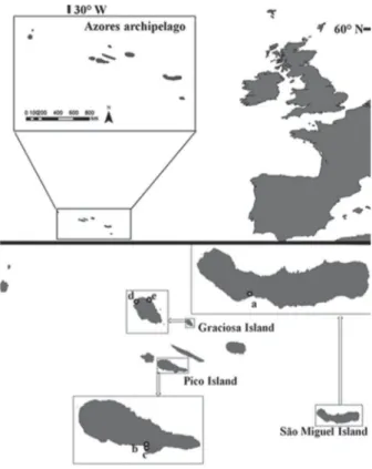
A total of 112 specimens were studied, including fresh specimens and historical collections: sporangial/game-tangial plants of Amphiroa beauvoisii J.V. Lamouroux (40 specimens) and A. vanbosseae Me. Lemoine (44 specimens); sporangial plants of A. misakiensis Yendo (10 specimens); and gametangial plants of A. cryptarthrodia Zanardini (10 specimens) and A. rigida J.V. Lamouroux (eight specimens). Fresh specimens were collected intertidally and subtidally, at a depth of 30 m, at 18 locations; 10 in the Gulf of California, in Mexico, three in the northeastern Mexican Pacific (Fig. 1) and five in the Azores archipelago of Portugal (Fig. 2). Collecting sites and voucher specimens are listed in Tab. 1. Algae were removed from the substrate with a hammer and chisel, brought to the laboratory and fixed in 4% formalin (v/v) in seawater. Specimens were stored in the Phycological Herbarium of the Autonomous University of Baja California Sur, in Baja California Sur, Mexico (code, FBCS) and in the Ruy Telles Palhinha Herbarium of the University of the Azores, in Ponta Delgada, Portugal (code, AZB). Histori cal collections encompass dried specimens of A. cryptarthrodia
and A. beauvoisii from the Dutch oceanographic CANCAP V expedition housed at the National Herbarium Nederland, Leiden University branch, in Leiden, the Netherlands (code, L). Herbarium abbreviations are as in Holmgren & Holmgren (1998).
Permanent microslides from longitudinal sections in the sagittal region of bi-tetrasporangial and gametangial conceptacles were obtained following the histological tech-nique of Riosmena-Rodríguez et al. (1999). The number of evaluated conceptacles was 130 for Amphiroa beauvoisii, 80 for A. vanbosseae, 50 for A. misakiensis, 30 for A. cryptar-throdia and 30 for A. rigida. Representative developmental
Figure 1. Location of the collecting sites: Gulf of California - (a) Puerto Peñasco, (b) Kino Nuevo, (c) Requesón, (d) San Juan de la Costa, (e) La Ballena, (f) I. Espíritu Santo, (g) Balandra, (h) Calerita, (i) El Sargento, (j) Isla Cerralvo; Nor-theastern Mexican Pacific - (k) Cerritos, (l) Isla Margarita, (m) Bahía Asunción.
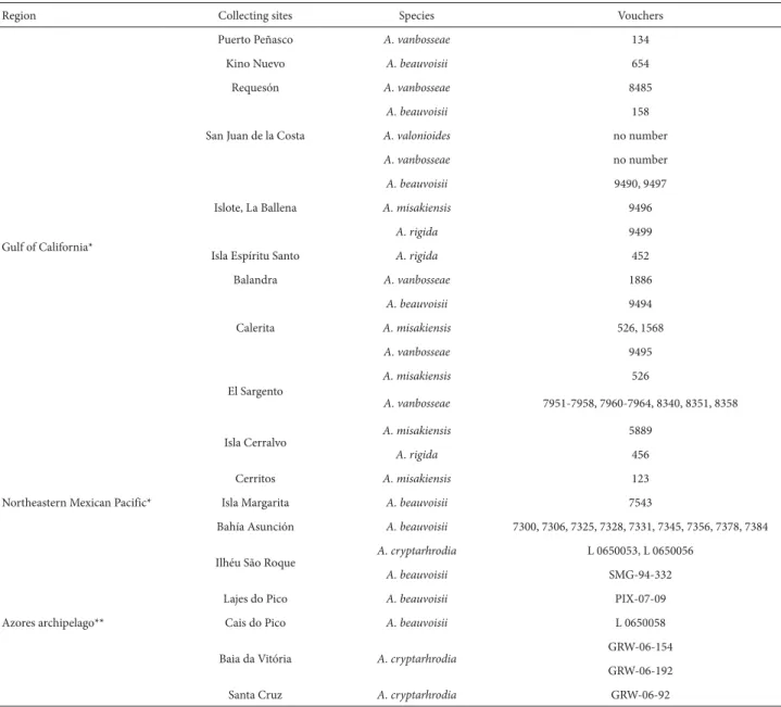
Table 1. Sample collection information for the studied species of Amphiroa from the Gulf of California, the Northeastern Mexican Pacific and the Azores archipelago.
Region Collecting sites Species Vouchers
Gulf of California*
Puerto Peñasco A. vanbosseae 134
Kino Nuevo A. beauvoisii 654
Requesón A. vanbosseae 8485
San Juan de la Costa
A. beauvoisii 158
A. valonioides no number
A. vanbosseae no number
Islote, La Ballena
A. beauvoisii 9490, 9497
A. misakiensis 9496
A. rigida 9499
Isla Espíritu Santo A. rigida 452
Balandra A. vanbosseae 1886
Calerita
A. beauvoisii 9494
A. misakiensis 526, 1568
A. vanbosseae 9495
El Sargento
A. misakiensis 526
A. vanbosseae 7951-7958, 7960-7964, 8340, 8351, 8358
Isla Cerralvo A. misakiensis 5889
A. rigida 456
Northeastern Mexican Pacific*
Cerritos A. misakiensis 123
Isla Margarita A. beauvoisii 7543
Bahía Asunción A. beauvoisii 7300, 7306, 7325, 7328, 7331, 7345, 7356, 7378, 7384
Azores archipelago**
Ilhéu São Roque A. cryptarhrodia L 0650053, L 0650056
A. beauvoisii SMG-94-332
Lajes do Pico A. beauvoisii PIX-07-09
Cais do Pico A. beauvoisii L 0650058
Baia da Vitória A. cryptarhrodia
GRW-06-154
GRW-06-192
Santa Cruz A. cryptarhrodia GRW-06-92
*Phycological Herbarium of the Autonomous University of Baja California Sur (code, FBCS). **Ruy Telles Palhinha Herbarium of the University of the Azores (code, AZB).
stages were described and photographed with a digital ca-mera (model C5060; Olympus, Tokyo, Japan) attached to a compound microscope (BX50F; Olympus) and edited with Photoshop 6.0.1 software (Adobe Systems Incorporated, San Jose, CA, USA). Additional data were obtained from Segawa (1940b), Ganesan (1968; 1971), Johansen (1968), Murata & Masaki (1978), Norris & Johansen (1981), Srimanobhas & Masaki (1987), Riosmena-Rodríguez & Siqueiros-Beltrones (1991; 1996), Womersley (1996), Moura & Guimarães (2002), Harvey et al. (2009), and Rosas-Alquicira et al. (2010). Vegetative anatomical terminology follows that of Woelkerling (1988), and reproductive terminology follows that of Johansen (1968; 1981), Murata & Masaki (1978) and Srimanobhas & Masaki (1987).
Results
Sporangial conceptacle development
elongate and divide to form rows of short overlying cells (Fig. 5). These layers of small cells, derived from the cavity cells, are pushed upward by further elongation of the cavity cells and eventually become the conceptacle roof (Fig. 6). Before cavity cells reach their full length, the destruction of some of them begins. Atrophy of the cavity cells forms a cavity, the so-called conceptacle chamber, followed by the development of the reproductive structures.
For all three of the species evaluated (Amphiroa beau-voisii, A. misakiensis and A. vanbosseae), atrophy of the cavity cells creates space for the sporangial initials (Fig. 7). Sporangial initials (Fig. 8-15) develop in the periphery of the chamber floor (A. beauvoisii, Fig. 8) or in the periphery and center (A. misakiensis and A. vanbosseae, Fig. 12 and 14). In all three species, while the initials are still very small, they elongate and divide transversally to form the sporangia mother cells and their stalk cells (Fig. 11 and 13). Autolysis continues until the adjacent cavity cells are largely des-troyed. Further growth mostly involves tetrasporangial or bisporangial maturation (Fig. 14 and 15, respectively) and cavity cell destruction. An exception occurs in A. beauvoisii
(Fig. 8, 10 and 15), in which cavity cells are still present in conceptacles with mature sporangia.
The conceptacle pore (Fig. 16-22) is initiated by an increase in the volume of subepithallial cells in the central region of the roof cavity (Fig. 16), to form large block-shaped cells (Fig. 19). The cells in the roof and immediately below the block-shaped cells elongate centripetally and migrate towards the future location of the pore (Fig. 17 and 18), compressing the cavity cells. Their continuous elongation causes the autolysis of the large block-shaped cells and the formation of the conceptacle pore (Fig. 19). A different pattern occurs in Amphiroa misakiensis, in which no large--block shaped cells appear. Instead, the cells located in the center of the roof elongate (Fig. 20) and those closest to the cavity show signs of atrophy (Fig. 21). The decalcification and atrophy of the cells immediately below the subepithallial cells results in the formation of a canal leading to the ex-terior (Fig. 22). This canal soon becomes lined with small, centripetally oriented elongated roof cells.
The final stage of development observed was senescence of the conceptacle. Two patterns were observed; the first, ob-served in the sporangial conceptacles of Amphiroa misakiensis
(Fig. 23 and 24), was initiated by the downward, lateral and upward growth of the cells of the conceptacle floor and the second pattern, observed in the sporangial conceptacles of
A. beauvoisii and A. vanbosseae, was initiated solely by the downward and lateral growth of the cells of the conceptacle floor (Fig. 25 and 26). For all conceptacle types, senescence occurred in the presence of tetrasporangia (Fig. 26).
Gametangial and carposporangial conceptacle development
We evaluated the development of male and female con-ceptacles in A. beauvoisii, A. cryptarthrodia, A. rigida and A. vanbosseae. Because conceptacle development was identical in all of those species, the various stages are illustrated with examples selected from among those taxa. The first stage in the development of male and female conceptacles is characterized by the anticlinal elongation and division of a group of peripheral cells in a subsurface stratum of the intergenicula. Those cells are designated cavity cells (Johansen 1968). The continued elongation and delayed cell division result in the formation of an initial conceptacle dome with a cellular cap (Johansen 1968), as shown in Fig. 27. The cavity cells secrete a mucilaginous material from their distal portion, the cap, formed by copious amounts of light, clear material (Fig. 27). Due to the presence of the cap, the epithallial cells and the central peripheral cells of the dome become detached (Fig. 28).
Cavity cell dissolution begins above the male gametangial initials and progresses upward toward the cavity cells and the conceptacle dome (Fig. 28). Following this destruction, the surrounding tissue grows over the fertile layer, forming the conceptacle roof (Fig. 29 and 30). Because the reproductive initials are still relatively small when this growth occurs, the lack of filling material within the cavity allows the roof to Figures 3-7. Early stages in the formation of sporangial conceptacles. Amphiroa
Figures 8-15. Development of sporangia in Amphiroa beauvoisii J.V. Lamouroux (8, 10 and 15), Amphiroa misakiensis Yendo (9, 11-13) and Amphiroa vanbosseae Me. Lemoine (14): (8). Undeveloped sporangia mother cells (arrow), interspersed cavity cells (double arrow) and peripheral mature tetrasporangia (arrowhead); (9). Sporangia mother cells and their stalk cell development on the periphery of the cavity floor (arrow); (10). Sporangium mother cell and its stalk cell development on the center of the cavity floor; (11). One stalk cell (arrows) and bisporangia mother cells (double arrow); (12). One stalk cell (arrows) and mature tetrasporan-gia (double arrow); (13). Single stalk cells (arrow) and sporantetrasporan-gia mother cell (double arrow); (14). Mature tetrasporangia with four zonately arranged spores (numbered); (15). Mature bisporangia with two zonately arranged bispores (numbered).
grow downward as well as centripetally. The terminal part of the roof elongates and divides transversely (Fig. 31), pro-viding new cells that repeat the process until the pore canal formation is completed. In the female gametangial initials, the process follows the same pattern, with cavity cell dissolution (Fig. 32 and 33), followed by the formation of a conceptacle roof and pore canal (Fig. 33 and 34).
Male gametangia were observed only for Amphiroa beauvoisii and A. vanbosseae. Their development begins with the formation of basal cells from the transversal basal division of the cavity cells (Fig. 35). After cavity cell dilution, each basal cell forms two spermatangial mother cells and the spermatangia continuously derive from the spermatangia mother cells (Fig. 36-38) to form simple and unbranched filaments. The spermatangia in turn, liberate spermatia that fill the chamber (Fig. 38).
Female gametangia were observed for Amphiroa beau-voisii, A.cryptarthrodia, A. rigida and A. vanbosseae. Their development initiates with the elongation and transverse di-vision of the basal region of the central cavity cells (Fig. 39). As a consequence, three new layers of cells cover the central region of the cavity floor; the elongation of cells from the terminal layer gives rise to the procarp initials (Fig. 39). Each of these, by elongation and transverse division, leads to the formation of a supporting cell and carpogonia branch initial (Fig. 40). The elongation and further division of the latter form the trichogyne (Fig. 41 and 42).
Figures 23-26. Senescence of sporangial conceptacles of Amphiroa misakien-sis Yendo (23 and 24) and Amphiroa beauvoisii J.V. Lamouroux (25 and 26): (23). Growth of the cells of the central region of the roof cavity (arrow) and the conceptacle floor (double arrow); (24). Complete senescence of the pore (arrow); (25 and 26). Downward growth of the cells of the central region of the roof cavity (arrow).
Figures 27-31. Early stages in the formation of the male conceptacles of Amphi-roa vanbosseae Me. Lemoine (27) and AmphiAmphi-roa beauvoisii J.V. Lamouroux (28-31): (27). Conceptacle cap (arrowhead) and formation of an initial conceptacle dome (arrow) above the developing conceptacle primordium (double arrow); (28). Cavity (double arrow) and protective dome cells (arrow) dissolution above the gametangial initials; (29 and 30). Surrounding cavity cells (arrow) grow over the fertile area forming the conceptacle roof; (31). Transverse division on the terminal part of surrounding cavity cells (arrow).
Figures 35-42. Development of male gametangia of Amphiroa vanbosseae Me. Lemoine (35-38), female gametangia of Amphiroa vanbosseae Me. Lemoine (40-42) and female gametangia of Amphiroa beauvoisii J.V. Lamouroux (39): (35). Anticlinal elongation and basal division of the central cavity cells giving rise to the basal cells (arrow); (36) Spermatangia mother cells on the cavity floor (arrow); (37). Basal cells (arrow) and spermatangia mother cells (double arrow); (38). Mature spermatangia branch reaching the cavity roof (arrow); (39). Elongation and successive transverse divisions of the basal region of the central cavity cells (arrow), forming the procarp initials (double arrow); (40). Supporting cell (arrow) and carpogonia branch initial (double arrow), scale bar, 10 µm; (41 and 42). Carpogonial branches with trichogynes (arrow).
The fact that the basic reproductive features are similar to those in other coralline red algae (Lebednik 1977) sug-gests that spermatia fuse with trichogynes and that male and female haploid nuclei then combine to form a diploid nucleus. The development of carposporangial filaments was observed for Amphiroa cryptarthrodia, A. rigida and A. van-bosseae. This phase initiated when the trichogynes were well developed (Fig. 43). The presence of a carpogonial branch and a discontinuous flat disc-like fusion cell, supporting a few residual carpogonia on its dorsal part (Fig. 44 and 45), provided evidence that fertilization had occurred. In A. vanbosseae,the carposporangialfilaments arose solely from the margins of the fusion cell (Fig. 46), whereas in A.
cryp-tarthrodia and A. rigida,they arose over the entire surface of the fusion cell (Fig. 47). The cells of the carposporangial filaments increase in size, the upper being relatively large and constituting the carposporangium (Fig. 48).
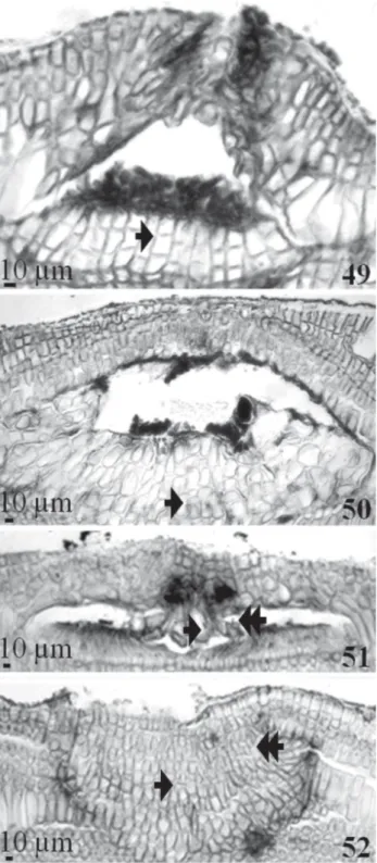
Two patterns were observed for the senescence of game-tangial/carposporangial conceptacles. The first, observed in carposporangial conceptacles (Fig. 49 and 50), was initiated by the downward, lateral and upward growth of the cells of the conceptacle floor. The second pattern, observed in the gametangial conceptacles (Fig. 51 and 52), was initiated only by downward and lateral growth. For all conceptacle types, senescence occurred in the presence of carposporangia (Fig. 49) and gametangia (Fig. 51).
between species within Amphiroa, the most diverse geni-culate genus of the subfamily Lithophylloideae.
The development of the conceptacle pore is described and illustrated here for the first time, as is conceptacle se-nescence, for which four patterns were observed: two for sporangial conceptacles, one for spermatangial conceptacles and one for carposporangial conceptacles. As first described for Amphiroa by Johansen (1972), Murata & Masaki (1978) and Srimanhobas & Masaki (1987), we confirmed that con-ceptacle roof development was by intersporangial growth for sporangial conceptacles and by vegetative filament growth up and over the chamber for the gametangial conceptacles. The development of the sporangial conceptacle cavity by elongation and atrophy of cavity cells described for the ge-nus by Johansen (1972) was also confirmed. Nevertheless, the presence of intact cavity cells among mature sporangia described and illustrated for Amphiroa zonata by Murata & Masaki (1978) was observed only for A. beauvoisii (cf. Fig. 15). The presence of cavity cells in mature sporangial conceptacles in this species was previously reported by Harvey et al. (2009).
The development of the carposporangial filament, previously suggested to be important for species identi-fication within the genus Fosliella (Chamberlain 1977), was also found to be important for the genus Amphiroa. In A. cryptarthrodia and A. rigida, we found that the carposporangial filament arose from the surface of the fusion cell. This finding was previously reported for A. rigida by Segawa (1940b); for A. foliacea by Ganesan (1968); for A. zonata for Murata & Masaki (1978); and for
A. beauvoisii and A. valonioides by Riosmena-Rodríguez & Siqueiros-Beltrones (1996). It is of note that Riosmena--Rodríguez & Siqueiros-Beltrones (1996) reported a different pattern for A. rigida from the Gulf of California. In A. vanbosseae, we found that the carposporangial fi-lament arose only from the margins of the fusion cell, as previously reported by Riosmena-Rodríguez & Siqueiros--Beltrones (1996) and Moura & Guimarães (2002) and by Riosmena-Rodríguez & Siqueiros-Beltrones (1996) for A. misakiensis (Tab. 2).
We confirmed the diagnostic importance of the sporan-gial pore canal anatomy in distinguishing between species within the subfamily Lithophylloideae, namely in Litho-phyllum (Woelkerling & Campbell 1992; Keats 1997; Harvey
et al. 2005) and Amphiroa (Harvey et al. 2009). The type of sporangial pore canal anatomy reported in this study for
Amphiroa beauvoisii and A. vanbosseae was also previously reported by Harvey et al. (2009), Norris & Johansen (1981) and Ganesan (1971). However, the present study reports and illustrates, for the first time, the complete occlusion of the pore canal by large block-shaped cells in both species (Fig. 8).
Based on the pore canal anatomy and the location of sporangia on the cavity floor, we created the following key, by which three of the five studied Amphiroa species can easily be distinguished:
Figures 49-52. Senescence of the gametangial/carposporangial conceptacle of Amphiroa vanbosseae Me. Lemoine (49-51) and Amphiroa beauvoisii J.V. Lamouroux (52): (49 and 50). Growth of the cavity floor cells (arrow); (51 and 52). Downward and lateral growth (arrow and double arrow respectively) of the cells of the conceptacle pore.
Discussion
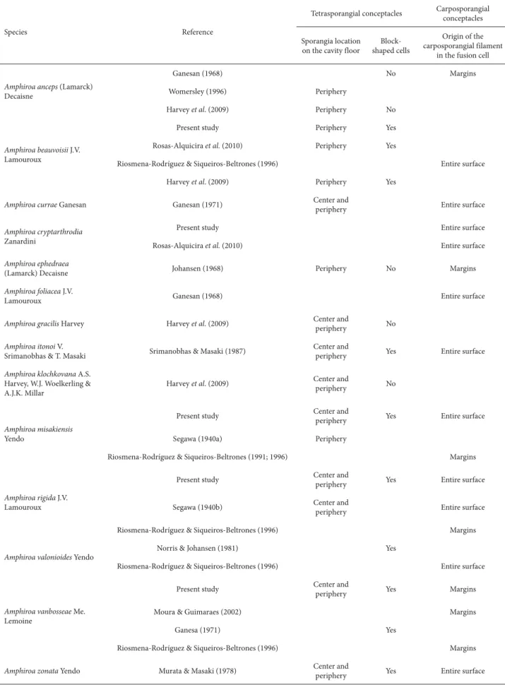
Table 2. Events in sporangial/carposporangial conceptacle development for Amphiroa species.
Species Reference
Tetrasporangial conceptacles Carposporangial conceptacles
Sporangia location on the cavity floor
Block-shaped cells
Origin of the carposporangial filament
in the fusion cell
Amphiroa anceps (Lamarck) Decaisne
Ganesan (1968) No Margins
Womersley (1996) Periphery
Harvey et al. (2009) Periphery No
Amphiroa beauvoisii J.V. Lamouroux
Present study Periphery Yes
Rosas-Alquicira et al. (2010) Periphery Yes
Riosmena-Rodríguez & Siqueiros-Beltrones (1996) Entire surface
Harvey et al. (2009) Periphery Yes
Amphiroa currae Ganesan Ganesan (1971) Center and
periphery Entire surface
Amphiroa cryptarthrodia Zanardini
Present study Entire surface
Rosas-Alquicira et al. (2010) Entire surface
Amphiroa ephedraea
(Lamarck) Decaisne Johansen (1968) Periphery No Margins
Amphiroa foliacea J.V.
Lamouroux Ganesan (1968) Entire surface
Amphiroa gracilis Harvey Harvey et al. (2009) Center and
periphery No
Amphiroa itonoi V.
Srimanobhas & T. Masaki Srimanobhas & Masaki (1987)
Center and
periphery Yes Entire surface
Amphiroa klochkovana A.S. Harvey, W.J. Woelkerling & A.J.K. Millar
Harvey et al. (2009) Center and
periphery No
Amphiroa misakiensis Yendo
Present study Center and periphery Yes Entire surface
Segawa (1940a) Periphery
Riosmena-Rodríguez & Siqueiros-Beltrones (1991; 1996) Margins
Amphiroa rigida J.V. Lamouroux
Present study Center and
periphery Yes Entire surface
Segawa (1940b) Center and
periphery Entire surface
Riosmena-Rodríguez & Siqueiros-Beltrones (1996) Margins
Amphiroa valonioides Yendo
Norris & Johansen (1981) Yes
Riosmena-Rodríguez & Siqueiros-Beltrones (1996) Entire surface
Amphiroa vanbosseae Me. Lemoine
Present study Center and
periphery Yes Margins
Moura & Guimaraes (2002) Margins
Ganesa (1971) Yes
Riosmena-Rodríguez & Siqueiros-Beltrones (1996) Margins
Amphiroa zonata Yendo Murata & Masaki (1978) Center and
Amphiroa vanbosseae exhibits the same pattern reported for A. zonata by Murata & Masaki (1978) and for A. itonoi
by Srimanobhas & Masaki (1987); A. beauvoisii and A. misakiensis are closely related to the Australian species A. gracilis and A. klochkovana (Harvey et al. 2009). A. cryptar-throdia and A. rigida are not included in this key, because no sporangial conceptacles were observed in those species.
The consistency of the observed reproductive deve-lopment patterns was found to be diagnostic for species within Amphiroa. It would be interesting to evaluate the importance of this feature in the closely-related genus from the Lithophyllum-Titanoderma complex included in the Lithophylloideae by Harvey et al. (2003), based on phylogenetic studies.
Acknowledgments
The authors are grateful to the staff of the Marine Botany Laboratory at the Universidad Autónoma de Baja California Sur (UABCS, Autonomous University of Baja California Sur) and the Phycology group of the Universidade dos Açores (UA, University of the Azores), for their assistance in the field; to Nuno V. Álvaro for the map of the studied areas; and to Ian Tittley, Lindsey Franger and Francisco Wallenstein for the English revision of the manuscript. We are also grateful to Dr. Phillip A. Lebednik, for providing references, and to the two anonymous reviewers for their helpful comments and sugges-tions. This work received financial support from the Portuguese
Centro de Investigação de Recursos Naturais (CIRN, Center for Research in Natural Resources), as well as from the Mexican
Consejo Nacional de Ciencia y Tecnología (CONACYT, Na-tional Council for Science and Technology) in conjunction with the Secretaría de Educación Pública (SEP, Department of Public Education) and the Secretaría de Medio Ambiente y Recursos Naturales (SEMARNAT, Department of the Environment and Natural Resources). Additional funding was provided by the European Regional Development Fund, through the Competi-tive Factors Thematic Operational Programme; the Portuguese
Fundação para a Ciência e Tecnologia (FCT, Science and Tech-nology Foundation; multiannual fund); CONACYT (masters and doctoral scholarship no. 176162 to EFR-A); ALBAN (the European Union Programme of High Level Scholarships for Latin America; scholarship no. E05D060221MX to EFR-A); and the Comisión de Operación y Fomento de Actividades Académicas (COFAA, Operation and Development Commit-tee for Academic Activities) and Estímulo al Desempeño de los Investigadores (EDI, Researcher Stimulus Fund) of the Mexican
Instituto Politécnico Nacional (IPN, National Polytechnic Insti-tute; fellowship grant to GH-C).
References
Chamberlain, Y.M. 1977. Observations on Fosliella farinosa (Lamour.) Howe (Rhodophyta, Corallinaceae) in the British Isles. British Phy-cological Journal 12: 343-58.
Ganesan, E.K. 1968. Studies on the morphology and reproduction of the articulated Corallines-III. Amphiroa Lamouroux emend. Weber van Bosse. Phykos 6: 7-28.
Ganesan, E.K. 1971. Amphiroa curare (Corallinaceae), a new species of marine algae from Venezuela. Phycologia 10: 155-68.
Harvey, A.S.; Broadwater, S.T.; Woelkerling, W.J. & Mitrovski, P. 2003.
Choreonema (Corallinales, Rhodophyta): 18S rDNA phylogeny and res-urrection of the Hapalidiaceae for the subfamilies Choreonematoideae, Austrolithoideae and Melobesioideae. Journal of Phycology 39: 988-98. Harvey, A.S.; Woelkerling, W.J.; Farr, T.; Neill, K. & Nelson, W. 2005.
Coralline algae of central New Zealand. An identification guide to common ‘crustose’ species. Wellington, NIWA.
Harvey, A.S.; Woelkerling, W.J. & Millar, A.J.K. 2009. The genus Amphiroa
(Lithophylloideae, Corallinaceae, Rhodophyta) from the temperate coasts of the Australian continent, including the newly described A. klochkovana. Phycologia 48: 258-90.
Holmgren, P.K. & Holmgren, N.H. 1998 [continuously updated]. Index Herbariorum: A global directory of public herbaria and associated staff. New York Botanical Garden’s Virtual Herbarium. http:// sweetgum.nybg.org/ih/ (Accessed on 10 Sept 2011).
Johansen, H.W. 1968. Reproduction of the articulated coralline Amphiroa ephedraeea. Journal of Phycology 9: 141-48.
Johansen, H.W. 1972. Conceptacles in the Corallinaceae. Proceedings of the 7th International Seaweed Symposium 114-119.
Johansen, H.W. 1981. Coralline Algae: A first Synthesis. Boca Raton, CRC Press.
Keats, D.W. 1997. Lithophyllum insipidum Adey, Townsend et Boykins and L. flavescens sp. nov.: two flat lithophylloid coralline algae (Coral-linales, Rhodophyta) abundant in shallow reef environments in Fiji. Phycologia 36: 351-365.
Lebednik, P.A. 1977. Postfertilization development in Clathromorphum,
Melobesia and Mesophyllum with comments on the evolution of the Coral-linaceae and the Cryptonemiales (Rhodophyta). Phycologia 16: 379-406. Moura, C.W. & Guimarães, S.M.P. 2002. Amphiroa van-boseae (Corallinales, Rhodophyta) no Atlántico tropical americano. Hoehnea 29: 267-273. Murata, K. & Masaky, T. 1978. Studies of reproductive organs in articulated
coralline algae of Japan. Phycologia 17: 403-412.
Norris, J.N. & Johansen, H.W. 1981. Articulated coralline algae of the Gulf of California, Mexico, I: Amphiroa Lamouroux.Smithsonian Contributions to the Marine Sciences 9: 1-29.
Riosmena-Rodríguez, R. & Siqueiros-Beltrones, D.A. 1991. First report of sexual conceptacles of Amphiroa misakiensis (Yendo) in the Gulf of California. Revista de Investigación Científica Serie Ciencias Marinas. Universidad Autónoma de Baja California Sur 2: 8-12. Riosmena-Rodríguez, R & Siqueiros-Beltrones D.A. 1996. Taxonomy of
the genus Amphiroa (Corallinales, Rhodophyta) in the southern Baja California Peninsula, México. Phycologia 35: 53-65.
Riosmena-Rodríguez, R.; Woelkerling, W.J. & Foster M.S. 1999. Taxonomic reassessment of rhodolith-forming species of Lithophyllum (Corallinales, Rhodophyta) in the gulf of California, Mexico. Phycologia 38: 401-17. Rosas-Alquicira, E.F.; Riosm ena-Rodríguez, R. & Neto, A.I. 2010. Segre-gating characters used within Amphiroa (Corallinales, Rhodophyta) and taxonomic reevaluation of the genus in the Azores. Journal of Applied Phycology 23: 475-488.
Segawa, S. 1940a. Systematic anatomy of the articulated corallines. I.
Amphiroa rigida Lamouroux. Journal of Japanese Botany 16: 219-25.
1. Large block-shaped cells in sporangial conceptacle canal (Fig. 8)
2. Sporangia location only in the periphery of the cavity floor (Fig. 8) ... 1. A. beauvoisii
2. Sporangia location in the center and periphery of the cavity floor (Fig. 14) ... 3. A. vanbosseae
Segawa, S. 1940b. Systematic anatomy of the articulated corallines. II.
Amphiroa misakiensis Yendo. Journal of Japanese Botany 16: 488-94. Srimanobhas, V. & Masaki, T. 1987. Amphiroa itonoi (Corallinales, Rho-dophyta), a new species of marine algae from Japan. Japanese Journal of Phycology 35: 1-9.
Woelkerling, W.J. 1988. The Coralline red algae: An analysis of the genera and subphamilies of nongeniculate Corallinaceae. London,
Oxford University Press. Woelkerling, W.J. & Campbell, S.J. 1992. An account of southern Australian species of Lithophyllum (Corallinaceae, Rhodophyta). Bulletin of the British Museum (Natural History). Botany series 22: 1-107.
Womersley, H.B.S. 1996. The marine benthic flora of southern Aus-tralia. Part III B. Canberra, Union Offset, Australian Biological Resources Study.