Brucellosis and
Infection in
Householders and Their Animals in Secure
Villages in Herat Province, Afghanistan: A
Cross-Sectional Study
Zarif Akbarian1, Ghulam Ziay2, Willy Schauwers3, Bashir Noormal1, Islam Saeed1, Abul Hussain Qanee4, Zabiullah Shahab1, Tania Dennison3, Ian Dohoo5, Ronald Jackson6*
1Afghan National Public Health Institute, Ministry of Public Health, Kabul, Afghanistan,2Central Veterinary Diagnostic & Research Laboratory, Kabul, Afghanistan,3Animal Health Development Program, Kabul, Afghanistan,4General Directorate of Animal Health and Production, Ministry of Agriculture, Irrigation and Livestock, Kabul, Afghanistan,5University of Prince Edward Island, Charlottetown, Canada,6EpiCentre, Massey University, Palmerston North, New Zealand
*Ron.Jackson@xtra.co.nz
Abstract
Background
Brucellosis and coxiellosis are known to be endemic in ruminant populations throughout Afghanistan, but information about their prevalence and factors that affect prevalence in householders and livestock under diverse husbandry systems and pastoral settings is sparse.
Methods/Principal Findings
We conducted a cross-sectional survey to investigate the seroprevalence of brucellosis and
Coxiella burnetiiin humans and livestock in six secure districts in Herat from 26thDecember
2012–17thJanuary 2013. A total of 204 households with livestock were surveyed in six Kuchi and five sedentary type villages. Blood samples from 1,017 humans, 1,143 sheep, 876 goats and 344 cattle were tested for brucellosis and Q fever. About one in six house-holds (15.7%) had at least oneBrucellaseropositive person, about one in eight households (12.3%) had at least oneBrucellaseropositive animal and about one in four (24.5%) had either seropositive animals or humans. Ninety-seven percent of households had at least oneC.burnetiiseropositive person and 98.5% of households had one or moreC.burnetii
seropositive animals. Forty- seven householders had serological evidence of exposure to bothC.burnetiiandBrucellaand eight animals were serologically positive for both dis-eases. Drinking unpasteurised milk (OR 1.6), treating animals for ticks (OR 1.4), milking sheep (OR 1.4), male gender (OR 1.4) and seropositivity toBrucella(OR 4.3) were identi-fied as risk factors for seropositivity toC.burnetiiin householders. Household factors OPEN ACCESS
Citation:Akbarian Z, Ziay G, Schauwers W, Noormal B, Saeed I, Qanee AH, et al. (2015) Brucellosis and Coxiella burnetiiInfection in Householders and Their
Animals in Secure Villages in Herat Province, Afghanistan: A Cross-Sectional Study. PLoS Negl Trop Dis 9(10): e0004112. doi:10.1371/journal. pntd.0004112
Editor:John A. Crump, University of Otago, NEW ZEALAND
Received:May 19, 2015
Accepted:September 2, 2015
Published:October 20, 2015
Copyright:© 2015 Akbarian et al. This is an open access article distributed under the terms of the Creative Commons Attribution License, which permits unrestricted use, distribution, and reproduction in any medium, provided the original author and source are credited.
Data Availability Statement:All relevant data are within the paper and its Supporting Information files.
associated with households having eitherBrucellaseropositive animals or humans were Kuchi households (OR 2.5), having4 rooms in the house (OR 2.9) and not owning land (OR 2.9).
Conclusions
The results from this study provide baseline information for the planning and monitoring of future interventions against these diseases. The implementation of this study greatly improved collaboration, coordination and capability of veterinary and public health profes-sionals from government, NGOs and donor funded projects.
Author Summary
Our study alerted authorities to a hitherto unrecognised high prevalence ofC.burnetii infections, acted as a catalyst for the introduction of a national vaccination programme for protection of sheep and goats from brucellosis using Rev1 vaccine, demonstrated the bene-fits of a coordinated approach and fostered a better understanding of the nature of infec-tion in different hosts and of the constraints for control faced by government services. A notable feature of the study was the enthusiasm and interest displayed by all of the partici-pants throughout, from heads of government services to field personnel and villagers. Livestock owners regard zoonoses as adversities that affect their livestock and members of their households and do not partition them separately as medical or veterinary problems. Control programs need to take that perception into account and wherever possible avoid vesting ownership separately into veterinary or public health agencies.
Introduction
Brucellosis and coxiellosis are known to be endemic in ruminant populations throughout Afghanistan but information about their prevalence and factors that affect prevalence in house-holders and livestock under diverse husbandry systems and pastoral settings is sparse. Brucel-losis in animals results in economic losses due to decreased productivity from abortions and reduced milk yield while the disease in humans can be severely debilitating, often with long-term adverse consequences for health [1].C.burnetiicauses abortions in domestic ruminants and fever, pneumonia, meningo-encephalitis and hepatitis in acute cases of Q fever in humans and endocarditis in chronic cases [2,3]. Little is known about the epidemiology ofC.burnetiiin animals and humans in Afghanistan although a United Nations Food and Agriculture (FAO) funded study found serological evidence of its occurrence in Bamyan province in 2011 [4]. Bru-cellosis and Q fever are likely to be severely under-diagnosed and reported as they are not spe-cifically included in the list of diseases in the donor-funded Essential Package of Hospital Services and Basic Package of Health Services. Until the start of this study there were no facili-ties for testing forC.burnetiiat the central veterinary and public health laboratories in Kabul or elsewhere in the Republic. It is highly likely that bothB.melitensisandB.abortusare present in Afghanistan but there is currently no in-country diagnostic capability for their culture or differentiation.
Union to Afghanistan for provision of technical assistance to the Afghan Ministry of Agriculture, Irrigation and Livestock. The funders had no role in study design, data collection and analysis, decision to publish, or preparation of the manuscript.
The objectives of the study were to
• Determine the prevalence of seropositivity for brucellosis andC.burnetiiin humans and live-stock in 20 randomly selected households with livelive-stock in each of 10 randomly selected secure villages in six of the 16 districts of Herat province;
• Identify factors associated with the risk of seropositivity to these diseases in households, members of households and their livestock;
• provide baseline information which may be used for design of control programs and evalua-tion of the efficacy of control program intervenevalua-tions.
Methods
A cross-sectional survey to investigate the seroprevalence of seropositivity to brucellosis andC. burnetiiin humans and livestock (cattle, sheep and goats) was conducted in six secure districts in Herat from 26thDecember 2012–17thJanuary 2013. Secure villages were located in districts considered to be secure because of no overt signs of anti-government activities therein, thus enabling public health and veterinary government agencies and DCA teams to conduct routine activities safely. The population of interest was humans and female livestock (cattle, sheep and goats) of breeding age in households in selected villages in Herat Province. The principal epide-miologic unit of interest was households with consideration given to both animals and humans within each household. A total of 204 households were surveyed in 11 villages, six of which were Kuchi [5] and five were sedentary type. Sedentary villages were selected from a list pro-vided by the Afghanistan Information Management System which listed all the villages in Afghanistan in 2006. Kuchi are nomadic or transhumant pastoralists. Their villages were selected from a list provided by the Dutch Committee for Afghanistan (DCA) and the veteri-nary field units supported by the DCA identified their locations as at October 2012. Up to date data on numbers of livestock in households were not available. The last livestock census, which was sample based, was conducted by FAO in 2002–2003. Villages were randomly selected using the Data Analysis Tool in Microsoft Excel 2007 and 20 plus five reserve households in each village were randomly selected from a list provided by village elders of all households with livestock. In each household blood samples were collected in sterile red top or serum separation vacutainers from up to five householders9 and60 years of age and up to 10 randomly selected female sheep and goats of breeding age and up to five female cattle of breeding age. The sample size of five householders was based on the average Afghan household size of 7.6 persons. Individual human data recorded at the time of sampling included age, sex, marital sta-tus, occupation and history of abortions (if married) and for animals were species, age and abortion history (seeS1 FileandS2 Filein Supplementary Information).“Small ruminants”is used throughout the paper as the collective term for sheep and goats.
was performed with the Microsoft Excel compare tool. Unique village and household identifi-ers were used for linkage of data among the three data sets.
Sera were separated at the Provincial Veterinary Laboratory in Herat and stored in duplicate cryovials before transport to the Central Veterinary Diagnostic and Research Laboratory and the Central Public Health Laboratory in Kabul. Human sera were considered brucellosis tive if they were competitive ELISA test positive. Animal sera were considered brucellosis posi-tive if they showed agglutination in the standard method Rose Bengal Test (RBT) and were competitive ELISA test positive. Competitive ELISA kits (COMPELISA) andB.abortusRose Bengal Test (standard) antigen were procured from the Animal and Plant Health Agency, United Kingdom. Brucellosis competitive ELISA samples giving an optical density (OD) equal to or below 60% of the mean of the OD of the 4 conjugate control wells were considered posi-tive. Human and animal sera were tested according to manufacturers’instructions with Coxiella burnetii(Q fever) Phase I and Phase II IgG ELISAs (IBL International Hamburg, Ger-many) and the Q fever LSI ELISA kit (LSI, Lissieu, France) respectively. The IBL human sera results were recorded as Units and the LSI animal ELISA results were expressed as optical den-sity Sample/Positive control ratios corrected for the negative control. Human sera were consid-ered positive if the result expressed as units was11 and animal sera were considered to be positive if the S/P ratio was40.
Data analysis
Estimates of prevalence (at the person and animal level) were calculated using robust standard errors (variance linearization) to account for clustering at the village and household levels. Variance linearization (also known as robust standard errors clustered on village) was used to estimate the variance of all prevalences and hence to derive the confidence intervals (CI). Infor-mation about the total sample size of the population at risk at each of the levels (village, house-hold and individual) was not recorded so neither sampling weights nor finite population correction factors were applied in the analyses.
Multivariable models were built to evaluate factors that influenced seropositivity toBrucella andC.burnetiiin humans, livestock and households. Data which could conceivably be associ-ated with disease status were first examined with descriptive statistics and tested for associa-tions between the outcome variables as a first screening process and variables significant at p<0.10 were entered into the multivariate models. Multivariable mixed logistic regression
models were built with random effects for village and household (except for the household-level analyses which just included a random effect for village). In some models, there was zero village-level variance once the household random effect was incorporated and in these cases the village-level random effect was dropped. Models were built using a manual backwards elim-ination process. Normality and heteroscedasticity of all random effects were evaluated graphi-cally and unless noted, were considered acceptable. Statistical analyses were performed using Epi Info version 7.1.2.0, and Stata 13 (StataCorp LP). Graphics were constructed in Excel (Microsoft).
Ethics statement
The study was approved by the Islamic Republic of Afghanistan Department of Public Health’s Institutional Review Board (Approval numbers 221665 and 221769). Study participants signed a consent form agreeing to participate prior to the survey and informed signed consent was obtained from all adult study participants and parents of children<18 years old at the time of
is a normal husbandry procedure and was supervised or conducted by veterinarians. This is in line with Animal Welfare legislation in New Zealand and current practice in the Ministry of Agriculture in Afghanistan.
Results
Human results
Brucellaserology in humans. The overall prevalence of seropositives in the 1,017 humans
tested in the 11 study villages was 5.2% (95% CI 1.8, 14.3) and seropositives were found in 10 villages. Humans with serological evidence of exposure to brucellosis were detected in 32 (15.7%) of the 204 study households and were strongly clustered in 3 villages (A, G and K); see
Fig 1A. The prevalence ofBrucellaseropositives was slightly lower in females (4.1%, 95% CI
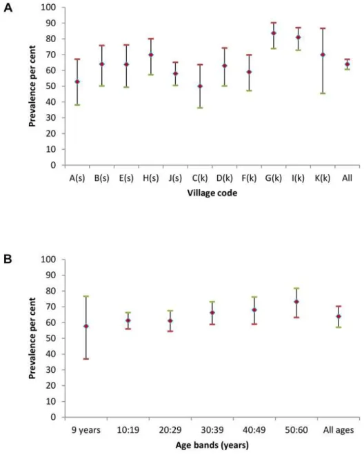
Fig 1. Human brucella seroprevalences (♦) with upper (-) and lower (-) 95% CI for each study village (A–K) in Fig 1A and categorised by age bands in Fig 1B.Sedentary villages in Fig 1A are identified by (s)
and Kuchi by (k).
1.1, 13.8) than in males (7.2%, 95% CI 2.9, 16.5) and higher in Kuchi householders (7.1%, 95% CI 1.2, 32.5) than in sedentary village householders (3.3%, 95% CI 0.5, 17.7). Prevalences were similar across age bands (seeFig 1B). Of the 53Brucellaseropositive humans, 47 were also seropositive forC.burnetii.
C.burnetiiserology in humans. The overallC.burnetiiseroprevalence in humans in the
11 study villages was 63.9% (95% CI 57.0, 70.3) and seropositives were found in all villages. Humans with serological evidence of exposure toC.burnetiiwere detected in 199 (97.5%) of the 204 study households. The prevalence was slightly higher in males (69.3%, 95% CI 61.1, 76.5) than in females (61.0%, 95% CI 57.0, 64.7). Village level prevalences were similar (Fig 2A) but overall prevalences were slightly higher in Kuchi (66.1%, 95% CI 51.0, 78.6) than in Fig 2. HumanC.burnetiiseroprevalences (♦) with upper (-) and lower (-) 95% CI for each study village (A–K) in Fig 2A and categorised by age bands in Fig 2B.Sedentary villages in Fig 2A are identified by (s)
and Kuchi by (k).
sedentary villages (61.7%, 95% CI 53.3, 69.5).C.burnetii seroprevalences categorised by 10 year age bands are shown inFig 2B. Seroprevalence increased with increasing age from an ini-tial high prevalence in the 26 persons, all aged 9 years, in the youngest age group.
Abortions and serological evidence of exposure toC.burnetiiorBrucella. Miscarriages
or abortions were reported by 146 (35.3%, 95% CI 28.7, 42.4) of the 414 married women for whom ages were recorded. Eight (5.5%, 95%CI 1.1, 23.7) of the women who reported that they had experienced an abortion wereBrucellatest positive and 101 (69.2%, 95%CI 58.0, 78.5) wereC.burnetiitest positive. Of the 414 married women of known age, the percentages of women reporting having had an abortion in three of theBrucella(B) byC.burnetii(C) catego-ries were B negative C negative 28.8%, B negative C positive 38.3%, B positive C positive 57.1%. There was only one woman who was C negative and B positive.
Animal results
Brucellaserology in animals. Sera were separated from 1,143 blood samples from sheep,
876 from goats and 344 from cattle. There were 33 RBT positives of which 30 were ELISA posi-tive. The seroprevalence was 1.4% (95% CI 0.7, 2.7) for sheep, 1.5% (95% CI 0.7, 3.0) for goats, 0.3% (95% CI 0.0, 3.2) for cattle and 1.3% (95% CI 0.7, 2.2) overall.
Brucellaseropositive animals were detected in 25 (12.3%) of the 204 study households and 10 of the 11 study villages. The prevalence in Kuchi villages was 1.8% (95% CI 0.8, 3.7) and 0.8% (95% CI 0.3, 2.3) in sedentary villages. The distribution of the prevalences ofBrucella seropositive animals in the 11 study villages is shown inFig 3A. There were no seropositive ani-mals in the 12–23 month age group and the prevalence was relatively stable across the older age groups (Fig 3B).
C.burnetiiserology in animals. The prevalence ofC.burnetiitest positives in animals
was 43.4% (95% CI 34.7, 52.5) for sheep, 52.7% (95% CI 43.8, 61.5) for goats, 5.2% (95% CI 3.1, 8.6) for cattle and 41.3% (95% CI 33.4, 49.6) overall.C.burnetiitest positive animals were detected in all of the study villages and in 201 (98.5%) of the 204 study households. The respec-tive prevalences for Kuchi and sedentary village animals were 44.3% (95% CI 31.0, 58.5) and 38.5% (95% CI 26.2, 52.6). Village levelC.burnetiiseroprevalences in animals are shown inFig 4A. Ten goats and 10 sheep were bothBrucellaandC.burnetiiseropositive. Age related sero-prevalences showed an increase from the 12–23 month age band to the 36–49 month band after which the prevalence was reasonably stable (seeFig 4B).
Multivariable models
Brucellaseropositivity in animals. The mixed-effects logistic regression models of
signifi-cant explanatory variables forBrucellaseropositivity in small ruminants with households incorporated as a random effect are shown inTable 1. The effect of Kuchi was primarily evi-dent in sheep where the OR was 5.3 if sheep and goats were separated out. The limited number of seropositive observations (n = 30), combined with the relatively small number of animals per household (N~ = 10) precluded a reliable evaluation of residuals at the household level (there was no evident variation at the village level). However, no obvious violations of assump-tions were noted.
C.burnetiiseropositivity in animals. Only age was significant in the mixed effects logistic
regression models of significant explanatory variables forC.burnetiiseropositivity in small ruminants, sheep and goats with households and villages incorporated as random effects. Vil-lage type was not significant.
the relatively small number of animals per household (N~ = 10) precluded a reliable evaluation of residuals at the household level (there was no evident variation at the village level). However, no obvious violations of assumptions were noted.
Risk of abortion in animals. Brucellaseropositivity and village type were associated with risk of abortions in small ruminants (seeTable 3) but there was no association withC.burnetii seropositivity. Separate models for sheep and goats showed the risk of abortion was similar for sheep (OR 5.7, 95%CI 1.1, 29.2) and goats (OR 6.4, 95%CI 2.3, 17.6) and for village type. Fig 3. Animal brucella seroprevalences (♦) with upper (-) and lower (-) 95% ci for each study village (a–
k) in fig 3a and categorised by age bands in fig 3b.Sedentary villages in fig 3a are identified by (s) and kuchi by (k).
Fig 4. AnimalC.burnetiiseroprevalences (♦) with upper (-) and lower (-) 95% ci for each study village (a–k) in fig 4a and categorised by age bands
in fig 4b.Sedentary villages in fig 4a are identified by (s) and kuchi by (k).
doi:10.1371/journal.pntd.0004112.g004
Table 1. Mixed-effects logistic regression results forBrucellaseropositivity in small ruminants with households incorporated as a random effect.
Variables Number OR (95% CI) P
Kuchi village 1,000 2.6 (1.1, 6.2) 0.03
Sedentary village 1,019 reference
Age continuous 1.03 (1.0, 1.1)
Household effect variance 0.9 (0.2, 4.6)
Households that reported abortions in their animals (n = 96) were more likely to have seropos-itive animals or humans (OR = 2.2 (95%CI 1.2, 4.2) than households which did not report abortions (n = 108).
Brucella
and
C
.
burnetii
seropositivity in householders
Brucellaseropositivity in householders. Multivariable modelling with village and
house-hold incorporated as random effects showed thatC.burnetiiseropositivity was the only signifi-cant predictor (OR = 4.3, P = 0.003) forBrucellaseropositivity in householders (village and household variance estimates 2.46 and 1.21 respectively)andvice versaforBrucella seropositiv-ity as a predictor forC.burnetiiseropositivity. As with the animal level data, there was limited ability to evaluate the distribution of residuals at the household level. Although no factors which could explain the variation in prevalence observed inFig 1A, fitting a null model with village as the random effect confirmed that the variation in village prevalences was statistically significant.C.burnetiiseropositivity in householders
Significant explanatory variables determined by multivariable modelling forC.burnetii seropositivity in householders with village and household incorporated as random effects are shown inTable 4.
Knowledge, attitude and practices
Household disease exposure. The analysis for associations between household disease exposure status was confined to brucellosis and not conducted for Q fever because 199 of the 204 households had evidence of exposure toC.burnetiiin their household members and 201 had evidence in their animals.
There were 32 (15.7%) households with serological evidence of exposure toBrucellain humans, 25 (12.3%) with evidence in animals, 25 with evidence in householders, 50 (24.5%) with evidence for animals or humans and 7 (3.4%) for both animals and humans. The
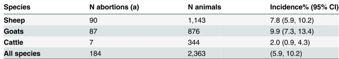
Table 2. Numbers of abortions, numbers of study animals and incidence of abortions with 95% confi-dence intervals in brackets.
Species N abortions (a) N animals Incidence% (95% CI)
Sheep 90 1,143 7.8 (5.9, 10.2)
Goats 87 876 9.9 (7.3, 13.4)
Cattle 7 344 2.0 (0.9, 4.3)
All species 184 2,363 (5.9, 10.2)
(a) refers to any occurrence during the animal’s life
doi:10.1371/journal.pntd.0004112.t002
Table 3. Mixed effects logistic regression results for risk of abortion inBrucellaseropositive small ruminants with households incorporated as a random effect.
Variable Number OR (95% CI) P
Brucella seropositive 29 6.4 (2,3, 17.6) <0.01
Brucella seronegative 1,990 reference
Kuchi village 1,000 0.6 (0.4, 1.1) 0.11
Sedentary village 1,019 reference
Household effect variance 1.8 (1.1, 2.8)
prevalence of households with serological evidence of exposure toBrucellain animals or humans categorised by number of rooms in households shows a downwards trend with increasing numbers of rooms (Fig 5).
The multivariable logistic regression model with households with serological evidence of exposure toBrucellain members of their household or in their animals as the outcome variable is shown inTable 5.
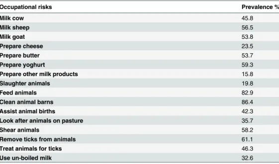
Prevalence of occupational risks forBrucellaandC.burnetii. The prevalence of
house-holder involvements with husbandry of animals and preparation of animal products which could entail risk of exposure toBrucellaandC.burnetiiare shown inTable 6. Most
Table 4. Mixed-effects logistic regression results forC.burnetiiseropositivity in householders with households and villages incorporated as random effects.
Variables Number OR (95% CI) P
Males 354 1.4 (0.96, 2.0) 0.06
Females 646 reference
Milk sheep 564 1.4 (1.0, 2.0) 0.04
Do not milk sheep 436 reference
Drink raw milk 326 1.6 (1.1, 2.3) <0.01
Do not drink raw milk 624 reference
Treat animals for ticks 609 1.4 (0.99, 1.9) 0.06
Do not treat animals for ticks 391 reference
Brucella test positive 53 4.3 (1.6, 11.4) <0.01
Brucella test negative 957 reference
Household effect variance 0.6 (0.3, 1.2), Village effect variance 0.1 (0.02, 0.5)
doi:10.1371/journal.pntd.0004112.t004
Fig 5. Point estimates (♦) and upper (-) and lower (-) 95% CI of prevalence of households with evidence of exposure toBrucellain animals or humans categorised by the number of rooms in the house.
householders were engaged in multiple activities with their animals; 29.8% with5, 44% with 6 to 10 and 26.2% with 11 to 16 activities. These risk factors did not have a statistically signifi-cant association with eitherBrucellaorCoxiellain our analyses, but this does not imply that they are not potentially important. Rather, it indicates that there was insufficient power in our study to detect such relationships.
Washing hands with soap and water after handling animals at birthing was practised by 73% of households but only 33% reported burying or burning afterbirths or aborted foetuses. Most were disposed of by feeding to dogs or in the open environment.
Discussion
Despite its conceptual appeal [6] and successful application over the past 20 years for infectious diseases such as bovine spongiform encephalopathy, highly pathogenic H5N1 avian influenza, C.burnetiiand leptospirosis, linked public health and veterinary field investigations of impor-tant zoonoses for which the reservoir hosts are farmed ruminants have been rarely conducted in low income countries. Serological studies involving humans and their livestock have been Table 5. Logistic regression results for risk of households having eitherBrucellaseropositive ani-mals or humans.
Variables Number OR (95% CI) P
Kuchi village household 101 2.5 (0.9, 6.6) 0.07
Sedentary village household 103 reference
4 rooms in house 87 2.9 (1.4, 6.1) <0.01
5 rooms in house 117 reference
Family not owning land 59 2.9 (1.3, 6.5) 0.01
Family owning land 145 reference
Village effect variance 0.3 (0.03, 2.5)
doi:10.1371/journal.pntd.0004112.t005
Table 6. Prevalence of occupational risks for exposure toBrucellaandC.burnetiiamong householders.
Occupational risks Prevalence %
Milk cow 45.8
Milk sheep 56.5
Milk goat 53.8
Prepare cheese 23.5
Prepare butter 53.7
Prepare yoghurt 59.3
Prepare other milk products 15.8
Slaughter animals 19.8
Feed animals 82.9
Clean animal barns 86.4
Assist animal births 42.3
Look after animals on pasture 35.7
Shear animals 58.2
Remove ticks from animals 61.1
Treat animals for ticks 46.3
Use un-boiled milk 32.6
reported for brucellosis in Kyrgyzstan [7], Mongolia [8] and Egypt [9], forC.burnetiiin Egypt [10] and for both brucellosis and Q fever in Chad [11] and the authors are aware of several unpublished investigations for brucellosis in Central Asia and China. Our study was made pos-sible by the terms of reference for the series of in-country studies in South Asia conducted under the Massey University European Commission funded and World Bank administered South Asia project which embodied veterinary and public health collaboration and allowed for investigator’s judgement for collection of information about the three zoonoses of interest which are characterised by multiple allied risk factors for transmission. Our inclusion of bru-cellosis and Q fever as the diseases of interest approach has obvious marginal cost advantages over study designs which are confined to one disease of interest.
Householder and animalBrucellaseroprevalences were 5.2% (95% CI 1.8, 14.3) and 1.3% (95%CI 0.7, 2.2) respectively with 15.7% of households having seropositive householders and 12.3% seropositive animals. There was evidence of clustering of infection in animals within a household and Kuchi village animals were more likely to be seropositive than those in seden-tary villages (Table 1).
The evidence of clustering is presented in the moderate level of household variance (Table 1)
The relatively constant prevalence over all age bands in householders (Fig 1B) indicates early age exposure to infection and contrasts with a finding of no seropositive animals in the 12–23 month age group of animals (Fig 3B). Humans are susceptible to infection at all ages, whereas with the exception of latent infection, successful infection in animals mainly occurs during pregnancy and most of the animals in the 12–23 month age band would have been either non-pregnant or in early first-parity pregnancy at the time of sampling. Seroprevalences of 1.4% in sheep and 1.5% in goats are highly indicative ofB.melitensisinfection for which the predominant reservoir hosts are small ruminants. Serology cannot distinguish Brucella species and the single test positive in cattle could be due to eitherB.melitensisorB.abortus.
The high householder and animalC.burnetiiseroprevalences of 63.9% (95%CI 57, 70.3) and 43.4% (95%CI 34.7, 52.5) respectively with almost all households (97.5% and 98.5%) hav-ing evidence of exposure to infection in householders and household livestock were unexpected findings. Prevalences varied among villages but not between Kuchi and sedentary villages or between sheep and goats. As withBrucellathe prevalence was markedly lower in cattle than in sheep and goats and there was evidence of early age exposure to infection in householders and animals.
[12] unlike humans where infection, apart from a small proportion of chronic relapsing cases, is usually self-limiting. The nature ofC.burnetiiinfection is probably similar; infections in ani-mals may persist for long periods [13,14] and most human infections are self-limiting.
We considered household prevalences derived by the study to be the most informative mea-sures of risk of exposure to infection. However, the prevalences of households with evidence of exposure toBrucellainfection in animals and householders are biased downwards to an unknown extent because only ten animals were tested in each household and the prevalence of Brucellatest positive animals was low. The bias was of no consequence forC.burnetiibecause of the high proportions of households with test positive householders and animals.
Confidence intervals of prevalence estimates were quite wide because multistage sampling with village as the primary sampling unit was done and this was accounted for in the analysis. True random sampling at each level (village, household and individual) with participation of 94% of selected households ensured that these are valid estimates. We also remind readers here that the study population consisted of households with animals so the estimates are only valid for that sub-population and not for the general population.
Many of the factors examined were village- or household-level factors for which the effective sample sizes were the numbers of villages and households. Despite this limitation and the study’s reliance on serology with no information as to when infection occurred, the multivari-able analyses were performed in an attempt to identify at least some of the risk factors for infec-tion in householders. The fact that the only factor identified forBrucellaseropositivity in householders wasCoxiellaseropositivity may have been due to its low prevalence which would have limited the power of the analysis for this infection. However, male gender, milking sheep, drinking raw milk, treating animals for ticks andBrucellaseropositivity were identified forC. burnetii(seeTable 4) and these are in accord with current understanding of the disease which considers the main routes of infection to be via infected milk and birth products and inhalation of contaminated dust. The nature ofC.burnetiiinfections has been well described in recent comprehensive reviews [15,16,17]. The organism can survive well for extremely long times in its desiccated spore-like form in dust; cattle sheep and goats are the main reservoirs for infec-tions and ticks may be the natural primary reservoir host[17].C.burnetiiinfection is asymp-tomatic in 50% to 60% of acute infections and its clinical polymorphism makes diagnosis difficult[17]. Abortions may occur in pregnant women and the chronic form which develops in about 5% of infections manifests as endocarditis, hepatitis and pneumonia. Diagnosis in rural communities in Afghanistan is particularly challenging because of poor access to competent and well-equipped health services and laboratories and competition from traditional healers in rural villages. A Disease Early Warning System (DEWS) for surveillance, investigation and reporting of infectious diseases in Afghanistan is operated by the surveillance department of the Afghanistan National Public Health Institute within the Ministry of Public Health. Q fever is not on the DEWS list of priority diseases and clinical syndromes but this study has greatly increased awareness of its importance among public health and veterinary authorities. AlthoughC.burnetiiinfections in animals may result in abortion and shedding in birth prod-ucts and milk, most infections are asymptomatic and differentiation from other causes of abor-tion which are known or suspected to be endemic in ruminants in Afghanistan is virtually impossible for reasons similar to those which prevail in the public health sector.
seropositivity to be interesting and worth reporting with the caution that the links with abor-tion should only be regarded as associaabor-tions and not as evidence of causality because the timing of infection with regard to abortion was not possible to determine in a cross-sectional study.
The KAP component of the study produced information about the prevalence of risky prac-tices for infection within households and the analysis of the evidence of exposure toBrucellain householders or in their livestock identified high risk households as Kuchi with4 rooms in their house and not owning land (Table 5). Activities which have been shown to be risky for zoonotic transmission ofBrucellain other resource-limited countries include rural residence, contact with infected animals at parturition, consumption of unpasteurised milk and dairy products, high risk occupations such as veterinarians and slaughterhouse workers
[19,20,21,22,23] and our study recorded disturbingly high prevalences of all of these activities among householders.
The study produced useful information which can be incorporated into the design of con-trol strategies and was a catalyst for a nation-wide serological survey of brucellosis in animals and the introduction of Rev1 vaccination of small ruminants by private veterinary units in high prevalence districts. Rev1 vaccination is the most important component of aB.melitensis con-trol program but its success depends on adequate coverage, identification of vaccinates, timely and adequate supply of quality assured vaccine, awareness in rural communities, movement controls and surveillance; all conditions which need to be maintained for many years for sus-tainability. These conditions are extremely difficult to achieve in the uneasy and unsafe envi-ronment which prevails in Afghanistan with almost total reliance on often poorly coordinated donor support from many NGOs and international organisations such as the FAO and the World Health Organisation. Control ofC.burnetiiinfections in humans and animals is diffi-cult even in well-resourced countries [24,25] but the outlook for its control in Afghanistan is particularly bleak. The success whichC.burnetiienjoys in the Afghanistan environment is tes-tament to the husbandry and livestock management practices which favour its long term sur-vival, exposure at a young age and highly efficient modes of transmission. Awareness campaigns aimed at educating householders and livestock owners about avoidance of risky practices offer some hope and benefits for brucellosis and echinococcosis but are likely to have little impact onC.burnetii. A high risk for Echinococcosis was identified by a high prevalence (62%) of feeding guts from slaughtered sheep or goats to dogs.
Supporting Information
S1 Checklist. STROBE Checklist.
(DOC)
S1 File. Animal test recording form.
(DOCX)
S2 File. Blood sample information form.
(DOCX)
S3 File. Householder KAP survey.
(DOC)
S4 File. National Multisectoral Assessment on Kuchi.
(DOC)
Acknowledgments
This study would not have been possible without the support received by the village elders and their communities. The support of the health and veterinary services of Herat province is greatly acknowledged in addition to the Dutch Committee of Afghanistan (DCA) which man-aged all logistics for implementation of the survey within the country and Peter Jolly, Jo McKenzie and Kurian Litty at Massey University for out-of-country support. Staff from DCA and the Health Protection and Research Organisation (HPRO) are thanked for the training, supervision and support provided to the interviewers. Special thanks go to Drs. Fakhri (DCA) and Khalil (Provincial Veterinary Office) for their coordination role in Herat and Drs. Sharifi, Rohullah Zekria and Wahid Bahir for laboratory oversight and technical support. Overall coor-dination and administrative support provided by Tania Dennison and the team at the Animal Health Development Project in Kabul greatly supported the successful and timely implementa-tion of this study.
Author Contributions
Conceived and designed the experiments: ZA WS BN IS AHQ TD RJ. Performed the experi-ments: ZA GZ WS AHQ ZS. Analyzed the data: ZA ID RJ. Wrote the paper: ZA ID RJ.
References
1. Corbel MJ (1997) Brucellosis: An overview. Emerging Infectious Diseases 3: 213–221. PMID:
9204307
2. Marrie TJ (1990) Q-fever—A review. Canadian Veterinary Journal-Revue Veterinaire Canadienne 31: 555–563.
3. Marrie TJ, Raoult D (1997) Q fever—A review and issues for the next century. International Journal of Antimicrobial Agents 8: 145–161. PMID:18611796
4. Saeed KMI, Jamalludin A, Ghiasi AF, Ashgar RJ (2013) Concurrent Brucellosis and Q fever Infection: A Case Control Study in Bamyan Province, Afghanistan. Central Asian Journal of Global Health 2. 5. Fd Weijer (2004) National Multisectoral Assessment on Kuchi 2004. Ministry for Rural Rehabilitation
and Development (See S4 File in Supplementary Information)
6. Zinsstag J, Schelling E, Roth F, Bonfoh B, de Savigny D, et al. (2007) Human benefits of animal inter-ventions for zoonosis control. Emerging Infectious Diseases 13: 527–531. PMID:17553265
8. Zolzaya B, Selenge T, Narangarav T, Gantsetseg D, Erdenechimeg D, et al. (2014) Representative Seroprevalences of Human and Livestock Brucellosis in Two Mongolian Provinces. Ecohealth 11: 356–371. doi:10.1007/s10393-014-0962-7PMID:25012215
9. El Sherbini A, Kabbash I, Schelling E, El Shennawy S, Shalapy N, et al. (2007) Seroprevalences and local variation of human and livestock brucellosis in two villages in Gharbia Governorate, Egypt. Trans-actions of the Royal Society of Tropical Medicine and Hygiene 101: 923–928. PMID:17604066
10. Nahed HG, Khaled AAM (2012) Seroprevalence ofCoxiella burnetiiantibodies among farm animals
and human contacts in Egypt. The Journal of American Science 8: 619–621.
11. Schelling E, Diguimbaye C, Daoud S, Nicolet J, Boerlin P, et al. (2003) Brucellosis and Q-fever sero-prevalences of nomadic pastoralists and their livestock in Chad. Preventive Veterinary Medicine 61: 279–293. PMID:14623412
12. Fensterbank R (1987) Some aspects of experimental bovine brucellosis. Annales De Recherches Veterinaires 18: 421–428. PMID:3132076
13. Berri M, Rousset E, Champion JL, Russo P, Rodolakis A (2007) Goats may experience reproductive failures and shedCoxiella burnetiiat two successive parturitions after a Q fever infection. Research in Veterinary Science 83: 47–52. PMID:17187835
14. Berri M, Souriau A, Crosby M, Rodolakis A (2002) Shedding ofCoxiella burnettiiin ewes in two preg-nancies following an episode of Coxiella abortion in a sheep flock. Veterinary Microbiology 85: 55–60. PMID:11792492
15. Guatteo R, Seegers H, Taurel A- F, Joly A, Beaudeau F (2011) Prevalence ofCoxiella burnetiiinfection in domestic ruminants: A critical review. Veterinary Microbiology 149: 1–16. doi:10.1016/j.vetmic. 2010.10.007PMID:21115308
16. Agerholm J S. (2013)Coxiella burnetiiassociated reproductive disorders in domestic animals-a critical review. Acta Veterinaria Scandinavica 55.
17. Porter SR, Czaplicki G, Mainil J, Guatteo R, Saegerman C (2011) Q fever: current state of knowledge and perspectives of research of a neglected zoonosis. International Journal of Microbiology 2011: Arti-cle ID 248418.
18. Corbel MJ (2006) Brucellosis in humans and animals; Corbel MJ, editor: World Health Organization in collaboration with the Food and Agriculture Organization of the United Nations and World Organisation for Animal Health. ix + 89 pp.-ix + 89 pp. p.
19. Havas KA, Ramishvili M, Navdarashvili A, Hill AE, Tsanava S, et al. (2013) Risk factors associated with human brucellosis in the country of Georgia: a case-control study. Epidemiology and Infection 141: 45–53. doi:10.1017/S0950268812000313PMID:22404868
20. John K, Fitzpatrick J, French N, Kazwala R, Kambarage D, et al. (2010) Quantifying Risk Factors for Human Brucellosis in Rural Northern Tanzania. Plos One 5.
21. Minas M, Minas A, Gourgulianis K, Stournara A (2007) Epidemiological and clinical aspects of human brucellosis in central Greece. Japanese Journal of Infectious Diseases 60: 362–366. PMID:18032835
22. Sofian M, Aghakhani A, Velayati AA, Banifazl M, Eslamifar A, et al. (2008) Risk factors for human bru-cellosis in Iran: a case-control study. International Journal of Infectious Diseases 12: 157–161. PMID:
17698385
23. Bahonar A, Dakhili M (2012) Determinants of human brucellosis in a region of Iran: a case-control study. International Journal of Infectious Diseases 16: E341–E341.
24. McCaughey C (2014) Q fever: a tough zoonosis. Veterinary Record 175: 15–16. doi:10.1136/vr.g4291
PMID:24993716