RESEARCH ARTICLE
Effect of Spaceflight on the Circadian
Rhythm, Lifespan and Gene Expression of
Drosophila melanogaster
Lingling Ma☯, Jun Ma☯, Kanyan Xu
*
Laboratory of Space Microbiology, Shenzhou Space Biotechnology Group, China Academy of Space Technology, Beijing, China
☯These authors contributed equally to this work.
Abstract
Space travelers are reported to experience circadian rhythm disruption during spaceflight. However, how the space environment affects circadian rhythm is yet to be determined. The major focus of this study was to investigate the effect of spaceflight on theDrosophila circa-dian clock at both the behavioral and molecular level. We used China’s Shenzhou-9 space-ship to carryDrosophila. After 13 days of spaceflight, behavior tests showed that the flies maintained normal locomotor activity rhythm and sleep pattern. The expression level and rhythm of major clock genes were also unaffected. However, expression profiling showed differentially regulated output genes of the circadian clock system between space flown and control flies, suggesting that spaceflight affected the circadian output pathway. We also in-vestigated other physiological effects of spaceflight such as lipid metabolism and lifespan, and searched genes significantly affected by spaceflight using microarray analysis. These results provide new information on the effects of spaceflight on circadian rhythm, lipid me-tabolism and lifespan. Furthermore, we showed that studying the effect of spaceflight on gene expression using samples collected at different Zeitgeber time could obtain different results, suggesting the importance of appropriate sampling procedures in studies on the ef-fects of spaceflight.
Introduction
Spaceflight has complex effects on the physiology of organisms from Earth. Major phenotypic changes include bone loss, muscle atrophy, altered energy metabolism, increased aging, circadi-an disruption, circadi-and sleep loss [1–5]. Among the physiological functions affected by spaceflight, the circadian clock system plays an important role in regulating various aspects of animal be-havior and physiology. Circadian clock malfunctions can lead to a variety of pathological con-ditions including sleep disorders, cardiovascular disorders, mental depression, metabolic syndromes, and inflammation [6–9]. Due to the important physiological functions of the circa-dian clock, its alteration during spaceflight could lead to serious health consequences. It is
a11111
OPEN ACCESS
Citation:Ma L, Ma J, Xu K (2015) Effect of Spaceflight on the Circadian Rhythm, Lifespan and Gene Expression ofDrosophila melanogaster. PLoS ONE 10(3): e0121600. doi:10.1371/journal. pone.0121600
Academic Editor:Henrik Oster, University of Lübeck, GERMANY
Received:August 14, 2014
Accepted:February 16, 2015
Published:March 23, 2015
Copyright:© 2015 Ma et al. This is an open access article distributed under the terms of theCreative Commons Attribution License, which permits unrestricted use, distribution, and reproduction in any medium, provided the original author and source are credited.
Data Availability Statement:All relevant data are within the paper and its Supporting Information files.
Funding:The author(s) received no specific funding for this work.
possible that some physiological phenotypic changes observed in astronauts and space flown animals may be caused by alteration of the circadian system. For example, various studies have reported that sleep, a well-known clock regulated behavior, changed in both duration and structure in space [10–14], and misalignment of sleep and circadian rhythm impaired the health, alertness and performance of astronauts [15]. Thus, understanding the influence of the space environment on the circadian clock system is of great importance for long-duration space exploration.
Although the potential impacts of space on the circadian clock system of animals and human beings were recognized shortly after spaceflight began, and the first report on its effect
onNeurosporacircadian rhythm was published in 1984 [16], little progress has been made
dur-ing the last 30 years. One important reason is the complexity of the space environment. Durdur-ing spaceflight, many environmental factors are changed dramatically, including a 24-h light-dark cycle, a gravitational force of 1 G, various stress stimuli, and altered feeding habits. All have been reported as possible Zeitgebers of the circadian clock. Thus, the altered circadian rhythm observed in both astronauts and test animals in space could be caused by the combined effects of environmental change. This environmental complexity has impeded research on the mecha-nism of how the space environment influences the circadian clock system, which still remains largely unknown.
The first question is whether and how gravity changes affect the circadian clock system. Un-like light, which is well accepted as the strongest Zeitgeber of the circadian clock, the relation-ship between gravity and the circadian clock is less clear. Several reports have shown that altered gravity can affect the amplitude of circadian rhythms in species from unicellular organ-isms to humans [17–21], and gravity changes can induce both phase-shift and synchronization of the circadian pacemaker [22,23]. However, these differences were not as severe as those caused by light changes, which suggests that gravity may be a weak Zeitgeber of the circadian clock. Since no proof exists at the molecular level to confirm that gravity changes affect the rhythm of major clock genes in a specific organ, the role of gravity as a circadian Zeitgeber remains controversial.
Another question about the mechanism by which spaceflight affects the circadian clock is whether there is any circadian change at the molecular level. Although investigations on circa-dian rhythm changes during spaceflight have continued for more than 30 years, most pub-lished results have focused on physiological or behavioral research, such as body temperature rhythm [10,24,25], free-running activity rhythm [26,27], and blood pressure rhythm [28,29]. There are few reports studying the effect of spaceflight on the expression of major circadian clock genes and circadian output genes at the molecular level. Thus, the possible mechanisms underlying circadian changes during spaceflight are still largely unknown.
Drosophilais a powerful model for circadian rhythm and sleep studies, exhibiting similar
circadian clock and sleep regulation mechanisms as mammals [30,31].Drosophilais also wide-ly used for studying the biological effect of the space environment [32–34]. Due to its small size and simple breeding conditions,Drosophilacan be loaded onto spaceships in large numbers even when astronaut participation is unlikely. In this study, we used the Shenzhou-9 spaceship to carryDrosophilasamples and performed various experiments to investigate the effects of spaceflight on circadian rhythm as well as on other physiological phenotypes.
Materials and Methods
Fly line and experimental conditions
Wild type male Canton-S flies were used in all experiments. Flies were grown on regular
hundred and forty male flies (1–3 days old) were collected and divided into three groups. Space flown group flies were sent into space, the lab control group (control-2) was maintained in an incubator in our lab, while the handling control group (control-1) underwent the same travel-ing processes as space flown flies before launchtravel-ing and a simulated launch process on June 6, 2012. Both control-1 and control-2 flies were used as ground controls. For each group, 180 flies were put into 10 regular food vials (15–20 flies in each vial), which were transferred into a trav-el box specifically designed for this study. An LED light with an adjustable on/off mode pow-ered by a lithium battery was placed in the travel box to maintain a 12 h:12 h light/dark cycle (LD) during spaceflight, with the light-on time set at 08:00 every day (GMT+8:00). Following instructions from the Shenzhou-9 spaceship project group, the travel box with space flown flies was sent to the China Jiuquan satellite launch site 10 days before the launch, where the box was put into the return cabin of the spaceship. The spaceship was launched on June 6, 2012, and stayed in orbit for 13 days. During the pre-launch and entire spaceflight period, the average temperature in both the return cabin and the incubator containing the two ground control boxes was 21 ± 3°C and the relative humidity was 35–60% (data from Shenzhou-9 spaceship project group).
Fly recovery and weight measurement
After the return cabin landed (10:03 on June 29, 2012), the travel box with space flown flies was brought back to the lab. All three travel boxes were opened at 10:00 on July 1, 2012, which was 48 h after the landing. Dead flies were excluded from sample collection and the surviving flies were anesthetized by carbon dioxide and weighed using an electronic balance. After weight measurement, the flies for behavioral and lifespan testing experiment were put into locomotor activity tubes; the remaining flies were evenly divided into six groups and were maintained in fresh food vials in LD conditions with the same phase as in the travel box before sample collec-tion. The first fly sample for the microarray and metabolic experiments was collected at 12:00, about 50 h after landing, and since the light-on time was set at 08:00, the Zeitgeber time of our first sample was ZT4. About 40% of space flown flies survived the spaceflight process, while the survival rates for control-1 and control-2 flies were 60% and 62%, respectively.
Behavior and lifespan tests
We used 15 space flown, 31 control-1 and 32 control-2 flies for behavioral and lifespan testing. Flies were first loaded into locomotor activity monitoring tubes containing 5% sucrose/1% agar food and incubated under LD conditions with the same phase as in the travel box for 5 days. After that, flies were transferred and maintained in constant darkness (DD) until all flies died. Every 5 days, flies were transferred into a new monitoring tube with fresh food in order to maintain the health of the flies during the lifespan test. Fly locomotor activity was monitored using a DAM system (Trikinetics, USA); total activity was calculated as the total bout number recorded by the DAM system; locomotor activity was analyzed using FaasX and Clocklab2 soft-ware (USA); sleep patterns and parameters were analyzed using Pysolo 0.9 softsoft-ware (USA), with sleep defined as an interval of 5 min or more of behavioral immobility; fly mortality was defined by the cessation of locomotor activity.
Fly sample collection and total RNA extraction
Flies for microarray and metabolic testing were incubated under LD conditions before collec-tion. Samples were collected every 4 h, starting from ZT4, for a total of six time points. At each time point, 10–15 flies were killed on dry ice, fly heads and bodies were separated and stored in
liquid nitrogen. Fly bodies were used for TAG and glycogen assays, and total RNA from fly heads was used for microarray and quantitative real time PCR (qPCR) experiments.
To collect total RNA, fly heads at each time point were divided into two parallel groups (at least five fly heads per group). Total RNA was extracted using Trizol reagent (Invitrogen, USA), and RNA quantity and quality were determined by Nanodrop 2000 (Thermo, USA) and electrophoresis.
Microarray and data analysis
For each fly group, 100 ng of total RNA was first converted to cDNA, then an overnightin
vitrotranscription reaction was performed to generate a pool of cRNA carrying a biotin tag
using a GeneChip3’IVT Express Kit (AmbionInc., USA). The labelled cRNA samples were hy-bridized withDrosophilaGenome 2.0 Array chips (Affymetrix, USA) following the protocol provided by the manufacturer. Data were acquired using a 7G GeneChip Scanner 3000 and processed by Expression Console software (Affymetrix, USA) using the RMA algorithm. Genes showing circadian expression patterns were identified using Short Time-series Expression Miner (STEM) software [35].
Primer Design and real-time PCR
For the qPCR experiment, the mRNA levels of different genes were measured using SYBR Green PCR mix (Applied Biosystems, USA) in an ABI 7500 Sequence Detection System (Ap-plied Biosystems). The following primers were designed using Beacon Designer and DNA-MAN software: fortimmRNA: 5’primer,CACTTCCGCAACAACAGAGT; 3’primer,
ACTCCGCAGGGTCAGTTTAA; forvrimRNA: 5’primer,CGGCTATGGAGATGGAATGATG; 3’
primer,GCTCTCGTCCTTCTTGTTGTC; formus209 mRNA: 5’primer, CAAGCCACCATCCT-GAAGAAG; 3’primer,GCGAGACAAGCGACACAT; forilp3 mRNA: 5’primer, AACGCAATGAC-CAAGAGAACT; 3’primer,TTGAGCATCTGAACCGAACTATC; forkif3c mRNA: 5’primer, CGCACTTAGGCACACCAA; 3’primer,TCCGCTCTCTCGCATTCT; forrp49 mRNA: 5’primer, CCGCTTCAAGGGACAGTATC; 3’primer,ATCTCGCCGCAGTAAACG. The level ofrp49 mRNA
was used as a control for the total RNA content in each sample. The values of other genes were normalized to those ofrp49.
TAG and glycogen measurement
Fly bodies were used for TAG and glycogen level tests. For each sample, fly bodies from all six time points were pooled together and divided into three parallel groups. After being washed twice using PBS, fly bodies were homogenized in homogenization buffer (20μl per fly body),
which was included in the Tissue triglyceride assay kit E1003-2 (Applygen Technologies Inc.). Protein concentration determination was performed using a BCA protein assay kit (Beyotime Institute of Biotechnology, China). Glycogen levels were measured using a glycogen assay kit KA0861 (Abnova). Protein concentrations were used to normalize glycogen and TAG levels in different samples.
Results and Discussion
Locomotor activity rhythm and sleep
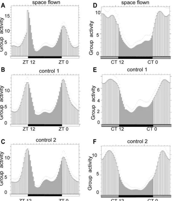
Drosophilaactivity rhythm. FaasX analysis showed that space flown flies exhibited normal lo-comotor activity rhythms as a group in both LD and DD conditions when compared with those of the two control groups (Fig. 1), suggesting that there was no significant change in
Dro-sophilalocomotor activity rhythm after spaceflight if flies were maintained in LD condition.
However, two facts must be considered. First, our behavior test was started 48 h after landing, during which time space flown flies might have re-adapted to the Earth’s environment. Second, our activity test was conducted in LD conditions in the first five days, and since light is a strong Zeitgeber, the effect of spaceflight on activity rhythm might be masked by the light/dark cycle. Previous research on the activity rhythm of a type of desert beetle during spaceflight reported
Fig 1. FaasX analysis showed that Drosophila locomotor activity rhythm was not changed after spaceflight.Space flown flies exhibited similar locomotor activity rhythm as the two control groups under both LD (A-C) and DD (D-F) conditions. White and black bars stand for daytime and nighttime, grey bar stands for subjective daytime in DD. Group activity was calculated by mean of five days. Two-way ANOVA was applied to the activity data for space flown and control flies at different Zeitgeber time, and the factor of treatment (space flown/control) was not significant by the conventional standard (p<0.05).
doi:10.1371/journal.pone.0121600.g001
tree-running period (tau) differences caused by gravitation field changes (μg and 2G) in
con-stant light conditions (LL and DD), but not in LD conditions, suggesting strong interference of light on the study of the effect of other Zeitgebers on the circadian clock [30]. Thus, due to the limitation in our experimental design, we cannot conclusively rule out the possibility that spaceflight might somehow affect the activity rhythm ofDrosophila. It would be better to
mon-itorDrosophilalocomotor activity rhythm in constant darkness during spaceflight. Clocklab2
analysis showed that the ratio of flies that exhibited rhythmic locomotor activity was relatively low in all three groups (42.86% for space flown flies, 21% for control-1 group flies, and 50% for control-2 group flies). Considering that the tested flies were more than 30 days old, our results were consistent with published findings that older flies exhibited weaker activity rhythms than younger flies [36].
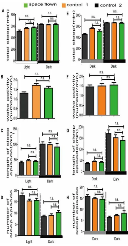
We also investigated sleep patterns and three major sleep parameters in space flown flies.
Drosophilasleep was defined as an interval of 5 min or more of behavioral immobility. We
ob-served no differences in total sleep time among space flown flies and the two control groups in either LD or DD conditions (Figs.2A, EandS1). Major sleep parameters, including length and number of sleep episodes, were also similar among the three groups (Fig. 2C, D, G, and H). Al-though there were slight differences in the length and number of nighttime sleep episodes be-tween space flown flies and the two control flies (Fig. 2C-D, 2G-H), they were statistically insignificant. The small sample number (15 space flown flies) and the re-adaption to the Earth’s environment could be the reason for the statistical insignificance. Further research is being conducted on the effect of gravity onDrosophilasleep using simulated microgravity in our lab.
Unlike astronauts, who usually have a busy schedule and live in an environment with a dis-rupted light/dark cycle during spaceflight, theDrosophiladid not experience intense labor pressure during spaceflight. Thus, the effect of a busy work schedule and disrupted circadian cycle on circadian rhythm and sleep could be excluded. Although no statistically significant re-sults were obtained in our behavior test, we believe that with careful experimental design and development of improved monitoring systems [37],Drosophilacould be a useful animal model for investigating the effect of space on animal behavior phenotypes such as sleep.
TAG level and lipid metabolism
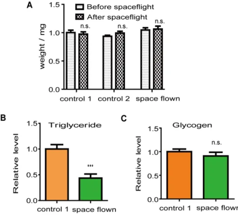
To study whether there was any change inDrosophilametabolism after spaceflight, we mea-sured the levels of two major energy stores (TAG and glycogen) in space flown and control-1 flies during a 24-h time period. The average TAG level of space flown flies was significantly lower than the average TAG level of control-1 flies (Fig. 3B). The difference in the TAG level was not caused by fly size differences since the weight between both groups was similar (Fig. 3A). In contrast, there was no difference in glycogen levels between the two fly groups (Fig. 3C).
Fig 2. Effect of spaceflight on major sleep parameters under both LD (A-D) and DD (E-H) conditions.
The average total sleep time (A, E), average length of sleep episode (C, G), and average sleep episode number (D, H) were calculated based on theDrosophilasleep definition. Green, orange and grey boxes represent data of space flown, control-1, and control 2 flies, respectively. White and black bars stand for daytime and nighttime, grey bar stands for subjective daytime in DD. Statistical significance was determined by two-tailed Student’s t-test with unequal variance. n.s. means no statistically significant differences. Error bars represent SEM.
doi:10.1371/journal.pone.0121600.g002
Another possibility is that increased activity during spaceflight led to the loss of energy stores in space flown flies. It has been reported that flies exhibit increased activity under a microgravi-ty environment [42]. Although our locomotor activity results showed that space flown flies were not more active than ground controls after they returned to Earth, they were monitored 48 h after landing, so we cannot rule out the possibility thatDrosophilalocomotor activity was increased during spaceflight, and the decreased TAG levels were caused by increased activity during spaceflight.
Lifespan
We also examined changes inDrosophilalifespan after spaceflight.Fig. 4presents the survival curves and average lifespan of the space flown and two control groups. Results showed that space flown flies lived slightly longer than the two control groups. The average lifespan of space flown flies was 51.82 ±0.93 days, while the average lifespan of control-1 and control-2 flies was 47.96 ±0.82 and 48.71 ±0.88 days, respectively. Although the small number of tested animals reduced the credibility of the results and further repeat experiments are necessary, the lifespan differences between space flown and control flies were statistically significant, indicating there might be a weak effect of spaceflight onDrosophilalifespan.
It was reported thatDrosophilalifespan can be regulated by both the insulin dependent pathway and insulin independent dietary restriction [43]. Considering that no mRNA level
Fig 3. Spaceflight affectedDrosophilalipid storage.(A), Space flown flies had similar weights as those of the two control groups. (B), Space flown flies had lower TAG levels compared with those of control-1 flies. (C), Space flown flies had similar body glycogen levels compared with those of control-1 flies. Both TAG and glycogen levels were normalized to the protein level. For each fly group, at least three independent groups of five fly bodies were analyzed.***indicates significant differences (p<0.001). Statistical significance was determined by two-tailed Student’s t-test with unequal variance.
changes in the insulin-pathway related genes were detected by microarray, except forilp3, and metabolism tests showed that TAG levels decreased in space flown flies, we hypothesized that the TAG level decrease indicated a dietary restriction during spaceflight, which, in turn, caused a lifespan extension through insulin independent mechanisms. However, further evidence is needed to confirm this hypothesis.
How spaceflight affects aging is still largely unknown. It was once widely accepted that the space environment accelerates the aging process [44,45]. However, there has been little evi-dence that directly proves whether the space environment influences animal lifespan. Recent research reported that genes involved in nematode lifespan extension were up-regulated after spaceflight [46], indicating that spaceflight might extend the life ofCaenorhabditis elegans. Our results, if they could be repeated, also suggested that spaceflight could extendDrosophila life-span. Thus, the opinion that space accelerates the aging process is becoming controversial and
Fig 4. Spaceflight affectedDrosophilalifespan.Space flown flies had significantly extended lifespan. (A): Survival curve for space flown, control-1 and control-2 flies; (B): Average lifespan comparison. Error bars represent SEM.***indicates significant differences (p<0.001). Statistical significance was determined by two-tailed Student’s t-test with unequal variance.
doi:10.1371/journal.pone.0121600.g004
needs more supporting evidence. With the increasing duration of spaceflight missions, such as human exploration of other planets or colonization of the moon, studies on the effect of the space environment on lifespan has become of increasing medical importance and will attract growing attention in the future.
Microarray analysis
We used microarray to profile gene expression in the heads of space flown flies as well as the two control groups. Samples from six time points during a 24-h period under LD conditions were hybridized with the Affymetrix GeneChipDrosophilaGenome Array 2.0 containing 18,500 probe sets. Each sample was hybridized to two chips to test reproducibility. The mean hybridization signal strength and the SEM for each probe set were calculated from the dupli-cate hybridizations.S1 Tableshows the expression levels of the 4483 genes obtained from the microarray experiment.
In the experimental design, we started sample collection for RNA extraction almost 50 h after landing. During this period of time flies underwent some re-adaptation to the Earth’s gravitational environment, which made it difficult to evaluate the role of spaceflight on gene expression changes. However, many previous studies have found that some physiological ef-fects of spaceflight might last much longer. For example, post flight swelling and elevation of human calf muscle transverse relaxation time persisted for several weeks after flight [47], and at two weeks post recovery, the hamstrings and intrinsic lower back muscles were still below baseline [48]. Studies onDrosophilaimmune response also found that the immune system of space flown flies function more effectively than that of ground control flies 3 days post landing [34]. Furthermore, the lipid store differences we detected also suggested that some physiologi-cal changes in space flown flies could persist, even 48h after landing. Hence, we expected that our microarray analysis would still detect some long-term effects of spaceflight onDrosophila
gene expression.
Effect of sampling time on the analysis of gene expression differences
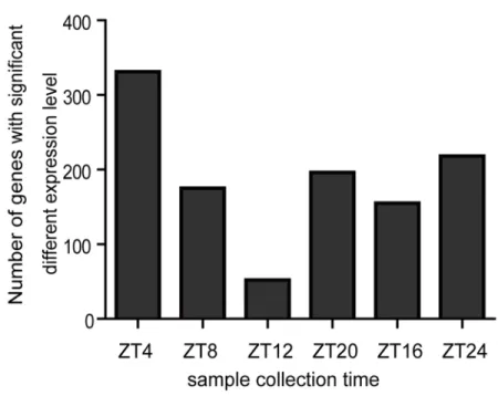
It is well known that many genes exhibit a circadian rhythmic expression pattern inDrosophila, thus gene expression comparison between two fly samples could vary according to the different sampling times over a 24-h time period under LD conditions. Here we searched for genes with significant expression level changes, defined as exhibiting more than a 1.5-fold change between space flown and control samples, at different time points, and then counted the number of genes with significant expression level changes at each time point (Fig. 5). The numbers of genes with significant expression level changes varied according to the different sampling times. At ZT4, for example, there were 330 genes with significant expression level change (up 127; down 203). At ZT12, however, the number decreased to only 52 genes (up 32; down 20). Our results showed that sampling time can greatly affect gene expression analysis results, sug-gesting the necessity to follow an appropriate sampling procedure in the investigation of the ef-fect of spaceflight on gene expression. Not only should space flown samples be collected as early as possible after they return to Earth, but samples should also be collected at either the same Zeitgeber time or multiple time points during a 24-h period. Otherwise, it is difficult to obtain repeatable data even if all other experimental conditions are strictly controlled.
the activity curve might be different, which might, in turn, result in expression level change dif-ferences of genes involved in such a physiological process. Secondly, spaceflight might cause phase changes in some physiological rhythms. Thus, genes involved in these processes might also experience phase changes in space flown samples. As a result, the expression of these genes at certain time points might show significant differences between space flown and control sam-ples. It is unlikely that the differences were caused by the alleviation of the spaceflight effect after landing, since the number of genes with significant expression level change increased again after reaching the trough at ZT12. However, as the number of genes with significant ex-pression level change at the last sampling time point were less than that at the first time point, it is still possible that the differences in time since landing could also contribute to
this phenomenon.
Fig 5. Effect of sampling time on the analysis of gene expression differences between space flown and control flies.(A) Gene expression levels in head samples collected at six time points during a 24-h period under LD conditions were compared between space flown flies and the two control groups. The number of genes that exhibited significant expression level change (>1.5 fold) at each time point were recorded. (B) Fisher’s exact test was used to determine the statistical significance of the differences between the data from every two time points. P<0.05 indicates significant differences.
doi:10.1371/journal.pone.0121600.g005
Spaceflight affected
Drosophila
gene expression
As shown above, gene expression level differences could vary according to the different sam-pling time in LD conditions, and measuring expression differences at only one single time point was unsatisfactory. Thus, we combined the gene expression data of all six time points and calculated the average expression level of each gene obtained from the microarray results. We used both statistical (unpaired Student’st-test, P<0.05) and fold change (more than
1.5-fold) thresholds as criteria for selecting genes with significant level change. We identified 54 genes with significant level change between space flown and control-1 flies (37 genes, up; 17 genes, down) (S2 Table) and 55 genes between space flown and control-2 flies (30 genes, up; 25 genes, down) (S3 Table). To reduce false positives, we next only selected genes that appeared on both lists, which reduced the number to 12. We listed these as genes with significant mRNA level change after spaceflight (Table 1).
We noticed a small overlap between the results obtained using the different controls. It is worth noting, however, that control-2 flies were maintained under ideal conditions in our lab incubator, while control-1 flies were maintained in similar conditions as the space flown flies. Furthermore, control-1 flies also underwent a simulated launching process and the same ground travel experience as space flown flies. Thus, the small overlap between the gene expres-sion data between the two control groups might reflect the effect of stress on gene expresexpres-sion.
Compared with other microarray reports investigating the space effect on gene expression [33,49], we detected far fewer genes with significant mRNA level change after spaceflight. This could be due to different experimental design. Firstly, we compared the average mRNA level of data from six time points in a 24-h period, not just data at a specific time point. As discussed above, our sampling design can exclude of sampling time on gene expression analysis. Second-ly, the short-term effect of spaceflight on gene expression was excluded in our study. Due to technical limitations, sample collection began 48 h after landing, much later than in the other reports. Thus, the expression level of many genes may have already recovered before we com-menced sample collection. Thirdly, unlike brain samples, there are many tissue and cell types
inDrosophilaheads, so some spaceflight effects in certain tissues or cells might be masked.
Fi-nally, for some reason, we only obtained reliable gene expression data for 4483 genes from our
Table 1. Genes with significantly changed expression level after spaceflight.
Representative Public ID
Gene Symbol
Spaceflight /control-1
Spaceflight /control-2
Function
CG18290-RA Act87E 1.58 1.68 Structural constituent of cytos- keleton, cytoskeleton organization
CG31149-RA CG42613 0.65 0.51 Low-density lipoprotein (LDL) receptor class A repeat
CG4784-RA Cpr72Ec 1.94 1.91 Structural constituent of chitin-based cuticle
CG14167-RA Ilp3 1.62 2.45 Insulin receptor binding, female mating behavior
CG17461-RA Kif3C 0.65 0.53 Microtubule motor activity, neuron projection morphogenesis
CG30179-RA Mlp60A 1.71 1.55 Zinc ion binding, muscle tissue development
CG2380-RA NfI 0.63 0.62 Sequence-specific DNA binding transcription factor activity
CG10806-RB Nha1 1.70 2.10 Sodium: hydrogen antiporter activity
CG8462-RA Obp56e 1.75 2.26 Odorant binding,multi cellular org- anism reproduction
CG1748-RA RhoGAP102A 0.63 0.56 Rho GTPase activator activity
CT36054 SK 1.84 1.76 Photoreceptor activity,regulation of membrane potential in photo-receptor cell
CG15427-RC tutl 1.66 1.79 Synaptic target recognition,regulation of dendrite morphogen- esis,axon guidance and defascicu- lation,larval and adult behavior regulation
microarray hybridization experiment, which also contributed to the shorter list of genes affect-ed by spaceflight.
Functional analysis of the 12 genes revealed one gene,ilp3, that might be involved in
Dro-sophilametabolism and lifespan regulation [50].Drosophilaregulates energy metabolism using
a system similar to the insulin/glucagon system used in mammals [51]. There are seven insu-lin-like peptides (ILPs) inDrosophila, among themilp2has the most potent function in regu-lating lifespan and metabolism [52]. Althoughilp3is not directly involved in lifespan
regulation, its expression is closely related toilp2mRNA level. Knock-down ofilp2has been re-ported to result in the up-regulation ofilp3via direct autocrine regulation through the insulin signaling pathway [50], indicating that the increase inilp3mRNA level might be caused by a compensation effect of decreasedilp2. Although we did not detect any mRNA level change of other ILPs or genes involved in the insulin signal transduction pathway, it is possible that changes in insulin related genes in certain cell types might be masked as we used a mixture of various cell types as the source of our mRNA sample for microarray. Alternatively, since the re-duction ofilp2cannot lead to lifespan extension, theilp3mRNA change in space flown flies might have nothing to do with the low lipid level and longer lifespan observed. Further re-search is needed to unveil the molecular mechanism and genes involved.
Major clock genes were unaffected after spaceflight
To determine whether there was any difference in theDrosophilacircadian clock after space-flight, we investigated the rhythmic expression pattern of major clock genes (per,tim,vri, and
cry) in space flown and control flies. As shown inS2 Fig, all four clock genes exhibited rhyth-mic patterns in both space flown and control flies at a similar phase, suggesting that the func-tion of the circadian oscillator in fly heads remained normal in LD condifunc-tions 48 h after landing. However, since 48 h is enough time for the re-adaption of theDrosophilacircadian clock from the space to Earth environment, we cannot exclude the possibility that the Drosoph-ilacircadian clock might be changed during spaceflight. Future microarray analysis using sam-ples collected during spaceflight is necessary to make such conclusions.
Identification of cyclic genes
Due to the insufficient number of space flown flies, we only collected samples at six time points in a 24-h period. However, most software used for detecting circadian expressed genes requires at least 12 samples in a 24- to 48-h period. Thus, we needed special analysis software to search circadian expressed genes from our microarray database. We chose STEM software developed for the analysis of short time series gene expression data [35]. STEM can identify statistically significant temporal expression profiles and associated genes. In our analysis, STEM first se-lected 25 distinct and representative temporal expression profiles as model profiles indepen-dent of the data; then assigned each of the 4483 genes passing the filtering criteria to the model profile that most closely matched the gene’s expression profile; and finally determined which model profiles had significantly more assigned genes compared to the average number assigned to the model profile, with significant model profiles highlighted in color. The analysis results are presented inS3A-S3C Fig.
STEM could not define the circadian expression profiles, so we established our own criteri-on and defined circadian expressicriteri-on profiles as profiles with expressicriteri-on patterns close to a co-sine wave. Using this criterion, we selected six model circadian expression profiles: profile 0, 4, 8, 16, 22, and 24. For space flown flies, only one circadian expression profile (profile 16) be-longed to the significant model profiles, but more circadian expression profiles bebe-longed to sig-nificant model profiles for the two control groups (S3A-S3C Fig.). Genes grouped into
circadian expression model profiles of the space flown flies and the two control groups are listed inS4 Tableand defined as circadian expressed genes. In total, there were 104, 90 and 335 circadian expressed genes in space flown, control-1 and control-2 flies, respectively.
From the circadian expressed gene lists, most circadian expressed genes in space flown flies were not on the circadian expressed gene lists of the two control groups. Only 21 genes were circadian expressed in all three samples (S3D Fig.), including the four circadian clock genes in-vestigated above. Since circadian expressed genes are generally accepted as output genes of the circadian clock system, our results indicated that although theDrosophilacircadian core oscil-lator was not changed after spaceflight, there were changes in the circadian clock output path-way after spaceflight.
We also investigated the phase differences of genes that showed circadian expression pattern in both space flown flies and at least one control group, which showed that some genes exhib-ited phase changes in space flown flies. For example, the peak level ofcpr60dmRNA in space flown flies was at ZT4, while in the two control groups, the peak level appeared at ZT20 and ZT16, respectively, suggesting an 8 to 12 h phase change (S4A Fig.). Furthermore,mus209 ex-hibited circadian expression pattern in both space flown and control-1 flies but had a 4 to 8 h phase advance in space flown flies (S4B Fig.). Since the four major circadian clock genes in fly heads did not show any phase shifts, it was unlikely that the phase of theDrosophilacentral clock system was changed after spaceflight. The circadian clock exists not only in the brain, but also in many other peripheral tissues [53–55], and some environmental stimuli can only entrain the peripheral clock with no effect on the central clock [56]. Thus, it is possible that during spaceflight certain peripheral clocks experienced a phase change, and in turn caused the phase changes of some circadian output genes highly expressed in these peripheral tissues.
In this paper, we tested the effect of spaceflight on the circadian output pathway using two different assays. The locomotor activity rhythm of space flown flies was normal, suggesting that either the effect of spaceflight onDrosophilalocomotor activity rhythm was too weak to be detected, or locomotor activity rhythm could quickly readapt to the Earth’s environment. However, gene expression analysis detected rhythmic changes of some circadian output genes in space flown flies, indicating that assays at the molecular level were more sensitive than be-havioral assays in detecting the effect of spaceflight on the circadian clock output pathway. However, with our current experimental design and results, we could not determine whether such changes in the circadian output pathway were caused directly by spaceflight or indirectly by the result of physiological changes after spaceflight. Finally, although we divided our sam-ples into two pools before RNA extraction, this was not a biological repetition. Repeat microar-ray experimentation is needed to verify our results.
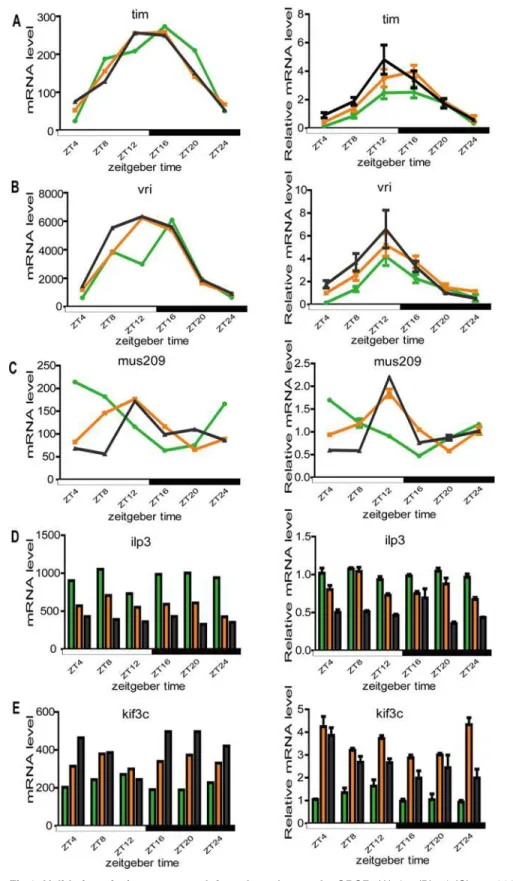
QPCR
Since microarray is a semi-quantitative tool for examining expression differences, we per-formed QPCR to confirm the observed gene expression differences and phase changes in rhythmic expressed genes from our microarray analysis. Two major circadian clock genes (tim
Fig 6. Validation of microarray result for selected genes by QPCR.(A)tim; (B)vri; (C)mus209; (D)ilp3; (E)kif3c. For each gene, the left plot is the microarray result and the right is the QPCR result. For each graph, the green, orange, and blank lines or histograms represent the space flown group, control-1 group and control-2 group, respectively.
doi:10.1371/journal.pone.0121600.g006
Supporting Information
S1 Fig. Spaceflight had no effect onDrosophilasleep profile.Space flown flies exhibited simi-lar sleep profiles as the two control groups under both LD (A) and DD (B) conditions.
Dro-sophilasleep was defined as an interval of 5 min or more of behavioral immobility. Sleep
profiles were analyzed using Pysolo 0.9 software. White and black bars stand for daytime and nighttime, grey bar stands for subjective daytime in DD. Error bars represent SEM.
(TIF)
S2 Fig. Gene expression curves from microarray data showed that the rhythmic expression of major circadian clock genes did not change after spaceflight.(A)vri; (B)tim; (C)per; (D)
cry.
(TIF)
S3 Fig. Model profile overview of gene expression analysis at different time points under LD conditions.(A), space flown flies; (B), control-1 flies; and (C) control-2 flies. The data was sampled at six time points: ZT0, ZT4, ZT8, ZT12, ZT16, and ZT20. The colored profiles had a statistically significant number of genes assigned. We defined six profiles (profile 0, 4, 8, 16, 22, 24) as circadian expression profiles, and genes assigned into these six profiles were identified as circadian expressed genes. (D) Rhythmic expression genes of space flown flies and two control groups. The blue, red and green circles represent the rhythmic expressed genes of space flown, control-1, and control-2 flies, respectively. The numbers in the non-overlapping part of the cir-cle represent the numbers of genes that showed circadian expression patterns only in one fly group. The numbers in the overlapping part of different circles represent the numbers of genes that showed circadian expression patterns in more than two fly groups.
(TIF)
S4 Fig. Phase change of some circadian expressed genes after spaceflight.(A)cpr60d(B)
mus209.
(TIF)
S1 Table. Microarray results of 4483 genes of space flown flies and the two control groups at six time points.
(XLS)
S2 Table. List of genes that showed significantly different expression levels between space flown and control-1 flies.
(XLS)
S3 Table. List of genes that showed significantly different expression levels between space flown and control-2 flies.
(XLS)
S4 Table. List of genes of space flown flies and the two control groups that belong to the six circadian expression profiles.
(XLS)
Author Contributions
References
1. Ohnishi K, Ohnishi T. The biological effects of space radiation during long stays inspace. Biol Sci Space. 2004; 18: 201–5. PMID:15858386
2. LeBlanc AD, Spector ER, Evans HJ, Sibonga JD. Skeletal responses to space flight and the bed rest analog: a review. J Musculoskelet NeuronalInteract. 2007; 7: 33–47. PMID:17396004
3. Morey-Holton ER, Hill EL, Souza KA. Animals and spaceflight: from survival to understanding. J Muscu-loskelet Neuronal Interact. 2007; 7: 17–15. PMID:17396002
4. Goel N, Dinges DF. Predicting Risk in Space: Genetic Markers for Differential Vulnerability to Sleep Re-striction. Acta Astronaut. 2012; 77: 207–13. PMID:23524958
5. Dayanandan P. Gravitational biology and space life sciences: current status and implications for the In-dian space program. J Biosci. 2011; 36: 911–9. PMID:22116289
6. Maury E, Ramsey KM, Bass J. Circadian rhythms and metabolic syndrome from experimental genetics to human disease. Circ Res. 2010; 106: 447–462. doi:10.1161/CIRCRESAHA.109.208355PMID: 20167942
7. Mavroudis PD, Scheff JD, Calvano SE, Androulakis IP. Systems biology of circadian-immune interac-tions. J Innate Immun. 2013; 5:153–62. doi:10.1159/000342427PMID:23006670
8. Campos Costa I, Nogueira Carvalho H, Fernandes L. Aging, circadian rhythms and depressive disor-ders: a review. Am J Neurodegener Dis. 2013; 2: 228–246. PMID:24319642
9. Paschos GK, FitzGerald GA. Circadian clocks and vascular function. Circ Res. 2010; 6: 833–41.
10. Gundel A, Polyakov VV, Zulley J. The alteration of human sleep and circadian rhythms during space-flight. J Sleep Res. 1997; 6: 1–8. PMID:9125693
11. Dijk DJ, Neri DF, Wyatt JK, Ronda JM, Riel E, Ritz-De Cecco A, et al. Sleep, performance, circadian rhythms, and light-dark cycles during two space shuttle flights. Am J Physiol Regul Integr Comp Phy-siol. 2001; 281: R1647–1664. PMID:11641138
12. Basner M, Dinges DF, Mollicone D, Ecker A, Jones CW, Hyder EC, et al. Mars 520-d mission simulation reveals protracted crew hypokinesis and alterations of sleep duration and timing. Proc Natl Acad Sci U S A. 2013; 110: 2635–40. doi:10.1073/pnas.1212646110PMID:23297197
13. Stampi C. Sleep and circadian rhythms in space. J Clin Pharmacol. 1994; 34: 518–34. PMID:8089264
14. Monk TH, Buysse DJ, Billy BD, Kennedy KS, Willrich LM. Sleep and circadian rhythms in four orbiting astronauts. J Biol Rhythms.1998; 13: 188–201. PMID:9615283
15. Schimmerling W, Cucinotta FA, Wilson JW. Radiation risk and human space exploration. Adv Space Res. 2003; 31: 27–34. PMID:12577903
16. Sulzman FM, Ellman D, Fuller CA, Moore-Ede MC, Wassmer G.Neurosporacircadian rhythms in space: a reexamination of the endogenous-exogenous question. Science. 1984; 225: 232–34. PMID: 11540800
17. Fuller CA, Murakami DM, Sulzman FM. Gravitational biology and the mammalian circadian timing sys-tem. Adv Space Res.1989; 9: 283–92. PMID:11537343
18. Fuller CA. The primate circadian timekeeping system in a hyper dynamic environment. Physiolo-gist.1984; 27: 93–94.
19. Murakami DM and Fuller CA. The effect of 2G on mouse circadian rhythms. J Gravit Physio. 2000; l7: 79–85. PMID:12124188
20. Holley DC, DeRoshia CW, Moran MM, Wade CE. Chronic centrifugation (hypergravity) disrupts the cir-cadian system of the rat. J Appl Physiol. 2003; 95: 1266–78. PMID:12794036
21. Fuller CA, Murakami DM, and Demaria-Pesce VH. Entrainment of circadian rhythms in the rat by one hour G pulses. Physiologist. 1992; 35(1 Suppl): 63–64.
22. Hoban-Higgins TM, Murakami DM, Tandon T, and Fuller CA. Acute exposure to 2G phase shifts the rat circadian timing system. J Gravi Physiol. 1995; 2: 58–59.
23. Casey T, Zakrzewska EI, Maple RL, Lintault L, Wade CE, Baer LA, et al. Hypergravity disruption of homeorhetic adaptations to lactation in rat dams include changes in circadian clocks. Biol Open. 2012; 1: 570–81. doi:10.1242/bio.2012687PMID:23213450
24. Fuller CA, Hoban-Higgins TM, Klimovitsky VY, Griffin DW, Alpatov AM. Primate circadian rhythms dur-ing spaceflight: results from Cosmos 2044 and 2229. J Appl Physiol. 1996; 81: 188–193. PMID: 8828664
25. Monk TH, Kennedy KS, Rose LR, Linenger JM. Decreased human circadian pacemaker influence after 100 days in space: a case study. Psychosom Med. 2001; 63: 881–5. PMID:11719625
26. Alpatov AM, Rietveld WJ, Oryntaeva LB. Impact of microgravity and hypergravity on free-running circa-dian rhythm of the desert beetleTrigonoscelis gigas Reitt. Biol Rhythm Res. 1994; 25: 68–177.
27. Hoban-Higgins TM, Alpatov AM, Wassmer GT, Rietveld WJ, Fuller CA. Gravity and light effects on the circadian clock of a desert beetle,Trigonoscelis gigas. J Insect Physiol.2003; 49: 671–675. PMID: 12837319
28. Karemaker JM, Berecki-Gisolf J. 24-h blood pressure in Space: the dark side of being an astronaut. Respir Physiol Neurobiol. 2009; 169S: S55–S58. doi:10.1016/j.resp.2009.05.006PMID:19481180
29. Agarwal R. Regulation of circadian blood pressure: from mice to astronauts. Curr Opin Nephrol Hyper-tens. 2010; 19: 51–8. doi:10.1097/MNH.0b013e3283336ddbPMID:19864947
30. Williams JA, Sehgal A. Molecular components of the circadian system inDrosophila. Annu Rev Physiol. 2001; 63: 729–55. PMID:11181974
31. Crocker A and Sehgal A. Genetic analysis of sleep. Genes Dev. 2010; 24: 1220–35. doi:10.1101/gad. 1913110PMID:20551171
32. Leandro LJ, Szewczyk NJ, Benguría A, Herranz R, Laván D, Medina FJ, et al. Comparative analysis of Drosophila melanogasterandCaenorhabditis elegansgene expression experiments in the European Soyuz flights to the International Space Station. Adv Space Res. 2007; 40: 506–12. PMID:18084631
33. Ikenaga M, Yoshikawa I, Kojo M, Ayaki T, Ryo H, Ishizaki K, et al. Mutations induced inDrosophila dur-ing space flight. Biol Sci Space.1997; 11: 346–50. PMID:11541768
34. Marcu O, Lera MP, Sanchez ME, Levic E, Higgins LA, Shmygelska A, et al. Innate immune responses ofDrosophila melanogasterare altered by spaceflight. PloS One. 2011; 6: e15361. doi:10.1371/ journal.pone.0015361PMID:21264297
35. Ernst J, Bar-Joseph Z. STEM: a tool for the analysis of short time series gene expression data. BMC Bioinformtaics. 2006; 7: 191
36. Rakshit K, Krishnan N, Guzik EM, Pyza E, Giebultowicz JM. Effects of aging on the molecular circadian oscillations inDrosophila. Chronobiol Int. 2012; 29: 5–14. doi:10.3109/07420528.2011.635237PMID: 22217096
37. Inan T, Etemadi M, Sanchez E, Marcu O, Bhattacharya S, Kovacs A. A miniaturized video system for monitoring the locomotor activity of walkingDrosophila melanogasterin space and terrestrial settings. IEEE Trans Biomed Eng. 2009; 56(2): 522–24. doi:10.1109/TBME.2008.2006018PMID:19272912
38. Leonard JI, Leach CS and Rambaut PC. Quantification of tissue loss during prolonged space flight. Am J Clin Nutr. 1983; 38: 667. PMID:6637859
39. Matsumoto A, Storch KJ, Stolfi A, Mohler SR, Frey MA, Stein TP. Weight loss in humans in space. Aviat Space EnvironMed. 2011; 82: 615–21. PMID:21702312
40. Stein TP, Leskiw MJ, Schluter MD, Hoyt RW, Lane HW, Gretebeck RE, et al. Energy expenditure and balance during spaceflight on the space shuttle. Am J Physiol. 1999; 276: R1739–48. PMID:10362755
41. Da Silva MS, Zimmerman PM, Meguid MM, Nandi J, Ohinata K, Xu Y, et al. Anorexia in space and pos-sible etiologies: an overview. Nutrition. 2002; 18: 805–13. PMID:12361771
42. Miller MS and Keller TS. MeasuringDrosophila(fruit fly) activity during microgravity exposure. J Gravit Physiol. 1999; 6: P99–100. PMID:11543046
43. Min K-J, Yamamoto R, Buch S, Pankratz M, Tatar M.Drosophilalifespan control by dietary restriction independent of insulin-like signaling. Aging Cell. 2008; 7: 199–206. doi:10.1111/j.1474-9726.2008. 00373.xPMID:18221413
44. Bourg EL. A review of the effects of microgravity and of hypergravity on aging and longevity. Exp Ger-ontol.1999; 34: 319–36. PMID:10433387
45. Vernikos J, Schneider VS. Space, gravity and the physiology of aging: parallel or convergent disci-plines? A mini-review. Gerontology. 2010; 56: 157–66. doi:10.1159/000252852PMID:19851058
46. Honda Y, Higashibata A, Matsunaga Y, Yonezawa Y, Kawano T, Higashitani A,et al. Genes down-regulated in spaceflight are involved in the control of longevity inCaenorhabditis elegans. Sci Rep. 2012; 2: 487. doi:10.1038/srep00487PMID:22768380
47. LeBlanc A, Lin C, Shackelford L, Sinitsyn V, Evans H, Belichenko O, et al. Muscle volume, MRI relaxa-tion times (T2) and body composirelaxa-tion after spaceflight. J. Appl. Physiol. 2000; 89(6): 2158–64. PMID: 11090562
48. LeBlanc A, Rowe R, Schneider V, Evans H, Hedrick T. Regional muscle loss after short duration space-flight. Aviat Space Environ Med. 1995; 66(12): 1151–4. PMID:8747608
50. Broughton S, Alic N, Slack C, Bass T, Ikeya T, Vinti G, et al. Reduction of DILP2 inDrosophilaTriages a metabolic phenotype from lifespan revealing redundancy and compensation among DILPs. PLoS One. 2008; 3: e3721. doi:10.1371/journal.pone.0003721PMID:19005568
51. Wu Q and Brown MR. Signaling and function of insulin-like peptides in insects. Annu Rev Entomol. 2006; 51: 1–24. PMID:16332201
52. Broughton SJ, Piper MD, Ikeya T, Bass TM, Jacobson J, Driege Y, et al. Longer lifespan, altered metab-olism, and stress resistance inDrosophilafrom ablation of cells making insulin-like ligands. Proc Natl Acad Sci USA. 2005; 102: 3105–10. PMID:15708981
53. Balsalobre A. Clock genes in mammalian peripheral tissues. Cell Tissue Res. 2002; 309: 193–99. PMID:12111549
54. Giebultowicz JM. Molecular mechanism and cellular distribution of insect circadian clock. Annu Rev Entomol. 2000; 45: 769–93. PMID:10761596
55. Xu K, Zheng X, and Sehgal A. Regulation of feeding and metabolism by neuronal and peripheral clocks inDrosophila. Cell Metab. 2008; 8: 289–300. doi:10.1016/j.cmet.2008.09.006PMID:18840359
56. Xu K, DiAngelo JR, Hughes ME, Hogenesch JB, Sehgal A. The circadian clock interacts with metabolic physiology to influence reproductive fitness. Cell Metab. 2011; 13: 639–54. doi:10.1016/j.cmet.2011. 05.001PMID:21641546