Drosophila Spaghetti and Doubletime Link
the Circadian Clock and Light to Caspases,
Apoptosis and Tauopathy
John C. Means1, Anandakrishnan Venkatesan1¤, Bryan Gerdes1, Jin-Yuan Fan1, Edward S. Bjes1, Jeffrey L. Price1,2*
1Division of Molecular Biology and Biochemistry, School of Biological Sciences, University of Missouri-Kansas City, Missouri-Kansas City, Missouri, United States of America,2Department of Neurology and Cognitive Neuroscience, School of Medicine, University of Missouri-Kansas City, Kansas City, Missouri, United States of America
¤ Current Address: Department of Physiology, University of Texas Health Science Center at San Antonio STRF- Greehey North Campus, San Antonio, Texas, United States of America
*pricejL@umkc.edu
Abstract
While circadian dysfunction and neurodegeneration are correlated, the mechanism for this is not understood. It is not known if age-dependent circadian dysfunction leads to neurode-generation or vice-versa, and the proteins that mediate the effect remain unidentified. Here, we show that the knock-down of a regulator (spag) of the circadian kinase Dbt in circadian cells lowers Dbt levels abnormally, lengthens circadian rhythms and causes expression of activated initiator caspase (Dronc) in the optic lobes during the middle of the day or after light pulses at night. Likewise, reduced Dbt activity lengthens circadian period and causes expression of activated Dronc, and a loss-of-function mutation inClkalso leads to expres-sion of activated Dronc in a light-dependent manner. Genetic epistasis experiments place Dbt downstream of Spag in the pathway, and Spag-dependent reductions of Dbt are shown to require the proteasome. Importantly, activated Dronc expression due to reduced Spag or Dbt activity occurs in cells that do not express thespagRNAi or dominant negative Dbt and requires PDF neuropeptide signaling from the same neurons that support behavioral rhythms. Furthermore, reduction of Dbt or Spag activity leads to Dronc-dependent Drosoph-ila Tau cleavage and enhanced neurodegeneration produced by human Tau in a fly eye model for tauopathy. Aging flies with lowered Dbt or Spag function show markers of cell death as well as behavioral deficits and shortened lifespans, and even old wild type flies ex-hibit Dbt modification and activated caspase at particular times of day. These results sug-gest that Dbt suppresses expression of activated Dronc to prevent Tau cleavage, and that the circadian clock defects confer sensitivity to expression of activated Dronc in response to prolonged light. They establish a link between the circadian clock factors, light, cell death pathways and Tau toxicity, potentially via dysregulation of circadian neuronal remodeling in the optic lobes.
OPEN ACCESS
Citation:Means JC, Venkatesan A, Gerdes B, Fan
J-Y, Bjes ES, Price JL (2015) Drosophila Spaghetti and Doubletime Link the Circadian Clock and Light to Caspases, Apoptosis and Tauopathy. PLoS Genet 11 (5): e1005171. doi:10.1371/journal.pgen.1005171
Editor:Paul H. Taghert, Washington University Medical School, UNITED STATES
Received:August 26, 2014
Accepted:March 25, 2015
Published:May 7, 2015
Copyright:© 2015 Means et al. This is an open access article distributed under the terms of the
Creative Commons Attribution License, which permits unrestricted use, distribution, and reproduction in any medium, provided the original author and source are credited.
Data Availability Statement:All relevant data are within the paper and its Supporting Information files.
Funding:The work was funded by NIH grant
R01GM090277 and a University of Missouri Research Board grant to JLP. The funders had no role in study design, data collection and analysis, decision to publish, or preparation of the manuscript.
Competing Interests:The authors have declared
Author Summary
Alzheimer’s disease is the most common cause of dementia in the aging population. It is a progressive neurodegenerative disorder that attacks the brain neurons, resulting in loss of memory, thinking and behavioral changes. One pathological hallmark is aggregation of the microtubule-associated protein Tau. A growing body of evidence highlights the impor-tance of caspase-dependent Tau truncation in initiation and potentiation of Tau aggrega-tion. Here we use the fruit fly Drosophila to examine the links between circadian rhythms, aging, apoptosis and Alzheimer’s Disease. We identified a regulator (spag)of the circadian kinase Dbt that functions to stabilize Dbt during the middle of the day. In addition, the caspase Dronc is regulated by Dbt and Spag and, when activated by reduction of either, targets Tau for cleavage, leading to behavioral deficits and shortened lifespans. The expres-sion of activated caspase occurs in several parts of the brain in a manner requiring signal-ing from a neuropeptide produced by circadian cells. Wild type flies with no genetic modifications eventually exhibit modified Dbt and expression of activated caspase at spe-cific times of day, further demonstrating the links between the circadian clock, light and apoptosis.
Introduction
Alzheimer’s disease (AD) is a neurodegenerative disorder that involves neuronal cell loss, ex-tracellular amyloid plaques, and inex-tracellular neurofibrillary tangles. During AD and other neurodegenerative diseases, neurons induce a series of proteases, including caspases, and a number of key proteins are cleaved by caspases including APP, Presenilin (PS1, PS2), Tau and Huntingtin [1–5]. This has led to the suggestion that the extensive neuronal loss observed in AD may result from the activation of apoptotic related pathways [6]. Caspases can be activated within a cell without immediately causing classical apoptosis, and there is evidence for pro-longed caspase activation without neuronal death [7]. For instance, in AD chronic caspase acti-vation may lead to cleavage of Tau and other essential cellular proteins and contribute to neuronal pathology prior to cell death [3]. Caspase-cleaved Tau is present in AD, but not con-trol brain [8,9]. Additionally, caspase-cleaved Tau is more fibrillogenic in vitro than full-length Tau [9].
Drosophila melanogasterhas been used as a model system for the analysis of AD [10]. The Drosophila Tau protein is similar to the mammalian one and accumulates in axonal processes [11]. When it is overexpressed in neurons or eyes, fly Tau induces apoptotic neuronal cell death [12]. In addition, synaptic dysfunctions induced by human or fly Tau have been pro-duced and analyzed in Drosophila larval motor neurons and neuromuscular junctions. Expres-sion of Tau within motor neurons generates altered morphology in the presynaptic terminals and defective synaptic transmission and microtubule-based axonal transport [13,14]. The fly mushroom body is the key locus for olfactory learning and memory in Drosophila, where human or fly Tau expression causes an impairment of associative learning and memory fol-lowed by neurodegeneration [15]. Finally, when expressed in the fly eye or brain, human Tau produces aspects of human tauopathies, including neurodegeneration [16,17].
Casein Kinase 1 and phosphorylates Per monomers, resulting in Per degradation [19,20]. In addition to its role in regulating circadian rhythms, Dbt has also been shown to be involved in regulating cell death pathways. For example, overexpression of Dbt in the fly eye has been shown to rescue the eye morphology defect caused by expression of proapoptotic proteins such as Reaper and Hid [21].
Circadian rhythm disturbances affect as many as 25% of AD patients during some stage of their disease [22]. As a consequence, sleep, the biological clock, and core body temperature are affected. Some common symptoms of AD that are related to disturbances in the circadian clock are insomnia, nocturnal behavioral changes, and excessive daytime sleepiness. A post-mortem study found significant differences in the expression pattern of circadian genes be-tween Alzheimer patients and controls [23]. In addition, the 3xTg (triple transgenic) mouse models of AD, which exhibit Tau neuropathology, showed deteriorated circadian organization of locomotor behavior [24]. However, it is not known if circadian components are directly linked to disease onset or if circadian dysfunction is just a consequence of AD.
Our search for proteins that interact with the Drosophila circadian kinase Dbt has led to re-sults demonstrating that Dbt and one of these interactors connect the circadian, cell death and neurodegenerative pathways. Initially, a screen was performed for effectors of Dbt’s circadian function; we screened a list of candidate genes (mostly phosphatase catalytic subunits) with dsRNAi lines crossed to the timGAL4 driver for those that would alter circadian periods and lead to changes in Dbt electrophoretic mobility, potentially indicative of those that would lead to autophosphorylation of Dbt. This screen identifiedspaghetti(CG13570, orspag).spag en-codes a tetratricopeptide repeat (TPR)-containing protein initially identified in a screen for modifiers of protein aggregation in Huntington disease and was reported to interact both with Huntingtin protein and the chaperone protein HSP90 [25]. Here, we show thatspag knock-down or reduced Dbt activity in circadian cells leads to longer circadian periods and expression of activated initiator caspase only during the middle of the day. Links to the circadian clock are shown by the effects of light, circadian mutants and circadian cells, while links to AD are shown by the resulting cleavage of Tau, enhancement of neurodegeneration, reduced health-span and effects of aging.
Results
Spaghetti
knockdown leads to circadian rhythm disruption, reductions in
Dbt, and Dbt-dependent caspase activation after extended light
treatment
We targeted the clock cells in flies withspagRNAi knock-down (timGAL4>UAS-dcr;
UAS-spagRNAi) and observed altered locomotor activity rhythms.spagknockdown flies exhibited long periods (25.5–26.5 h) or arrhythmic locomotor activity (S1 FigandTable 1), depending on the specific RNAi transgene, the inclusion ofdcrin the genotype and the duration of trans-gene expression after eclosion. Null mutations ofspagand ubiquitous knock-down ofspag
were lethal, so all of our analysis has employed knock-downs ofspagin circadian cells with the
timGAL4 orpdfGAL4 drivers.
In addition to locomotor defects, knockdown ofspagled to a decrease in Dbt protein levels and an increase in the levels of activated initiator caspase Dronc, detected with an antibody that only detects the cleaved (and thereby activated) form of Dronc [26] (Figs1AandS2A; See
ZT6 (Fig 1B). In addition, the levels of activated Dronc were reduced after ZT9 and gone by ZT11 (Fig 1B). These effects ofspagknock-down were further confirmed in Drosophila S2 cells, which lack a circadian rhythm, and knockdown ofspagwith two different non-overlap-ping dsRNAi’s led to a decrease in Dbt levels, followed by accumulation of activated Dronc (Figs1CandS3A). The loss of Dbt from Drosophila S2 cells or fly heads was preceded by a post-translational modification of Dbt that produced a mobility shift with SDS-PAGE (S2B–
S2E Fig), and in fly heads the loss was not always complete but was enhanced at higher temper-atures, longer times post-eclosion and by light rather than darkness (S2C, S2D, and S2E Fig). Light is a strong trigger, as Dronc was activated 7 hrs after light periods starting at ZT13 in a number of lines (includingspagRNAi) that produced activated Dronc during the day (S2F Fig). A variable amount of Dbt reduction was also observed in the middle of the night (ZT19; Figs1AandS3B), although activated Dronc was not detected. In summary, reducedspaglevels in circadian cells triggers post-translational modification of Dbt, reduced Dbt levels and accu-mulation of activated caspase at specific times of day.
To address whether the caspase activation was a consequence of loss of Dbt function we ex-pressed the kinase-dead (DbtK/R) form of Dbt in circadian cells with atimGAL4 driver. The DbtK/Rprotein acts as a dominant negative to antagonize endogenous Dbt [27]. DbtK/Rflies also showed elevated caspase activity at ZT7, while flies expressing DbtWTdid not (Fig 1D). The results suggest that the kinase activity of Dbt negatively regulates expression of activated Dronc, and that reductions in this activity can cause expression of activated Dronc.
Table 1. Locomotor activity rhythms of flies withspagRNAi knockdown.
Week Genotype Avg Period (h)±SEM % Rhythmicity (n)
2 UAS-dcr2;timGAL4>/CyO 24.5±0.2 61 (28)
Wild type (Canton S) 23.6±0.06 87 (15)
UAS-dcr2;timGAL4>/23896 26.3±0.1** 67 (43)
UAS-dcr2;timGAL4>/31253 0 (5)1
UAS-dcr2;timGAL4>/103353KK 0 (0)2
timGAL4>/31253 24.2±0.4 20 (15)
timGAL4>/103353KK 25.2±0.3* 45 (20)
1 Wild type (Canton S) 23.6±0.2 97 (32)
103353KK 23.8±0.3 91 (11)
31253 23.6±0.1 80 (10)
UAS-dcr2;timGAL4>/31253 24.8±0.5 56 (9)
UAS-dcr2;timGAL4>/103353KK 25.0±0.5 20 (10)
timGAL4>/31253 23.4±0.08 40 (15)
timGAL4>/103353KK 24.3±0.2 47 (15)
111 of 16
flies died during the locomotor assay. 216 of 16
flies died during the locomotor assay.
ThetimGAL4 or UAS-dcr;timGAL4 lines were crossed to the indicated responder line (stock number from VDRC or Bloomington Stock Center indicated), entrained to LD 12hr:12hr, and male progeny hemizyous for UAS-dcr(where indicated) and heterozygous fortimGAL4 and the responder were collected after 18 days. These were immediately placed into activity assays that were monitored for one week in DD (1 week assays) or aged for 1 week and then monitored for another week in DD (2 week assays). The periods were determined by chi-square periodogram analysis, and rhythmicflies produced a single strong peak above the line of statistical significance (p<0.01) and clear rhythmicity by visual inspection of the actograms. The average periods of rhythmicflies are tabulated here, along with the % rhythmicity (n is the number offlies tested). The effect of group on period was statistically significant by one-way ANOVA (F(10, 129) = 34.342, p<0.001). The difference from all groups (**) or from the relevant controls (*, Canton S and responders only) was significant by post-hoc Tukey (p<0.05).
To determine if other clock mutants showed activated caspase expression we collected heads ofper0andClkJrkmutant flies and analyzed for activated caspase expression.ClkJrkflies showed activated caspase expression at ZT7 like flies expressing DbtK/R, butper0did not (Fig 1E), and more extensive analysis ofClkJrkflies showed activated caspase only at ZT7-9 inClkJrk
flies (S4 Fig).timGAL4>UAS-dbtK/Rflies express high levels of PER [27], which should repress the CLK/CYC transcription factor and thereby produce a condition like that found inClkJrk
flies, which lack CLK-dependent transcription. Therefore, activated caspase is produced in two different circadian mutants with similar effects on the circadian transcription cycle. In order to determine if the time of the circadian clock (e.g., ZT7) is needed for production of activated Dronc, we examined the timing of Dronc induction inperS(orperL);timGAL4>UAS-dbtK/R flies, along with its activation inClkJrkflies and wild type flies, and in all mutant flies activation was detected from ZT7-9 (S4 Fig), despite the fact that theperSmutation significantly
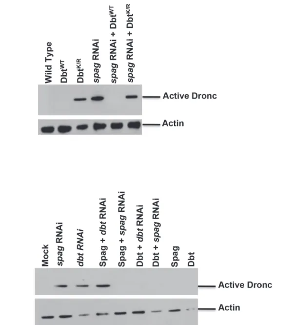
Fig 1. Knock-down ofspaghettireduces Dbt and causes accumulation of activated initiator caspase at ZT7.(A) Wild type Canton S or
timGAL4>UAS-spagRNAi fly heads were harvested at various times as indicated (ZT: hours after lights on in a 12hr:12hr light:dark cycle) and
immunoblotted for Dbt, active Dronc and actin as a loading control. (B)timGAL4>spagRNAi fly heads were harvested at the indicated times in LD (ZT) and immunoblotted for Dbt or active Dronc. (C) S2 cells were harvested 24 and 48 hours afterspagdsRNA addition (two different nonoverlapping dsRNAis were used) and immunoblotted for Dbt, active Dronc and actin. (D)timGAL4>UAS-dbtK/Rand-dbtWTfly heads were harvested at the indicated times and immunoblotted for active Dronc and actin. (E)ClkJrkflies express activated Dronc at ZT 7. Fly heads from various clock mutants or wild type flies were
collected at the indicated times and analyzed for activated caspase expression. (F) Reduced Dbt levels caused by RNAi knock-down ofspagare caused by the proteasome. S2 cells were untreated or treated withspagdsRNA +/- MG132 (a proteasome inhibitor) and immunoblotted for Dbt and actin.
shortened the period of locomotor activity for thetimGAL4>UAS-dbtK/Rgenotype (31.0 ± 0.4, n = 14 vs 33.7 ± 0.9, n = 15 for theperSandper+;dbtK/Rgenotypes respectively; allperL; dbtK/R
flies, n = 16, were arrhythmic). Taken together with the ZT7 time of Dronc activation for the largely arrhythmictimGAL4>UAS-dbtK/RandClkJrkgenotypes, the absence of Dbt reductions in DD (S2E Fig), and the production of activated Dronc in light-pulsed flies at night (S2F Fig), these results suggest that the production of activated caspase is a transient response to pro-longed light exposure that also involves reductions in several circadian genes (e.g.,spag,dbt
andclk).
Dbt is targeted for degradation via the proteasome
S2 cells were treated withspagdsRNA with and without the proteasome inhibitor MG132 and immunoblotted for Dbt. Endogenous Dbt levels were stabilized in the presence of the protea-some inhibitor in the presence ofspagRNAi, which led to complete absence of Dbt in the ab-sence of proteasome inhibitor, but higher Dbt levels in the preab-sence of proteasome inhibitor andspagRNAi were obtained than with proteasome inhibitor only (Fig 1F). The higher levels of Dbt in the presence ofspagRNAi and proteasome inhibitor than with proteasome inhibitor only, coupled with the lower levels of Dbt withspagRNAi only, suggest that Spag may have both proteasome-dependent positive and proteasome-independent negative effects on Dbt lev-els. This result also demonstrates that Dbt is degraded by the proteasome in response tospag
knock-down. The transient accumulation of forms of Dbt with slow electrophoretic mobility (S2B–S2E Fig) suggests that phosphorylation and ubiquitination of Dbt is the initial conse-quence of thespagknock-down (See [28,29] for evidence of phosphorylation.), with proteaso-mal degradation of Dbt a subsequent consequence.
Dbt
WTcan rescue
spag
RNAi-induced caspase activation
When DbtWTis expressed in fly head circadian cells in aspagRNAi background accumulation of activated Dronc is blocked (Fig 2A), suggesting that the effects ofspagknock-down are me-diated by reductions in Dbt. In addition, when Dbt is overexpressed in S2 cells it is able to block the accumulation of activated caspase associated withspagordbtRNAi (Fig 2B). Howev-er, Spag overexpression was not able to block activated caspase accumulation brought on by
dbtRNAi but did block activated caspase accumulation byspagRNAi, suggesting that Spag is upstream of Dbt and confirming the specificity of the dsRNAi knock-down forspag(Fig 2B).
Lowered Spag and Dbt activity in circadian cells lead to accumulation of
activated Dronc in the optic lobes via PDF signaling
Fly brains fromtimGAL4>UAS-spagRNAi flies collected at ZT7 showed elevated levels of ac-tive caspase not found in control brains, but not at ZT19 (Fig 3A and 3B). This was also ob-served using thepdfGAL4 driver, which is expressed in the PDF-secreting brain neurons that drive circadian rhythms of locomotor activity in constant darkness, and with expression of the dominant negative DbtK/Rwith both drivers (Figs3Dand4Afor ZT7 andS5Afor ZT19).tim -GAL4 clock cells expressing DbtK/Rexpressed both activated caspase and Dbt-MYC signal, but activated caspase was expressed in other cells as well (Fig 3D). Taken together, these results suggest both autonomous and non-autonomous effects of the transgene expression.
caspase in large areas of the brain inpdfGAL4>UAS-spagRNAi (or—dbtK/R) flies, the results demonstrate that signaling by PDF is required for this broad activation pattern, and in fact it is even needed in an autocrine manner for expression in the PDF+cells that also express PDF re-ceptor [30], because no activation is seen in these cells in the absence of the PDF receptor (Fig 4B). The CM1 antibody, a marker for Caspase-9 like Dronc activity, was also used to detect Dronc activity, and showed a similar pattern as the anti-Dronc antibody used (Fig 3C). In addi-tion, RNAi to Dronc eliminated most of the signal, confirming that the caspase signal we detect is indeed from Dronc (Fig 3C). Since all Dronc signal is eliminated with expression ofdronc
RNAi with the timGAL4 driver, the results suggest that broader Dronc activation requires Fig 2. Overexpression of DbtWTcan rescue the caspase activation associated withspagRNAi.(A) Fly heads expressing DbtWT, DbtK/R,spagRNAi (31253),spagRNAi+DbtK/RorspagRNAi+DbtWTwith thetimGAL4 driver were collected at ZT7 and analyzed for active Dronc or actin. (B) S2 cells stably overexpressing Spag or DbtWTwere treated withspagordbtdsRNA and immunoblotted for active Dronc or actin.
Dronc activation in TIM+cells, which then signal a corresponding increase in the surrounding tissue via PDF signaling.
Tau is a substrate of Dronc when Dbt or Spag are knocked down
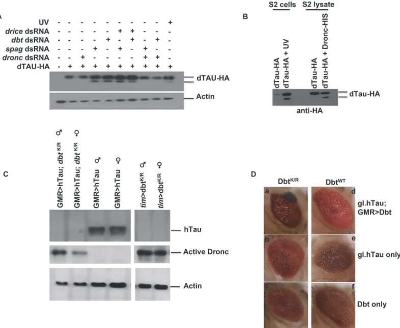
Sincespagwas initially found in a screen involving neurodegeneration and caspases have been implicated in the cleavage of key proteins associated with diseases such as AD, we examined whether Tau was a substrate for Dronc. First, we expressed HA-tagged Drosophila Tau (dTau) in S2 cells and either treated the cells with UV irradiation to induce widespread caspase activa-tion or used RNAi to knock down either Spag or Dbt. When either Spag or Dbt was knocked down cleavage of dTau-HA was detected (Fig 5A). To confirm that Dronc was the main cas-pase involved in this cleavage we used RNAi and targeted either Dronc or Drice, the other main caspase involved in cell death. When Dronc was silenced along with Spag or Dbt, cleavage was inhibited, but when we targeted Drice the cleavage was still detected (Fig 5A).
To further confirm that Dronc was targeting dTau we incubated the dTau lysate with active recombinant Dronc. Samples from cells that were UV-irradiated showed a cleavage product for dTau. In addition, when active Dronc was used we also detected the same cleavage product (Fig 5B), which was not detected with lysates of S2 cells not treated with UV or active Dronc. This suggests that dTau is indeed a substrate for Dronc.
Dbt
K/Rexpressed with hTau leads to activation of Dronc, cleavage of
Tau and an enhanced disrupted eye phenotype
Expression of human Tau (hTau) in fly eyes produces neurodegeneration and has been used as a fly model for tauopathies [16,17]. Therefore, we expressed DbtWTor DbtK/Ralong with hTau in the fly eye using the eye specific GMR driver to determine if Dbt might enhance eye neuro-degeneration. When DbtK/Rwas expressed activated Dronc was detected, both with and with-out hTau expression, while expression of hTau alone was not sufficient to activate Dronc (Fig 5C). Expression of DBTK/Rand hTau with GMR-GAL4 led to significant reductions in Tau lev-els at ZT7 (Fig 5C). Moreover, when DbtK/Rwas expressed along with hTau there was signifi-cantly increased disruption in the eye (Fig 5DandS1 Table). The enhanced disruption of the eye was manifested by the appearance of melanized patches in the eye (Fig 5D) and a decreased average surface area of the eye (S1 Table). Expression of DbtWTdid not lead to enhancement of the eye phenotype (Fig 5DandS1 Table).
Cell death-independent expression of activated Dronc in young flies and
circadian effects on the cell death pathway in older flies
To determine whether Dronc activation led to classical apoptosis we looked at another marker of apoptosis. Diap1 is an antiapoptotic protein that regulates cell death in flies by binding to and inhibiting the activity of Dronc. When cell death occurs Diap1 is targeted for degradation/ cleavage, thereby freeing Dronc [31]. We examined Diap1 levels in fly heads and observed no difference between young wild type andtimGAL4>UAS-spagRNAi ordbtK/Rflies in which Dronc activation occurs (Fig 6A). This lack of effect on Diap1 levels was also observed in S2 cells treated withspagordbtRNAi or expressing DbtK/R(Fig 6D). However, when flies aged a
image of each column shows the average intensity of the Z-stack image, followed by individual slices from the given Z-stack. Wild type controls showed no activated Dronc. (B) Whole brains were collected at ZT19 and PDF (red) but no active caspase (green) were detected in either genotype. (C) The caspase signal detected in fly brains is Dronc specific. Active caspase detected using anti-CM1 or anti-active Dronc at ZT7 in DbtK/Ror DbtK/RandDroncRNAi fly brains. (D) Caspase activation is detected in clock cells and non-clock cells. Whole brains were collected at ZT7 and imaged for Dbt-MYC (green) and active caspase (red), which was detected only intim-GAL4>UAS-dbtK/Rbrains in both cells expressing Dbt-MYC and surrounding cells.
Diap1 cleavage product was detected in thespagRNAi anddbtK/Rflies (Fig 6B) and activated caspase expression was also detected. Interestingly, agedspagRNAi flies showed activated cas-pase expression at all time points examined (Fig 6B). To determine if this pathway occurred naturally in aging flies we collected wild type Canton S (C.S.) fly heads at 60 days. Aged flies showed a mobility shift of Dbt at ZT 7 and 19 that was similar to what we observed inspag
RNAi flies (Fig 6C). In addition, increased active caspase expression was detected at ZT7 and 19 for the aged flies (Fig 6C) and Diap1 cleavage was detected from ZT7 to 19 for the aged W. T. flies (Fig 6C). These results demonstrate that wild type flies exhibit the same circadian-de-pendent activation of apoptotic pathways that are produced inspagRNAi,dbtK/RandClkJrk
flies at younger ages, and suggest that reductions in activity of these circadian genes accelerate an age-dependent pathway that leads to activation of the apoptotic pathway.
spag
RNAi flies have reduced climbing and lifespan
Sincespagregulates a pathway that leads to the activation of caspases by reduction of Dbt, and this then leads to the cleavage of Tau by these activated caspases and ultimately to expression of apoptotic markers, we wanted to determine if there were behavioral and lifespan manifesta-tions. The circadian locomotor assays initially suggested that there were effects as flies age. Lo-comotor behavior is preserved in young flies. However, as transgenic flies with reduction of
spagby RNAi age, they tend to die during the locomotor assay (Table 1). The loss of the climb-ing response has been used to monitor age-related changes in Drosophila. Normal Drosophila show a strong negative geotactic response. When tapped to the bottom of a vial they rapidly climb to the top of the vial, and most flies remain there. Flies with knockdown ofspaginitially climb as well as control flies. However, over time they decline in performance more rapidly than controls (Fig 7A). The progressive, accelerated decline in climbing ability inspagRNAi flies demonstrates a functional deficit produced by knockdown ofspagin clock cells. In addi-tion,spagRNAi flies had a reduced lifespan compared to control flies (Fig 7B). By contrast, ex-pression of DbtK/Rin circadian cells did not produce accelerated death and loss of climbing ability in comparison with wild type Canton S flies, indicating that the effects of Dbt activity re-duction are not as severe as those ofspagreduction (See figure legend for discussion of
statistics).
Discussion
We have identified a new player (spag) that links circadian signaling, cell death and tauopathies together. Orthologs of Spag in yeast and humans function as co-chaperones of Hsp90 to regu-late its activity and recruit client proteins for Hsp90. They are part of multiprotein complexes that contribute to biogenesis of cellular machineries like RNA polymerase, ribonucleoproteins and Phosphatidyl-Inositol 3-kinase-related kinases [32,33]. Recently, Drosophila Spag has been shown to associate with Hsp90 and Hsp70 to likewise contribute to assembly of several of Fig 4. Pdf signaling from circadian neurons is required for accumulation of activated caspase in optic lobes.(A) Whole brains were collected at ZT7 frompdfGAL4>UAS-spagRNAi (or-dbtK/R, -dbtWT) flies and wild type control (pdfTm3; progeny inheriting a balancer chromosome rather than the UAS responder) and processed for detection of PDF (red) and active caspase (green). Knock-down ofspagor reduction of Dbt activity both produced activated caspase expression in the optic lobes in a region innervated by the PDF+ axons. (B) Whole brains were collected at ZT7 fromtimGAL4>UAS-dbtK/R(ordbtWT) flies and processed for detection of PDF (red) and active caspase (green). Flies that were heterozygous for thepdfreceptor deletion (pdfrKO/+) expressed activated Dronc with DbtK/R
, while those that were hemizgous for the mutation (pdfrKO
males) did not. The first image of each column is the average intensity of the Z-stack image, followed by individual slices from the Z-stack. SeeS5 Figfor ZT19 analysis, which showed no expression of activated caspase under any condition.
these factors [34]. The human ortholog of Spag (RPAP3) is a binding partner for a WD40 re-peat protein that is involved in apoptosis. RPAP3 contains TPR domains and regulates apopto-sis induced by several stimuli [35,36].
When we knock downspagin circadian cells using RNAi we observed reduction in Dbt lev-els and an increase in the activated caspase Dronc in fly heads. Interestingly, this mostly oc-curred during the day (ZT7) or after extended light treatment at night, increased as flies age Fig 5. Spag or Dbt reduction leads to Dronc-dependent Tau cleavage and neurodegeneration in eyes overexpressing hTau.(A) S2 cells were transfected with Drosophila HA-tagged Tau +/- RNAi tospag,dbtalone or with eitherdroncordriceRNAi. Samples were then immunoblotted for dTau using anti-HA antibody.“UV”cells were treated with UV light to induce caspase activation, and this also produced cleavage of Tau.droncRNAi inhibited Tau cleavage, whiledriceRNAi did not. (B) Drosophila HA-taggedtauwas transfected in S2 cells and either UV treated to induce caspase activation or harvested and incubated with active recombinant Dronc. Samples were then immunoblotted for dTau using anti-HA. (C) Male and female flies were collected at ZT7 and immunoblotted for hTau, active Dronc and actin. Adult flies expressing DbtK/Rin the eye with the indicated drivers in the presence or absence of hTau have activated Dronc, while expression of hTau alone did not produce activated Dronc. Flies expressing both hTau and DbtK/Rshow reduced Tau expression. (D) Adult flies expressing both DbtK/Rand hTau produce an enhanced disrupted eye phenotype. Adult eyes of various genotypes were imaged and representative examples are shown. (a) gl.htau; GMRGAL4>UAS-dbtK/R(b) GMRGAL4>/+; gl.htau(c) GMRGAL4>UAS-dbtK/R(d) gl.htau;
GMRGAL4>UAS-dbtWT(e) GMRGAL4>/+; gl.htau(f) GMRGAL4>UAS-dbtWT. See alsoS1 Tablefor quantification of eye size differences.
and did not occur in constant darkness. Accumulation of activated Dronc was also observed with expression of the kinase dead form of Dbt (DbtK/R) in circadian cells. This suggests that some factor present or active during the light might regulate the Spag-Dbt pathway and confer a transient sensitivity to caspase activation after extended light treatment.
One feature that is common to various neurodegenerative diseases is the acceleration of the age-related disruption of the daily cycle of sleep and wake. Our work suggests that the most im-mediate way for the clock to influence neurodegeneration is by circadian gene-dependent con-trol over the expression of pro-neurodegenerative factors. While these factors are not
expressed in young flies with normal circadian clocks, the mutant clocks that we have produced Fig 6. Apoptosis is not detected in young flies but is in olderspagRNAi and wild type flies.(A) Fly heads were harvested from 7-day old or (B) 30-day old wild type flies Canton S flies (C.S.) or flies expressing the indicated transgene with thetimGAL4 driver at various times indicated and immunoblotted for Diap1, active Dronc or actin. Only 30-day old flies with reducedspagordbtactivity produced cleaved Diap1 (indicated by*), andspagRNAi flies produced active Dronc at all times at 30 days. (C) Fly heads from C.S. flies were collected at 7 and 60 days and immunoblotted for Dbt, active Dronc, Diap1 and actin. 60 day old C.S. flies showed Dbt mobility shift, active Dronc expression and Diap1 reduction at ZT7 and 19. Note that there were nospagRNAi flies still alive at this time. (D) S2 cells treated withspag,dbt,diap1dsRNA or stably expressing DbtK/Rwere immunoblotted for Diap1, demonstrating reduced Diap1 only with dsRNAi fordiap1.
in flies resemble those produced in aging wild type flies, in which Dbt modification, activated caspase expression and cleaved Diap1 are detected at ZT7 and ZT19. Prior work has shown the circadian function is blunted along with reduced healthspan in aging flies [37–39]. Circadian dysfunction would enhance their susceptibility to light-dependent neurodegeneration. In a pre-vious report, theperiodgene was mutated in flies withsniffergene mutations causing neurode-generation. The flies with both mutations displayed faster neurodegeneration and had shorter lifespans compared to flies with single mutations. This suggests that disrupted circadian rhythms can accelerate the process of neurodegeneration [40].
What is the nature of this mechanism? Previous work linked Spag to Huntington’s disease and Hsp90 and a possible role in aggregation [25]. In addition, caspases have been shown to be involved in the cleavage of Tau, a protein associated with AD, and this caspase-mediated cleav-age of Tau is associated with its aggregation. This led us to examine whether Tau was cleaved by the caspase Dronc in the Spag-Dbt pathway. HA-dTau was cleaved in S2 cells when we knocked down either Dbt or Spag, and this cleavage was prevented when we knocked down Dronc, but not the effector caspase Drice. This result is consistent with cleavage of Tau by Dronc in response to lowered Dbt and Spag activity.
We examined whether long-term reductions of Spag and Dbt activity in circadian cells have any detrimental effects on the fly. While younger flies with reduced Spag or Dbt activity did not exhibit expression of cell death marker Diap1, older flies with chronic reductions in Spag or Dbt activity exhibited elevated levels of Diap1 cleavage—a marker of the apoptotic pathway. These results suggest that chronic reductions in Spag or Dbt activity eventually produce deleterious effects.
Flies with reducedspaglevels had more cleaved Diap1 compared todbtK/Rflies, higher levels of active Dronc and more rapid decline of climbing proficiency and lifespan. This puzzled us since both lines activate Dronc and lead to dTau cleavage, so we expected identical phenotypes. One possibility is that since Spag has been shown to interact with Hsp90, Spag might regulate Hsp90 and control the level of aggregation that occurs. In such a model, if Spag is eliminated and can no longer regulate Hsp90, Hsp90 might no longer interact with dTau and therefore no longer function to reduce dTau aggregates. The defect in Hsp90 function is not predicted for
dbtK/Rflies, which retain Spag. In addition, Hsp90 is a known regulator of cell death and has been shown to inhibit apoptosis [41]. Removal of Spag might lead to dysregulation of Hsp90, preventing it from regulating components of the cell death pathway and causing a higher level of activated caspase than observed with Dbt inhibition alone.
Is this pathway evolutionarily conserved? To address this we used the fly eye and expressed human Tau along with DbtWTor DbtK/R. When hTau and DbtK/Rwere coexpressed together
function of age. Flies of each genotype were scored in three replicate groups of 10 for each genotype, and all showed the same trends. Genotypes: UAS-dcr; timGAL4>/UAS-spag RNAi(31253),timGAL4>dbtWT, -dbtK/R, Canton S wild type flies. A Kruskal-Wallis nonparametric ANOVA indicated a significant effect of genotype on the time at which flies lost their climbing ability (H[3, N = 120] = 61, p<0.0001); the multiple comparisons p values indicated that thespagRNAi genotype differed significantly from all the others (p<0.0001), while the other three did not differ significantly. (B) Survival curves for the same genotypes as in (A). Flies were maintained at 25°C, transferred every 10 days in three replicate batches for each genotype, and scored for survival. All three replicates showed the same trend. A Kruskal-Wallis nonparametric ANOVA indicated a significant effect of genotype on lifespan (H3[N = 400] = 197, p<0.001). The multiple comparisons p values indicated that thespagRNAi genotype differed significantly from the other three while thedbtK/R genotype differed only from thespagRNAi and thedbtWT(p<0.002); no other comparisons differed significantly. (C) Model of Spag and Dbt regulation of caspases. Dbt regulates caspases by phosphorylation, which inhibits caspase activity in normal conditions. Whenspagis removed Dbt is targeted for
phosphorylation and degradation by the proteasome, freeing the caspase and allowing for cleavage of Tau and possibly other substrates (“?”) and production of tauopathy
an enhanced disrupted eye phenotype was produced together with Donc activation and Tau cleavage, and the phenotype is less severe when hTau is coexpressed with DbtWT. Since loss of Dbt kinase activity leads to Dronc activation, Dbt may be inhibiting Dronc by direct phosphor-ylation of Dronc, or alternatively there may be another intermediate target of Dbt. Prior work in mammals has demonstrated a link between reduced circadian clock function and neurode-generation, as well as a link between CKIδ/εand apoptosis [42–45]. Reduced clock function
has been produced by alterations to circadian transcriptional regulators, with increased neuro-degeneration produced in response to reactive oxygen species and induction of apoptosis. CKI regulation of cell death and cell cycle arrest has been linked to effects on the mitotic spindle, p53 and cell surface receptors involved in cell death. It is likely that circadian clocks and CKI affect apoptosis and neurodegeneration at multiple steps in addition to the ones outlined in this manuscript. However, the direct effects of the clock components on Dbt levels and the con-sequent expression of an activated initiator caspase suggest that these events may be upstream and global mediators of circadian effects on apoptosis and neurodegeneration.
It has been shown that these cell death components that are normally involved in destruc-tion can also play critical roles in nonapoptotic events such as dendrite pruning, which occurs during development to create proper neural circuits. In Drosophila, caspase activity is detected locally in the degenerating dendrites and mutation of Dronc preserves most of the dendrite morphology [46]. In this instance caspases are not activated in the context of apoptosis, but in cell survival processes.
A possible role for the Spag-Dbt-Dronc pathway in dendritic or axonal pruning/remodeling is intriguing in light of the existing literature on the role of the circadian clock and light in these processes[47–50]. There is circadian remodeling of lateral neuron (PDF+) axon branch-ing patterns as well as the size and synapses of several noncircadian neurons in the optic lobes, which are extensively innervated by the PDF+axons. These circadian changes require a func-tional circadian clock, are enhanced by light, and in the case of some optic lobe changes require signals from the lateral neurons. Intriguingly accumulation of activated Dronc in the optic lobe at ZT7 intimGAL4>UAS-spagRNAi (or UAS-dbtK/R) flies also required light and was pro-duced by PDF signaling from the lateral neurons; therefore, this activation may in fact be due to hyperactivation of the same pathways that trigger normal circadian neuronal remodeling in the optic lobes. In wild type flies, some of the optic lobe neurons exhibit largest axon size in the morning and the evening, suggesting that caspases (presumably below the level of detection) might contribute to pruning during the middle of the day, at times when activated caspases are detected here. It is not certain whether expression of activated Dronc in the optic lobe cells not expressing thespagRNAi (ordbtK/R) also involves reductions in Dbt activity in those cells or is produced by a different pathway in response to PDF signaling, but it is likely that Dbt reduc-tions in these cells also occur, as the reducreduc-tions in Dbt detected in immunoblots of total head extracts can be quite complete. We would argue that PDF signaling is important for global DBT reductions, casapse activation and Tau cleavage, and that these are produced cell autono-mously in S2 cells byspagRNAi or DBTK/Rexpression. Reductions inspagmay also trigger the non-cell autonomous reductions in Dbt levels and caspase activation, or these may be triggered by aspag-independent mechanism in response tospagdecreases in the PDF+cells. Caspase in-volvement in synapse degeneration has previously been suggested to contribute to AD patholo-gies [51].
context of AD and other tauopathies and sheds new light on the underlying mechanism that regulates the disease state and its connection to the circadian clock. Recently, we identified an-other TPR-containing protein (Bride of Dbt, or Bdbt) that interacts with Dbt to enhance Dbt activity and regulate its phosphorylation state [29]. It will be interesting to determine if this protein is part of any Spag-Dbt complexes that might regulate AD and apoptosis. Furthermore, it will be important to establish the genetic, environmental or aging processes that could inter-act with the mechanism to inter-activate it during normal aging to produce AD or potentially other outcomes that negatively impact health and lifespan.
Materials and Methods
Fly stocks
Drosophila RNAi stocks for spaghetti (CG13570) were obtained from the Bloomington Dro-sophila Stock Center (stock number: 31253, targeting nucleotides 778–1200 of the transcript) and the Vienna Drosophila RNAi Center (VDRC stock numbers: 23896, targeting nucleotides 886–1233 of the transcript; 103353KK, targeting nucleotides 780–1200). In addition, thedronc
RNAi line 23033 was obtained from the VDRC. The expression of hTau was under the control of theglass(gl) promoter provided by George Jackson [17] forFig 5DandS1 Table, or alterna-tively a UAS-hTau fly line from Mel Feany [16] was used to express human Tau with the GMR-GAL4 driver on the X chromosome (Bloomington stock center). The UAS-dbt-myclines and thetimGAL4 and UAS-dcr;timGAL4 driver lines have been described previously [27,28]. ThepdfGAL4 driver line (stock number 6899) and thePdfreceptor (CG13758) null mutant line (Pdfr5304; line 33068) were obtained from the Bloomington Drosophila Stock Center.ClkJrk
[52],pero,perSandperLmutations [53] were used alone or together with thedbtK/Rconstructs as described in the text.
Locomotor activity analysis
UAS-spag RNAilines were crossed with UAS-dcr2;timGal4 lines. Progeny were then continu-ously reared at 23°C in a 12 hr:12 hr LD cycle for one more week after collection of adults (or longer where indicated) to insure complete RNAi knock-down effects, and loaded onto moni-tors (Trikinetics, Waltham, MA) for behavioral monitoring in constant darkness (DD) for at least 5 days. Actogram activity records and periodogram analysis to determine periods were employed as previously described with ClockLab [27].
Immunoblot analysis
Adult brain immunofluorescence
Brains were dissected from flies at ZT7 and ZT19, fixed, permeablized and incubated with anti-Dronc (1:100), anti-CM1 (Cell Signaling #9661), anti-PDF (1:5000) (PDF C7, Developmental Studies Hybridoma Bank) or anti-Myc (1:1000) (Santa Cruz sc-789-G) antibody overnight at 4°C. The following day samples were incubated with fluorescently-labeled secondary antibod-ies (anti-rabbit IgG Alexa fluor 488 or anti-mouse IgG Alexa fluor 568, 1:1000). The brains were examined using an Olympus Fluoview confocal, and Z stacks were obtained.
Drosophila Tau cleavage assay
Drosophila S2 cells were transiently transfected with dTau-HA and 24h later cells were treated with dsRNA corresponding todronc,dbt,spagordrIceand harvested 48h later. For UV treat-ment, cells were harvested 24h later. Samples were analyzed by immunoblot using anti-HA antibody.
For treatment with active Dronc-His, S2 cells were harvested 24h after dTau-HA transfec-tion, resuspended in Buffer A (25mM TrisCl pH 8.0, 50mM NaCl, 10mM DTT) and lysed by
4x freeze-thaw. After lysis, cells were spun down at 13000rpm and the supernatant was collect-ed and incubatcollect-ed with active Dronc-His [54] for 4h at 30°C. The reaction was stopped by the addition of 5X SDS loading buffer and analyzed for dTau-HA by immunoblot analysis. As a positive control, extracts were analyzed from S2 cells expressing dTau-HA and treated with UV light.
Quantification of eye size in flies expressing hTau and/or Dbt-MYC
Fly eye size was measured from photomicrogaphs using the ImageJ program (open source pro-gram, NIH). A circumference was drawn around the eye and the area was obtained from the measure command. The values (number of pixels) were then tabulated and averaged and statis-tics were performed using the Statistica (Statsoft OK) software.RT-PCR analysis
Fly heads (50–100) or S2 cells were collected and frozen in liquid nitrogen. Total RNA was iso-lated using the Trizol Reagent (Invitrogen), and cDNA synthesis was performed using the Taq-man Reverse Transcription Kit (Life Technologies). PCR was performed using gene specific primers forspagand actin for 20 cycles, and the products were analyzed on an agarose gel.
Spag and Dbt rescue or overexpression experiments
S2 cells stably expressing Spag or Dbt were untreated or treated with dsRNA corresponding to
spagordbt. Forty eight hours after dsRNA addition cells were harvested and analyzed for active Dronc, Dbt or actin. Plasmids used for this experiment were the following: pMT-dbt-myc, pMT-dbtK/R-myc, pAC-spag-ha.
Primers used to generate dsRNA and the procedure for treatment of S2
cells with dsRNAi
dbt(nt 321–674):
forward 5’-TAATACGACTCACTATAGGGGCGCGTGGGTAACAAATATC-3’, reverse 5’-TAATACGACTCACTATAGGGTGTATGTAATCGATGCGGGA-3’,
dronc(nt 939–1454):
drIce(nt 653–968):
forward 5’- TAATACGACTCACTATAGGGACTGCCGCTACAAGGACATT-3’, reverse 5’- TAATACGACTCACTATAGGGGCGTGCACTGGAATCTTGTA-3’,
diap1(nt 491–1019):
forward 5’-TAATACGACTCACTATAGGGCCGGCATGTACTTCACACAC-3’, reverse 5’-TAATACGACTCACTATAGGGTTCTGTTTCAGGTTCCTCGG-3’,
spagset 1 (nt 735–1242):
forward 5’-TAATACGACTCACTATAGGGCAAAAGTGGGCCAAACTTTAC-3’ reverse 5’TAATACGACTCACTATAGGGTTCTGGGCTGCGTTCTAT-3’,
spagset 2 (nt22-227):
forward 5’-TAATACGACTCACTATAGGGGGAGCGCTAGCAACAGAAAT-3’ reverse 5’-TAATACGACTCACTATAGGGGCGACTTCTGGAGCTCTTTC-3’, PCR fragments were produced from Drosophila genomic DNA with these primer sets. dsRNAi was produced and transfected into S2 cells by the procedures of the Perrimon lab (http://www.flyrnai.org/DRSC-HOME.html), using the T7 promoters encoded by the primers. The cells were harvested at the times indicated in the figure legends. In one experiment, the proteasome inhibitor MG132 was added to a concentration of 50–100μM, and in another MYC-tagged Dbt was overexpressed.
Generation of
spag
expression clones
A cDNA clone for CG13570 (RE03224) was obtained from the Drosophila Genomics Resource Center (DGRC, Bloomington, IN). We used the DGRC Gateway collection in order to clone
spaginto vectors allowing expression in S2 cells. A full-lengthspagORF was generated by PCR (Phusion, cat# F-553S, NEB) and cloned into pENTR/SD/D-TOPO (Cat# K240020, Life Tech-nologies) by TOPO-mediated cloning. This clone was used to generate plasmids containing an HA tag. The clonase enzymatic mixture (Cat# 11791–019) was purchased from Life Technolo-gies.spagwas cloned into the pGateway vector pAHW (Cat#1095) to generate an N-terminal 3xHA-tag plasmid. The plasmid were driven by an Act5c promoter allowing for constitutive expression in S2 cells.
Generation of dTau-HA plasmid
For the expression of Drosophila Tau (dTau) in S2 cells, the pFLC1-TAU cDNA (Drosophila Genomics Resource Center, Clone # RE16764) was cloned into the pAc S2 cell expression vec-tor. The N-terminal HA tag was added using the following primers:
Tau N-TER Forward, 5’ -GCGCGAATTCGCGACCCTATGGCGTACCCGTAC-GACGTGCCGGACTACGCGATGGCGGATGTCCTGGAGAAAAGCTCACTG 3’,
Tau N-TER DRA Reverse, 5’—GGCGCATGTCCGACTTGTACC 3’
The DNA was digested with DraIII and EcoRI and swapped for the original fragment in the pAc-Tau.
Climbing and lifespan assays
Flies were maintained at 24°C on a 12 hr:12hr light/dark cycle. For aging studies, virgin male flies were isolated and maintained in 20 per vial and transferred to a fresh vial every 10 days. The number of dead flies was recorded.
the vial. We transferred a group of ten flies into the lower vial and allowed the flies to acclima-tize to the new setting for 1 minute before conducting the assay. Flies were gently tapped down to the bottom of the vial and the number of flies that climbed above the 8 cm mark by 10 sec-onds after the tap was measured[55]. This was repeated with the same group ten times, allow-ing for a 1 minute rest period between each trial. The average number of flies per group that passed the 8-cm mark as a percentage of total flies was recorded.
Supporting Information
S1 Fig.spagis required in circadian cells to produce rhythmic behavior.Representative DD
actogram records of progeny with the indicated genotypes are presented (left), along with peri-odogram analysis to determine the period of the rhythm (right). Knock-down ofspagled to a longer period (B, D) or arhythmicity (C) when compared to control (A). Flies were assayed starting two weeks after collection of adults, except for panel D, for which the record started one week after collection.
(PDF)
S2 Fig. Analysis of the parameters affecting Dbt post-translational modification and degra-dation triggered byspagRNAi.This analysis demonstrated that Dbt reduction is variable and
accentuated by higher temperatures and light, with a post-translational modifications of Dbt observed with shorter periods ofspagRNAi treatment or in younger flies. (A) The specificity of the purified anti-activated Dronc antibody (SeeMaterials and Methods) for the active cleaved form of Dronc using recombinant full length and active cleaved Dronc. (B) S2 cells were treated withspagdsRNA, harvested at 1 and 2 hours after dsRNA addition and immunoblotted for Dbt. (C)timGAL4>spagRNAi fly heads from young (3 days) and old (7 days) collections were immunoblotted for Dbt. (D)timGAL4>spagRNAi flies were reared at 24 or 27°C, and fly heads were harvested at 7 days or 14 days after collection at the indicated times and immuno-blotted for Dbt. (E) Constant darkness blocks Dbt reduction caused by knock-down ofspagin fly heads. Flies expressingspagRNAi with thetimGAL4 driver were entrained in a light/dark cycle or kept in constant darkness, collected at the indicated days and times (ZT7 in LD or CT7 in DD) and examined for Dbt levels. (F) Flies were light pulsed for 7 hours at night (starting at ZT13) and Dronc activation was determined by immunoblot.
(PDF)
S3 Fig. Confirmation ofspagknock-down and effects in various lines.(A) RT-PCR analysis
ofspagtranscript in variousspagRNAi lines and in S2 cells. (B) Analysis of differentspag
RNAi lines for effects on Dbt and caspase activation. Fly heads fromtimGAL4>UAS-spag RNAi lines with different RNAi constructs were isolated at the indicated times of day, and immunoblotted for Actin, Dbt and activated Dronc. Dbt was consistently reduced and Dronc was consistently activated at ZT7, while Dbt was variably reduced at ZT19 without detection of activated Dronc. The numbers above the lanes are the numbers for the relevant line from the Vienna Drosophila Resource Center (VDRC).
(PDF)
S4 Fig. Time course analysis of Dronc activation in various mutant genotypes.Heads of adult wild type Canton S flies (C.S.) and of the indicated mutants were collected at the indicat-ed times and immunoblottindicat-ed for active Dronc and actin.
(PDF)
S5 Fig. Brain confocal images collected at ZT19 showing no expression of activated caspase.
(green) and PDF (red) were detected. The top image for each column depicts the average inten-sity of the Z-stack image with each additional image representing an individual slice from the Z-stack. (B) Pdf receptor mutants at ZT19 expressing wild type or catalytically inactive Dbt (DbtK/R) collected at ZT19. Whole brains were collected and active caspase (green; not detected at this time) and PDF (red) were assayed. The first image of each column depicts the average intensity of the stack image and each image below that depicts individual slices from the Z-stack. (C) A single optical section magnified to show detection of activated caspase and PDF in the optic lobes oftimGAL4>UAS-spagRNAi flies at ZT7. Most of the activated caspase is de-tected in areas surrounding the PDF+axons rather than in the axons themselves.
(PDF)
S1 Table. Average Areas of Fly Eyes Expressing hTau and Dbt.
(DOCX)
Acknowledgments
We would like to thank the Bloomington Drosophila Stock Center and Vienna Drosophila RNAi Center for fly stocks. The Drosophila Genomics Resource Center provided the Gateway vectors, and the Developmental Hybridoma Bank the anti-actin antibody.
Author Contributions
Conceived and designed the experiments: JCM JLP. Performed the experiments: JCM JLP AV BG JYF ESB. Analyzed the data: JCM JLP AV BG JYF. Contributed reagents/materials/analysis tools: JCM JLP AV BG JYF ESB. Wrote the paper: JCM JLP.
References
1. Kim TW, Pettingell WH, Jung YK, Kovacs DM, Tanzi RE (1997) Alternative cleavage of Alzheimer-as-sociated presenilins during apoptosis by a caspase-3 family protease. Science 277: 373–376. PMID: 9219695
2. Gervais FG, Xu D, Robertson GS, Vaillancourt JP, Zhu Y, et al. (1999) Involvement of caspases in pro-teolytic cleavage of Alzheimer's amyloid-beta precursor protein and amyloidogenic A beta peptide for-mation. Cell 97: 395–406. PMID:10319819
3. de Calignon A, Fox LM, Pitstick R, Carlson GA, Bacskai BJ, et al. (2010) Caspase activation precedes and leads to tangles. Nature 464: 1201–1204. doi:10.1038/nature08890PMID:20357768
4. Sanchez Mejia RO, Friedlander RM (2001) Caspases in Huntington's disease. Neuroscientist 7: 480– 489. PMID:11765125
5. Khurana V, Elson-Schwab I, Fulga TA, Sharp KA, Loewen CA, et al. (2010) Lysosomal dysfunction pro-motes cleavage and neurotoxicity of tau in vivo. PLoS Genet 6: e1001026. doi:10.1371/journal.pgen. 1001026PMID:20664788
6. Marx J (2001) Neuroscience. New leads on the 'how' of Alzheimer's. Science 293: 2192–2194. PMID: 11567120
7. McLaughlin B, Hartnett KA, Erhardt JA, Legos JJ, White RF, et al. (2003) Caspase 3 activation is es-sential for neuroprotection in preconditioning. Proc Natl Acad Sci U S A 100: 715–720. PMID: 12522260
8. Rohn TT, Rissman RA, Davis MC, Kim YE, Cotman CW, et al. (2002) Caspase-9 activation and cas-pase cleavage of tau in the Alzheimer's disease brain. Neurobiol Dis 11: 341–354. PMID:12505426
9. Gamblin TC, Chen F, Zambrano A, Abraha A, Lagalwar S, et al. (2003) Caspase cleavage of tau: link-ing amyloid and neurofibrillary tangles in Alzheimer's disease. Proc Natl Acad Sci U S A 100: 10032– 10037. PMID:12888622
11. Heidary G, Fortini ME (2001) Identification and characterization of the Drosophila tau homolog. Mech Dev 108: 171–178. PMID:11578871
12. Chen X, Li Y, Huang J, Cao D, Yang G, et al. (2007) Study of tauopathies by comparing Drosophila and human tau in Drosophila. Cell Tissue Res 329: 169–178. PMID:17406902
13. Chee FC, Mudher A, Cuttle MF, Newman TA, MacKay D, et al. (2005) Over-expression of tau results in defective synaptic transmission in Drosophila neuromuscular junctions. Neurobiol Dis 20: 918–928. PMID:16023860
14. Mudher A, Shepherd D, Newman TA, Mildren P, Jukes JP, et al. (2004) GSK-3beta inhibition reverses axonal transport defects and behavioural phenotypes in Drosophila. Mol Psychiatry 9: 522–530. PMID: 14993907
15. Mershin A, Pavlopoulos E, Fitch O, Braden BC, Nanopoulos DV, et al. (2004) Learning and memory deficits upon TAU accumulation in Drosophila mushroom body neurons. Learn Mem 11: 277–287. PMID:15169857
16. Wittmann CW, Wszolek MF, Shulman JM, Salvaterra PM, Lewis J, et al. (2001) Tauopathy in Drosophi-la: neurodegeneration without neurofibrillary tangles. Science 293: 711–714. PMID:11408621
17. Jackson GR, Wiedau-Pazos M, Sang TK, Wagle N, Brown CA, et al. (2002) Human wild-type tau inter-acts with wingless pathway components and produces neurofibrillary pathology in Drosophila. Neuron 34: 509–519. PMID:12062036
18. Hardin PE (2011) Molecular genetic analysis of circadian timekeeping in Drosophila. Adv Genet 74: 141–173. doi:10.1016/B978-0-12-387690-4.00005-2PMID:21924977
19. Price JL, Blau J, Rothenfluh A, Abodeely M, Kloss B, et al. (1998) double-time is a novel Drosophila clock gene that regulates PERIOD protein accumulation. Cell 94: 83–95. PMID:9674430
20. Kloss B, Price JL, Saez L, Blau J, Rothenfluh A, et al. (1998) The Drosophila clock gene double-time encodes a protein closely related to human casein kinase Iepsilon. Cell 94: 97–107. PMID:9674431
21. Guan J, Li H, Rogulja A, Axelrod JD, Cadigan KM (2007) The Drosophila casein kinase Iepsilon/delta Discs overgrown promotes cell survival via activation of DIAP1 expression. Dev Biol 303: 16–28. PMID:17134692
22. Weldemichael DA, Grossberg GT (2010) Circadian rhythm disturbances in patients with Alzheimer's disease: a review. Int J Alzheimers Dis 2010.
23. Cermakian N, Lamont EW, Boudreau P, Boivin DB (2011) Circadian clock gene expression in brain re-gions of Alzheimer 's disease patients and control subjects. J Biol Rhythms 26: 160–170. doi:10.1177/ 0748730410395732PMID:21454296
24. Sterniczuk R, Dyck RH, Laferla FM, Antle MC (2010) Characterization of the 3xTg-AD mouse model of Alzheimer's disease: part 1. Circadian changes. Brain Res 1348: 139–148. doi:10.1016/j.brainres. 2010.05.013PMID:20471965
25. Zhang S, Binari R, Zhou R, Perrimon N (2010) A genomewide RNA interference screen for modifiers of aggregates formation by mutant Huntingtin in Drosophila. Genetics 184: 1165–1179. doi:10.1534/ genetics.109.112516PMID:20100940
26. Yoo SJ, Huh JR, Muro I, Yu H, Wang L, et al. (2002) Hid, Rpr and Grim negatively regulate DIAP1 levels through distinct mechanisms. Nat Cell Biol 4: 416–424. PMID:12021767
27. Muskus MJ, Preuss F, Fan JY, Bjes ES, Price JL (2007) Drosophila DBT lacking protein kinase activity produces long-period and arrhythmic circadian behavioral and molecular rhythms. Mol Cell Biol 27: 8049–8064. PMID:17893330
28. Fan JY, Preuss F, Muskus MJ, Bjes ES, Price JL (2009) Drosophila and vertebrate casein kinase Idelta exhibits evolutionary conservation of circadian function. Genetics 181: 139–152. doi:10.1534/ genetics.108.094805PMID:18957703
29. Fan JY, Agyekum B, Venkatesan A, Hall DR, Keightley A, et al. (2013) Noncanonical FK506-binding protein BDBT binds DBT to enhance its circadian function and forms foci at night. Neuron 80: 984– 996. doi:10.1016/j.neuron.2013.08.004PMID:24210908
30. Im SH, Taghert PH (2010) PDF receptor expression reveals direct interactions between circadian oscil-lators in Drosophila. J Comp Neurol 518: 1925–1945. doi:10.1002/cne.22311PMID:20394051
31. Muro I, Means JC, Clem RJ (2005) Cleavage of the apoptosis inhibitor DIAP1 by the apical caspase DRONC in both normal and apoptotic Drosophila cells. J Biol Chem 280: 18683–18688. PMID: 15774476
33. Eckert K, Saliou JM, Monlezun L, Vigouroux A, Atmane N, et al. (2010) The Pih1-Tah1 cochaperone complex inhibits Hsp90 molecular chaperone ATPase activity. J Biol Chem 285: 31304–31312. doi: 10.1074/jbc.M110.138263PMID:20663878
34. Benbahouche NE, Iliopoulos I, Torok I, Marhold J, Henri J, et al. (2014) Drosophila Spag is the homolog of RNA Polymerase II Associated Protein 3 (RPAP3), and recruits the Heat Shock Proteins 70 and 90 (Hsp70 and Hsp90) during the assembly of cellular machineries. J Biol Chem 289:6236–6247. doi:10. 1074/jbc.M113.499608PMID:24394412
35. Itsuki Y, Saeki M, Nakahara H, Egusa H, Irie Y, et al. (2008) Molecular cloning of novel Monad binding protein containing tetratricopeptide repeat domains. FEBS Lett 582: 2365–2370. doi:10.1016/j.febslet. 2008.05.041PMID:18538670
36. Yoshida M, Saeki M, Egusa H, Irie Y, Kamano Y, et al. (2013) RPAP3 splicing variant isoform 1 inter-acts with PIH1D1 to compose R2TP complex for cell survival. Biochem Biophys Res Commun 430: 320–324. doi:10.1016/j.bbrc.2012.11.017PMID:23159623
37. Umezaki Y, Yoshii T, Kawaguchi T, Helfrich-Forster C, Tomioka K (2012) Pigment-dispersing factor is involved in age-dependent rhythm changes in Drosophila melanogaster. J Biol Rhythms 27: 423–432. doi:10.1177/0748730412462206PMID:23223368
38. Rakshit K, Krishnan N, Guzik EM, Pyza E, Giebultowicz JM (2012) Effects of aging on the molecular cir-cadian oscillations in Drosophila. Chronobiol Int 29: 5–14. doi:10.3109/07420528.2011.635237PMID: 22217096
39. Luo W, Chen WF, Yue Z, Chen D, Sowcik M, et al. (2012) Old flies have a robust central oscillator but weaker behavioral rhythms that can be improved by genetic and environmental manipulations. Aging Cell 11: 428–438. doi:10.1111/j.1474-9726.2012.00800.xPMID:22268765
40. Krishnan N, Rakshit K, Chow ES, Wentzell JS, Kretzschmar D, et al. (2012) Loss of circadian clock ac-celerates aging in neurodegeneration-prone mutants. Neurobiol Dis 45: 1129–1135. doi:10.1016/j. nbd.2011.12.034PMID:22227001
41. Arya R, Mallik M, Lakhotia SC (2007) Heat shock genes—integrating cell survival and death. J Biosci 32: 595–610. PMID:17536179
42. Tamaru T, Hattori M, Ninomiya Y, Kawamura G, Vares G, et al. (2013) ROS Stress Resets Circadian Clocks to Coordinate Pro-Survival Signals. PLoS One 8: e82006. doi:10.1371/journal.pone.0082006 PMID:24312621
43. Beyaert R, Vanhaesebroeck B, Declercq W, Van Lint J, Vandenabele P, et al. (1995) Casein kinase-1 phosphorylates the p75 tumor necrosis factor receptor and negatively regulates tumor necrosis factor signaling for apoptosis. J Biol Chem 270: 23293–23299. PMID:7559483
44. Musiek ES, Lim MM, Yang G, Bauer AQ, Qi L, et al. (2013) Circadian clock proteins regulate neuronal redox homeostasis and neurodegeneration. J Clin Invest 123: 5389–5400. doi:10.1172/JCI70317 PMID:24270424
45. Li C, Macdonald JI, Hryciw T, Meakin SO (2010) Nerve growth factor activation of the TrkA receptor in-duces cell death, by macropinocytosis, in medulloblastoma Daoy cells. J Neurochem 112: 882–899. doi:10.1111/j.1471-4159.2009.06507.xPMID:19943845
46. Schoenmann Z, Assa-Kunik E, Tiomny S, Minis A, Haklai-Topper L, et al. (2010) Axonal degeneration is regulated by the apoptotic machinery or a NAD+-sensitive pathway in insects and mammals. J Neu-rosci 30: 6375–6386. doi:10.1523/JNEUROSCI.0922-10.2010PMID:20445064
47. Fernandez MP, Berni J, Ceriani MF (2008) Circadian remodeling of neuronal circuits involved in rhyth-mic behavior. PLoS Biol 6: e69. doi:10.1371/journal.pbio.0060069PMID:18366255
48. Mehnert KI, Cantera R (2011) Circadian rhythms in the morphology of neurons in Drosophila. Cell Tis-sue Res 344: 381–389. doi:10.1007/s00441-011-1174-xPMID:21562943
49. Damulewicz M, Pyza E (2011) The clock input to the first optic neuropil of Drosophila melanogaster ex-pressing neuronal circadian plasticity. PLoS One 6: e21258. doi:10.1371/journal.pone.0021258 PMID:21760878
50. Sivachenko A, Li Y, Abruzzi KC, Rosbash M (2013) The transcription factor Mef2 links the Drosophila core clock to Fas2, neuronal morphology, and circadian behavior. Neuron 79: 281–292. doi:10.1016/j. neuron.2013.05.015PMID:23889933
51. D'Amelio M, Cavallucci V, Cecconi F (2010) Neuronal caspase-3 signaling: not only cell death. Cell Death Differ 17: 1104–1114. doi:10.1038/cdd.2009.180PMID:19960023
53. Konopka RJ, Benzer S (1971) Clock mutants of Drosophila melanogaster. Proc Natl Acad Sci U S A 68: 2112–2116. PMID:5002428
54. Means JC, Muro I, Clem RJ (2006) Lack of involvement of mitochondrial factors in caspase activation in a Drosophila cell-free system. Cell Death Differ 13: 1222–1234. PMID:16322754