Arthritis at Molecular Level
Vesa-Petteri Kouri1,2., Juri Olkkonen1., Emilia Kaivosoja1
, Mari Ainola1,3, Juuso Juhila4, Iiris Hovatta4,5, Yrjo¨ T. Konttinen1,3,6,7, Jami Mandelin1,3,6*
1Department of Medicine, Institute of Clinical Medicine, University of Helsinki, Helsinki, Finland,2Department of Anatomy, Institute of Biomedicine, University of Helsinki, Helsinki, Finland,3Department of Medicine/inva¨rtes medicin, Helsinki University Central Hospital, Helsinki, Finland,4Research Program of Molecular Neurology and Department of Medical Genetics, Haartman Institute, University of Helsinki, Helsinki, Finland,5Department of Mental Health and Substance Abuse Services, National Institute for Health and Welfare, Helsinki, Finland,6ORTON Orthopedic Hospital of the ORTON Foundation, Helsinki, Finland,7COXA Hospital for Joint Replacement, Tampere, Finland
Abstract
Introduction:Patients with rheumatoid arthritis (RA) have disturbances in the hypothalamic-pituitary-adrenal (HPA) axis. These are reflected in altered circadian rhythm of circulating serum cortisol, melatonin and IL-6 levels and in chronic fatigue. We hypothesized that the molecular machinery responsible for the circadian timekeeping is perturbed in RA. The aim of this study was to investigate the expression of circadian clock in RA.
Methods:Gene expression of thirteen clock genes was analyzed in the synovial membrane of RA and control osteoarthritis (OA) patients. BMAL1 protein was detected using immunohistochemistry. Cell autonomous clock oscillation was started in RA and OA synovial fibroblasts using serum shock. The effect of pro-inflammatory stimulus on clock gene expression in synovial fibroblasts was studied using IL-6 and TNF-a.
Results:Gene expression analysis disclosed disconcerted circadian timekeeping and immunohistochemistry revealed strong cytoplasmic localization of BMAL1 in RA patients. Perturbed circadian timekeeping is at least in part inflammation independent and cell autonomous, because RA synovial fibroblasts display altered circadian expression of several clock components, and perturbed circadian production of IL-6 and IL-1bafter clock resetting. However, inflammatory stimulus disturbs the rhythm in cultured fibroblasts. Throughout the experiments ARNTL2and NPAS2appeared to be the most affected clock genes in human immune-inflammatory conditions.
Conclusion:We conclude that the molecular machinery controlling the circadian rhythm is disturbed in RA patients.
Citation:Kouri V-P, Olkkonen J, Kaivosoja E, Ainola M, Juhila J, et al. (2013) Circadian Timekeeping Is Disturbed in Rheumatoid Arthritis at Molecular Level. PLoS ONE 8(1): e54049. doi:10.1371/journal.pone.0054049
Editor:Oliver Frey, University Hospital Jena, Germany
ReceivedAugust 3, 2012;AcceptedDecember 6, 2012;PublishedJanuary 15, 2013
Copyright:ß2013 Kouri et al. This is an open-access article distributed under the terms of the Creative Commons Attribution License, which permits unrestricted use, distribution, and reproduction in any medium, provided the original author and source are credited.
Funding:This work was supported by the Academy of Finland, Finnish Society for Rheumatology, Finska La¨karesa¨llskapet, ORTON Orthopaedic Hospital of the ORTON Foundation, Danish Council for Strategic Research, Scandinavian Rheumatology Research Foundation, Paulo Foundation, University of Helsinki, National Graduate School of Musculoskeletal Disorders and Biomaterials and Orion-Farmos Research Fund. The funders had no role in study design, data collection and analysis, decision to publish, or preparation of the manuscript.
Competing Interests:The authors have declared that no competing interests exist.
* E-mail: [email protected]
.These authors contributed equally to this work.
Introduction
Endogenous circadian rhythm is maintained by the central circadian timekeeper, the suprachiasmatic nuclei of the hypothal-amus. The clock machinery is composed of heterodimeric transcription factors and transcription regulatory proteins. The expression of these factors oscillates rhythmically over 24-hour cycles [1–3]. The rhythm of the central master clock is transferred to cells in the peripheral organs through hormonal and neuronal connections [2,4]. The de-synchronization of endogenous and geophysical time leads to fatigue as in jet lag. Altered circadian rhythm can cause e.g., cancer and obesity [5,6].
The molecular core of the circadian machinery is composed of transcription factors Aryl hydrocarbon Receptor Nuclear Translocator-Like (ARNTL/BMAL1) and Circadian Locomotor
positive limb of circadian machinery. RORs bind into the promoter region and drive the expression of BMAL1. By competing of this binding site REV-ERBs inhibit ROR activity. This network of feedback loops affecting on the positive and negative sites around the core drives the usually extremely accurate self-sustained rhythmic expression of the circadian clock genes. Basic helix–loop–helix (bHLH) transcription factors BHLHE40 (DEC1) and BHLHE41 (DEC2) directly inhibit BMAL1:CLOCK by competing in binding of E-box elements in DNA. Thus, they form still another layer of the control mechanisms. This complex system of transcriptional regulation forms the molecular clockwork of the circadian timekeeper and creates the rhythmic expression of its output genes [1,2,4].
Rheumatoid arthritis (RA; MIM 180300) is a chronic inflam-matory joint disease, a progressive destructive and debilitating arthritis caused by an invasive infiltrate composed of inflammatory cells and synoviocytes. Patients suffering from RA as well as other autoimmune diseases display blunted hypothalamic-pituitary-adrenal (HPA) axis reactions to inflammation [7], altered rhythm of circulating cortisol, melatonin and IL-6 levels [8,9]. Remark-ably, up to 80% of patients experience chronic fatigue [10,11].
The function of T- and B-cells is perturbed in RA. This is seen e.g., in autoantibody production and in the fact that therapeutic targeting of either T- or B-cells is an effective treatment for RA. Interestingly, the same molecules that function in the intact clock controlling circadian rhythm e.g., rhythmic cortisol production [12] also control the development and function of immune cells. For example,Dec1deficient animals develop systemic autoimmu-nity [13],Dec2affects the development of Th2 cells [14],Rorcthe development of Th17 cells [15] andRorais a negative regulator of NF-kB signaling pathway [16].
It also appears that there are bidirectional links between the circadian time keeping and TNF-a production. For example, Cry1/Cry2 deficiency in mice affects TNF-a production and exacerbates arthritis [17]. Conversely, TNF-a can alter the expression of clock genes and inflammation modifies the endogenous rhythm [18,19].
Because RA patients suffer from chronic fatigue, have immune cell dysfunction, and pathogenic production of TNF-a, all of which are affected by the molecular clock, we hypothesized that the circadian timekeeping is perturbed in RA. Thus, we analyzed the function of the circadian clock in RA patients and compared that to the function of clock in osteoarthritis (OA) patients, who have cartilage deterioration without autoimmunity and chronic fatigue. By doing so, we revealed clearly abnormal molecular clockwork in the synovial membrane tissue of the patients as well as in the cells derived from the synovial membrane.
Methods
Subjects
The research plan was approved by the ethical committee of the Helsinki University Central Hospital. Guidelines of the Declara-tion of Helsinki were followed. RA patients fulfilled the 1987 revised criteria of the American College of Rheumatology. Tissue samples (n = 10 for both RA and OA patients) were taken at 10 a.m. 62 h during synovectomy or operation for total joint replacement. Samples were divided so that one half was formalin fixed and embedded in paraffin and the other half was immediately snap-frozen in isopentane precooled by dry ice and stored at270uC.
Immunohistochemistry
Serial 3mm thick paraffin tissue sections from the formalin fixed synovial membrane samples were stained using Bond MaX fully automated IHC and ISH Staining System (Leica Microsystems) using programs and Bond reagents for Dewax solution for deparaffinization, Epitope Retrieval solution 1 for antigen retrieval and Bond Polymer Refine kit for antigen detection. Briefly, slides were deparaffinised followed by one wash in absolute ethanol. Antigens were retrieved following endogenous peroxidase quenching. Sections were incubated in 1mg/ml rabbit anti-MOP3 (anti-Bmal1; AB4140, Chemicon). Rabbit IgG at the same concentration was used for negative staining control. After secondary antibody containing the HRP polymer, color was developed using H2O2 and DAB. Between each step slides were washed at least three times. Finally, slides were dehydrated, cleared and coverslips were mounted using Mountex (Histolab).
RNA Isolation, cDNA Synthesis and Quantitative Real-time PCR
RNA was isolated using RNeasy Mini Kit according to the manufacturer’s instructions. RNA concentrations were measured using NanoDrop ND-1000 instrument (Thermo Fisher Scientific). The cDNA synthesis was performed using 500 ng of total RNA and iScriptTMcDNA Synthesis Kit (Bio-Rad) in a 20ml reaction volume. Quantitative real-time PCR was performed from 1:5 diluted cDNA in iQTMSYBRHGreen Supermix (Bio-Rad) using gene specific primers (Table 1) in 20ml reaction volume. The PCR was performed in iQ5 real-time PCR detection system (Bio-Rad).
Cell Culture
Synovial fibroblasts from RA (n = 3) and OA (n = 3) patients were established using explant culture method. Tissue samples were cut into pieces with a sterile scalpel in a laminar flow hood. The explants were left overnight in RPMI-1640 medium (Lonza) containing 10% fetal bovine serum (Lonza) with 1000 U/ml penicillin and 0.1 mg/ml streptomycin (106) solution. The next
day, the media were changed to basal RPMI with 10% FBS media and 100 U penicillin and 0.1 mg streptomycin (16solution). The medium was changed twice a week for 3 weeks. After roughly 80% monolayer confluence was reached, the explants were removed, and the cells were subcultured 1:3 until confluent. Passages 2–4 were used for subsequent experiments.
Clock Resetting with Serum Shock
The clock resetting in RA and OA fibroblasts was performed as reported earlier [20]. Briefly, cells were cultured in RPMI including supplements until ready for the serum shock. For the serum shock 150 000 cells were plated on 6-well plates. The cells were grown to confluence. At TIME = -2, the medium was replaced to RPMI containing penicillin/streptomycin and 50% horse serum (GIBCO). Samples to determine baseline were also collected at this point. After 2 hours, the medium was replaced with serum-free RPMI containing the antibiotics (TIME = 0). Exactly at the indicated times, the wells were washed with PBS and lyzed with 350ml RLT lysis buffer (Qiagen). The lysates were stored at270uC until RNA isolation was performed.
TNF-aand IL-6 Stimulation
18 hours serum starvation using RPMI containing penicillin/ streptomycin and 1% fetal bovine serum. At TIME = 0, the media were replaced with RPMI media containing penicillin/ streptomycin, 1% fetal bovine serum and TNF-a (10 ng/ml; R&D Systems), IL-6 (10 ng/ml; R&D Systems) or no cytokines (negative control). At the indicated times, the wells were washed with PBS and lyzed with 350ml RLT lysis buffer (Qiagen). The lysates were stored at 270uC until RNA isolation was performed.
Statistical Analysis
The data from tissues were analyzed using Student’s t-test and Pearson correlation test with SPSS 15.0 for Windows. The data from clock reset experiment were fitted to a sine wave equation, y(x) = BaseLine+Amplitude sin (frequency x+PhaseShift) in Matlab (The MathWorks Inc., Natick, MA, USA). The frequency was fixed to 24 hours. From IL-1b, IL-6 and Per1, the first time point (TIME = 0) was excluded from the fit, to avoid errors generated by the initial high expression peak caused by the stimulus. A Student’s t-test was applied to determine, whether or not the phase, baseline (general expression level) or amplitude (peak expression) was shifted between RA and OA samples.
Results
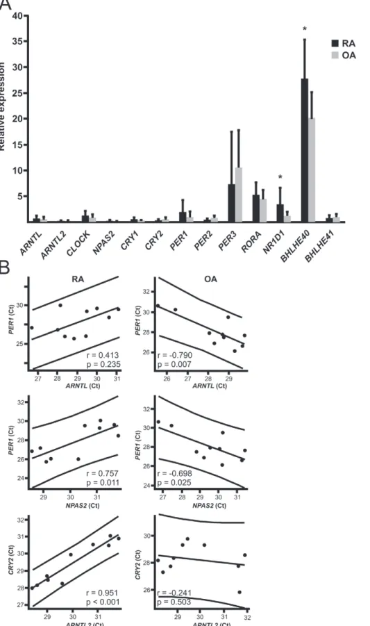
Clock Gene Expression is Disturbed in RA Synovium Driven by our hypothesis that the circadian timekeeping is perturbed in RA, we analyzed expression levels of clock genes in the synovial membrane of RA patients in comparison to OA patients. The expression levels of BHLHE40 and NR1D1( REV-ERBa) were significantly increased in RA synovium compared to OA synovium (Figure 1a).
To overcome the variation of sampling time between individuals we reasoned that the best indication of the subjective (endogenous) time would be the correlation of the expression of BMAL1 and PER1, which are normally in antiphase against each other. Due to the negative feedback loop, the peak expression of BMAL1 and PER1 should definitely not occur at the same time. Indeed, this was true in the control synovial membranes obtained from OA patients: highBMAL1expression coexisted with lowPER1expression andvice versa(Figure 1). RA patients, in contrast, did not have this kind of anti-phase BMAL1 and PER1 expression (Figure 1). Similarly, high ARNTL2 and NPAS2 expression occurred simultaneously with the inhibitory clock components only in RA. Most striking was the significant positive correlation of NPAS2 and PER1 expression in RA (Figure 1), while it as expected was significantly negative in OA (Figure 1). Further, ARNTL2 expression correlated positively with the expression of CRY1 (.715; p,0.05), CRY2 (.951; p,0.001), PER2 (.862; p,0.01), BHLHE40 (.936; p,0.001) and BHLHE41 (.785; p,0.01) only in RA.
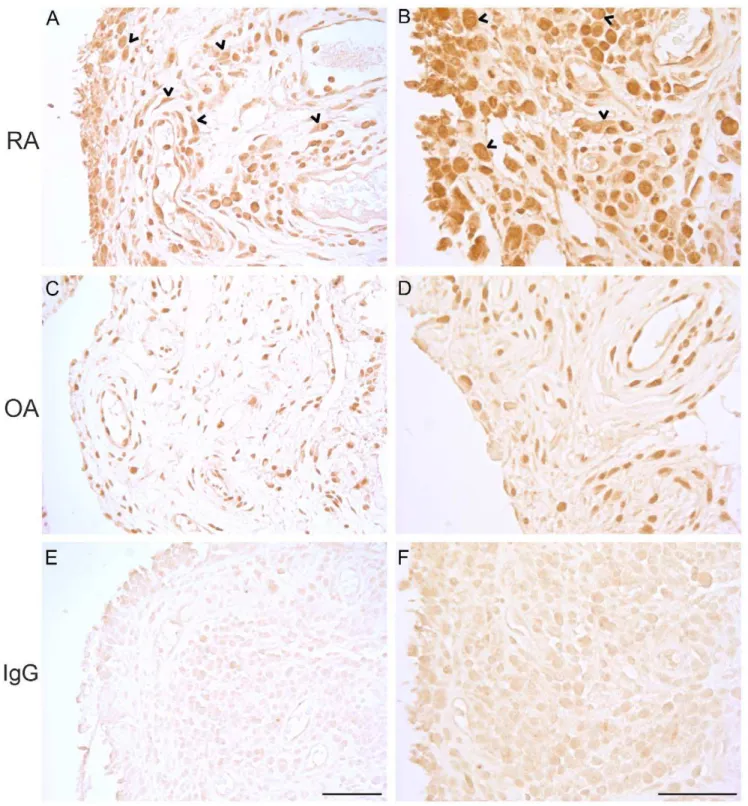
Ectopic Sub-cellular BMAL1 Protein Localization in RA Synovium
After revealing that the expression of clock genes is disturbed in the RA patients, we sought for confirmation to this observation on the protein level. We used immunohistochemistry to localize the limiting core clock component BMAL1 in the synovial tissues of RA and OA patients. The intense cytoplasmic BMAL1 protein staining in RA tissues draws attention to another major aberration. Not only are the phases ofARNTLs andNPAS2relative toCRYs Table 1.Primers.
Gene GeneBank Accession 59Primer 39Primer Length
BMAL1 NM_001178 CTGGAGAAGGTGGCCCAAAGAG CCACTGGAAGGAATGTCTGGAGTC 250
ARNTL2 NM_020183 GCTAGAGGCTACCAGGCAAAACC GGTCCACTGGATGTCACTGAAGTC 193
CLOCK NM_004898 TTCTGCCTCTTCTCGGAGTTCAAG CCTGGGTGGAGTGCTCGTATC 103
NPAS2 NM_002518 CTTCCCTGCCTCCCAACCATC GGTCCCTGGCTGTTGTGAGTAG 151
BHLHE40 NM_003670 TCAGCAGCAGCAGAAAATCATTGC GTGGGTGACAAGCTGCGAAGAC 187 BHLHE41 NM_030762 TGCTTTACAGAATGGGGAGCGATC CCCTGGGTGTCCAGCTCTCAAAC 134
PER1 NM_002616 CTCCAATCAGGACGCACTTTC GCTGCCAAAGTATTTGCTTGTG 211
PER2 NM_022817 TGTAGGGGCGGACTGCAAAC TGCTGGTATGACTTGTGTCACTAC 251
PER3 NM_016831 TGAAGAATCCATCCCATCCTACTG TATACTGCTGTCGCTGCTTCC 218
CRY1 NM_004075 TCTGGCATCAGTACCTTCTAATCC CTGTGTGTCCTCTTCCTGACTAG 226
CRY2 NM_021117 GGTGAAGAACTCAGCAAACGG ACACACATGCTCGCTCTATCTC 189
RORA NM_134262.2 CCAGCCCCGACGTCTTCAAAT GCCATGAGCGATCTGCTGACA 150
RORB NM_006914.3 ACCGTTGCCAACACTGCCGA GCTGGTGCTTCTGCACCTCA 126
RORC NM_001001523.1 GGGCTGCAGCGAGCTCATCA TCTCTTGGAGCCCTGGCCGA 134
NR1D1 NM_021724 CTTGGCTGCCCAGCGTCATAAC CCAGATCTCCTGCACCGTTCG 274
DBP NM_001352 CTTAAGCCCCAGCCAATCATGAAG CCGCCCGCACCGATATCTG 160
IL-1b NM_000576 TGGCAATGAGGATGACTTGT GGAAAGAAGGTGCTCAGGTC 237
IL-6 NM_000600.3 AGGAGACTTGCCTGGTGAAA GAGGTGCCCATGCTACATTT 329
b-act NM_001101.3 TCACCCACACTGTGCCCATCTACGA CAGCGGAACCGCTCATTGCCAATGG 295
PBGD NM_000190.3 ACATGCCCTGGAGAAGAATG AGATGCGGGAACTTTCTCTG 237
RPLP0 NM_001002 GGCGACCTGGAAGTCCAACT CCATCAGCACCACAGCCTTC 149
Figure 1. The expression of circadian genes in RA and OA patients.A. Synovial membrane samples were collected from RA and OA patients (n = 10 for each) and analyzed for circadian gene expression. The relative expression of each gene is expressed as mean6SD. B. The expression of
BMAL1andPER1should be in anti-phase. Pearson correlation and the linear fit for the expression ofBMAL1andPER1in the synovial membrane of RA
patients reveals that high expression ofBMAL1correlates with highPER1expression andvice versa, lowBMAL1expression correlates with lowPER1
expression was coupled with highPER1expression in the synovial membrane of OA patients. Similar findings were observed withNPAS2andPER1as
well as withARNTL2andCRY2. Each dot indicates the Ct value ofBMAL1,NPAS2orARNTL2in the x-axis andPER1orCRY2in the y-axis in the synovial
membrane sample of an individual patient. *p,0.05. doi:10.1371/journal.pone.0054049.g001
andPERs disturbed in RA, but this is also accompanied by ectopic localization of a core clock component (Figure 2).
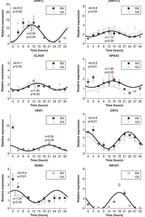
Cell Autonomous Defect of Clock Function in RA To rule out the possibility that inflammation, medication or some endocrine factors in the patients is the reason for the perturbed expression of circadian genes in RA synovium, we induced clock resetting with serum [20] in synovial fibroblasts isolated from RA and OA tissue samples. Immediately after resetting the expression pattern ofBMAL1was delayed 0.5 hours (p = 0.05) in RA fibroblasts compared to OA fibroblasts, i.e. the expression ofBMAL1in RA cells peaked 30 minutes later that OA cells. In addition to this, the expression level ofBMAL1in RA cells never reached the level of OA (p = 0.05). The expression peak of ARNTL2 was delayed 1.1 hours (p,0.05) and the level was throughout the experiment significantly lower (p,0.05) in RA cells compared to OA cells. Similarly,CLOCKexpression was delayed 1.4 hours (p,0.05) in RA fibroblasts and the relative expression was constantly lower (p,0.05) in RA cells andNPAS2expression was delayed 1.9 hours (p = 0.05) but was constantly higher in RA fibroblast (p = 0.01) than in OA fibroblasts. Based on the delayed expression of the core clock, one would expect that the expression of clock driven circadian genes would lag in RA cells compared to OA cells. In contrast, the expression ofPER1increased 0.5 hours earlier (p = 0.05), and the expression of CRY2 was constantly higher in RA than OA cells after serum shock. Also CRY1 expression increased earlier in RA cells than in OA cells, indicating that expression of the negative feedback loop is activated faster in RA than in OA. On the positive limb of the circadian machinery, RORA expression was 1.2 hours (p,0.05) ahead and constantly higher (p = 0.01) in RA cells than in OA cells, and NR1D1expression was exactly the same in both cell types (Figure 3).RORBandRORCwere not expressed.
Altered Circadian IL-1band IL-6 Expression in RA Fibroblasts
RA patients display altered circadian hormonal and cytokine profiles. For example, the circulating concentration of IL-6 in RA patients differs from that of healthy individuals [8]. We tested the possibility that the perturbed clock function is also reflected in disturbedIL-6expression after clock resetting. Fibroblasts derived from RA displayed significantly weaker rhythmic expression of IL-6 after clock resetting than cells from OA synovium (Figure 4). Both the general expression as well as the peak expression was higher in OA cells (p,0.05 for both). Because of this observation, we analyzed if this is the case also forIL-1borTNF-aexpression, which are also rhythmically produced pro-inflammatory cytokines [21,22]. Similar to IL-6 expression, RA fibroblasts displayed significantly weaker rhythmic expression of IL-1b than OA fibroblasts (p,0.05) Also TNF-a expression was rhythmic over time. In contrast to the expression ofIL-1bandIL-6, the general expression level ofTNF-awas higher (p = 0.05) in RA cells than in OA cells. Like in the expression ofIL-1b, both the phase (p,0.05) and the amplitude (p,0.05) in TNF-aexpression were different between the cells derived from RA and OA synovium (Figure 4) confirming the near absent and misaligned self-sustained rhythmic expression of pro-inflammatory cytokines in RA cells.
Inflammation Changes the Expression of Clock Genes In Vitro
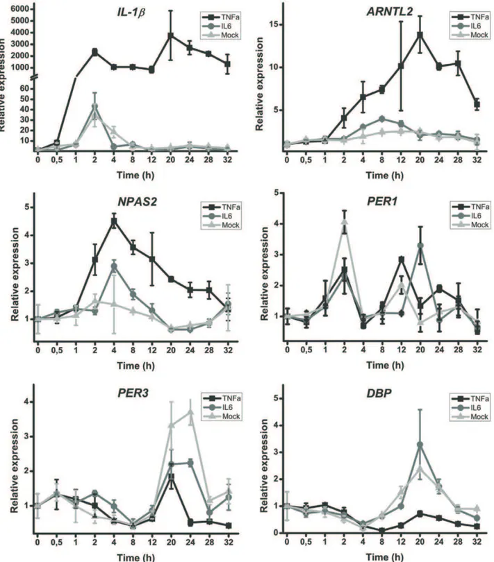
Due to the bidirectionally maintained control of clock and inflammation, we investigated the effect TNF-a on human fibroblasts. A strong response in the expression of IL-1b
(Figure 5) was evident confirming the effect of TNF-a on the cells. The most significantly affected clock genes after stimulation with TNF-a were ARNTL2and NPAS2. Unlike earlier observa-tions in mouse cells [18], TNF-adid not affect the expression of PER1. However, the inhibitory (anti-phase) effect of TNF-aon the expression of clock controlled genes DBP and PER3 was clear (Figure 5). IL-6 did not markedly alter the expression of clock genes.
Discussion
Abnormal fatigue, morning stiffness and altered circadian hormone and cytokine production suggest that the internal neuroimmunoendocrine milieu and the activity of daily living may be desynchronized in RA patients [7,8,10] i.e., the body is adjusted for activity (subjective day) during sleep (geophysical night), but meets the challenges of the day when geared for sleep and rest. Central molecular clock in the hypothalamus normally synchronizes body functions and entrains the circadian clocks in all cells of the peripheral tissues [2,4]. According to our findings the clock is dysfunctional in the joints of RA patients. Our results suggest that inflammation may initially disturb circadian time-keeping and that a cell autonomous defect prevents accurate clock function in the synovial fibroblasts of RA patients.
The variation in the expression of circadian genes at a defined and very narrow time point used for sample collection was evident between different individuals. Due to the characteristic oscillation of the expression of clock genes [1–3] leading to individual sleeping habits of each subject this was expected. To overcome the problem, we utilized the characteristic property of the clock and analyzed the endogenous (subjective) time using the expected anti-phase expression of inhibitory circadian genes in RA synovium and in OA synovium, i.e. checked the circadian time. This anti-phase expression of inhibitory circadian components, which forms the very basis for the circadian timekeeping, was totally lost in RA synovium i.e., the endogenous circadian time is clearly perturbed in the synovial membrane tissue of RA patients.
The perturbed expression of the machinery is also reflected in the amount and sub-cellular localization of BMAL1 protein. Apparently, there is much higher levels of BMAL1 protein in RA than OA while at RNA levels the difference is not significant. Of the core clock genes the expression of BMAL1 varies the most during the day. Due to this oscillation of the expression, however, we may have not detected the highest differences in the gene expression. The most dramatic differences in the gene expression may well be seen at different time points whereas the amount of protein in the tissue may reflect the differences in the expression or different kinetics in the posttranslational mechanisms that regulate the degradation of the core clock proteins [23,24]. This may well explain why the gene expression and protein amounts do not correlate. However, the result reinforces the finding of a disturbed clock in RA tissue.
Figure 3. Relative clock gene expression over time after serum shocked primary fibroblasts.Fitted sin graphs of the expression and mean6SD of the mean from three individual patients per group are shown. The time of serum removal is 0 in the graphs and the relative expression is normalized to the expression before serum (at time =22).Db indicates the general difference in the expression i.e., the baseline of the expression. Phase differences are marked as red solid lines and the difference is indicated in hours.
we observed near two-hour phase differences between RA and OA in the circadian gene expression already short after clock resetting. This is remarkable, because normally the self-sustained rhythm varies only 0.32 hours per day [26] and patients from whom these fibroblasts were isolated were randomly selected i.e., they were not
selected by chronotype or any other factor that may impact the results. Thus, our results most likely reflect true differences between RA and OA patients. Importantly, the expression of PER1, for example, increased equally in RA and OA cells immediately after clock resetting, but the self-sustained rhythm was divergent. This excludes the effect of the initial serum shock, inflammation or other endocrine factors as the sole reasons for the disturbed clock gene expression in RA synovium and underlines a primary, cell autonomous defect in the control of the biorhythm in RA, i.e. a defect in the circadian clockwork itself.
Because RORs drive the expression ofBMAL1[27,28], lag of BMAL1 expression suggests delayed or diminished expression of RORgenes or a low ratio between the expression ofRORgenes and their competitorREV-ERBain RA cells. However, early and increased RORA expression in RA cells and equal REV-ERBa
expression in RA and OA cells exclude this possibility. The expression ofARNTL2andNPAS2were also different in RA and OA. This is particularly important based on the fact thatARNTL2 andNPAS2,notPER1,react to inflammation in human fibroblasts and some mouse tissues (data not shown).
RA patients have HPA-axis dysfunction in response to inflammation [7,8]. Due to the bidirectional links between inflammation and clock, we wanted to investigate the effect of inflammation on clock gene expression. IL-6 did not affect significantly the expression of clock genes. Thus, the purpose of robust oscillation of IL-6 levels in the blood most likely is not to synchronize peripheral clocks. However, TNF-a affected clock gene expression in the cultured synovial fibroblasts, indicating that inflammation can disturb normal circadian rhythm rapidly in the peripheral tissues.
Accurate circadian clock allows orchestrated proactive rather than reactive bodily functions. The data presented here clearly show that RA patients have a deficiency in the function of the clock. It must be emphasized that it is not the synovium or synovial fibroblasts that may cause these disturbances. Rather this cell autonomous phenomenon reflects the behavior of all molecular clocks in the body including the central clock, because circadian clock has virtually the same molecular makeup in the central timekeeper and different peripheral cells [29]. Based on this, our in vitroresults suggest that RA patients, despite obtaining resetting stimuli every day, tend to lose the rhythm relatively fast due to a cell autonomous defect in their circadian clock. This may explain why the patients experience the fatigue as if they would be in a constant jet lag. The clock not only controls the circadian rhythm but also affects the development and function of immune cells. Thus, the immunological, neuronal and endocrine problems observed in the RA patients may be caused by cell autonomous dysfunction of the clock. This is further reinforced by the fact that novel biotherapies (anti-TNF, anti-B cell, anti T-cell, or anti-IL-6) have only limited impact on chronic fatigue in RA [30] at least partly excluding the possibility that inflammatory factors directly cause this severe problem in RA. Notably,ARNTL2and NPAS2 are the clock genes that throughout different experiments were the most disturbed.
Conclusion
Herein, we show that RA patients display cell autonomous disconcerted circadian timekeeping. Throughout the analysis ARNTL2andNPAS2were the genes revealed to be most associated with human inflammatory conditions. Strikingly, PER1 did not react to inflammatory stimulation in human fibroblasts. It appears that RA patients may experience the fatigue as if they would be in constant jet lag. Thus, chronotherapy that takes this altered
Figure 4. The oscillation ofIL-1b,IL-6andTNF-ain RA and OA cells after serum shock. Relative gene expression over time is displayed. Fibroblasts derived from RA synovium display weaker rhythmic expression ofIL-1b,IL-6andTNF-aafter clock resetting than
cells from OA synovium. Fitted sin graphs of the expression and mean
6SD of the mean from three individual patients per group is shown. The time of serum removal is 0 in the graphs and the relative expression is normalized to the expression before serum (at time =22). Db indicates the general difference in the expression i.e., the baseline of the expression. DA indicates the differences in the peak amplitude. Phase differences are marked as red solid lines and the difference is indicated in hours.
rhythm into account and/or tries to correct it could be beneficial for the patients.
Acknowledgments
We thank Mr. Erkki Ha¨nninen for expert technical help.
Author Contributions
Conceived and designed the experiments: VPK JO JJ IH JM. Performed the experiments: VPK JO JJ JM. Analyzed the data: VPK JO MA EK YTK JM. Contributed reagents/materials/analysis tools: EK MA YTK JM. Wrote the paper: IH YTK JM.
Figure 5. TNF-aincreasedARNTL2andNPAS2, and suppressesPER3andDBPexpression but IL-6 did not.Relative gene expression over time is displayed. Representative data from one experiment is shown. The experiment was repeated five times using fibroblasts from different OA patients. The time of TNF-aor IL-6 addition is 0 in the graphs.
References
1. Welsh DK, Takahashi JS, Kay SA (2010) Suprachiasmatic nucleus: Cell autonomy and network properties. Annu Rev Physiol 72: 551–577. 2. Takahashi JS, Hong HK, Ko CH, McDearmon EL (2008) The genetics of
mammalian circadian order and disorder: Implications for physiology and disease. Nat Rev Genet 9: 764–775.
3. Stetson MH, Watson-Whitmyre M (1976) Nucleus suprachiasmaticus: The biological clock in the hamster? Science 191: 197–199.
4. Dibner C, Schibler U, Albrecht U (2010) The mammalian circadian timing system: Organization and coordination of central and peripheral clocks. Annu Rev Physiol 72: 517–549.
5. Turek FW, Joshu C, Kohsaka A, Lin E, Ivanova G, et al. (2005) Obesity and metabolic syndrome in circadian clock mutant mice. Science 308: 1043–1045. 6. Fu L, Pelicano H, Liu J, Huang P, Lee C (2002) The circadian gene Period2 plays an important role in tumor suppression and DNA damage response in vivo. Cell 111: 41–50.
7. Chikanza IC, Petrou P, Kingsley G, Chrousos G, Panayi GS (1992) Defective hypothalamic response to immune and inflammatory stimuli in patients with rheumatoid arthritis. Arthritis Rheum 35: 1281–1288.
8. Cutolo M, Villaggio B, Otsa K, Aakre O, Sulli A, et al. (2005) Altered circadian rhythms in rheumatoid arthritis patients play a role in the disease’s symptoms. 4: 497–502.
9. Kowanko IC, Pownall R, Knapp MS, Swannell AJ, Mahoney PG (1981) Circadian variations in the signs and symptoms of rheumatoid arthritis and in the therapeutic effectiveness of flurbiprofen at different times of day. Br J Clin Pharmacol 11: 477–484.
10. Hewlett S, Cockshott Z, Byron M, Kitchen K, Tipler S, et al. (2005) Patients’ perceptions of fatigue in rheumatoid arthritis: Overwhelming, uncontrollable, ignored. 53: 697–702.
11. Repping-Wuts H, van Riel P, van Achterberg T (2009) Fatigue in patients with rheumatoid arthritis: What is known and what is needed. Rheumatology (Oxford) 48: 207–209.
12. Ishida A, Mutoh T, Ueyama T, Bando H, Masubuchi S, et al. (2005) Light activates the adrenal gland: Timing of gene expression and glucocorticoid release. Cell Metab 2: 297–307.
13. Sun H, Lu B, Li RQ, Flavell RA, Taneja R (2001) Defective T cell activation and autoimmune disorder in Stra13-deficient mice. Nat Immunol 2: 1040–1047. 14. Yang XO, Angkasekwinai P, Zhu J, Peng J, Liu Z, et al. (2009) Requirement for the basic helix-loop-helix transcription factor Dec2 in initial TH2 lineage commitment. Nat Immunol 10: 1260–1266.
15. Ivanov II, McKenzie BS, Zhou L, Tadokoro CE, Lepelley A, et al. (2006) The orphan nuclear receptor RORgammat directs the differentiation program of proinflammatory IL-17+T helper cells. Cell 126: 1121–1133.
16. Delerive P, Monte D, Dubois G, Trottein F, Fruchart-Najib J, et al. (2001) The orphan nuclear receptor ROR alpha is a negative regulator of the inflammatory response. EMBO Rep 2: 42–48.
17. Hashiramoto A, Yamane T, Tsumiyama K, Yoshida K, Komai K, et al. (2010) Mammalian clock gene cryptochrome regulates arthritis via proinflammatory cytokine TNF-alpha. J Immunol 184: 1560–1565.
18. Cavadini G, Petrzilka S, Kohler P, Jud C, Tobler I, et al. (2007) TNF-alpha suppresses the expression of clock genes by interfering with E-box-mediated transcription. Proc Natl Acad Sci U S A 104: 12843–12848.
19. Durum SK, Schmidt JA, Oppenheim JJ (1985) Interleukin 1: An immunological perspective. Annu Rev Immunol 3: 263–287.
20. Balsalobre A, Damiola F, Schibler U (1998) A serum shock induces circadian gene expression in mammalian tissue culture cells. Cell 93: 929–937. 21. Gudewill S, Pollmacher T, Vedder H, Schreiber W, Fassbender K, et al. (1992)
Nocturnal plasma levels of cytokines in healthy men. Eur Arch Psychiatry Clin Neurosci 242: 53–56.
22. Lange T, Dimitrov S, Born J (2010) Effects of sleep and circadian rhythm on the human immune system. Ann N Y Acad Sci 1193: 48–59.
23. Lee C, Etchegaray JP, Cagampang FR, Loudon AS, Reppert SM (2001) Posttranslational mechanisms regulate the mammalian circadian clock. Cell 107: 855–867.
24. Kwon I, Lee J, Chang SH, Jung NC, Lee BJ, et al. (2006) BMAL1 shuttling controls transactivation and degradation of the CLOCK/BMAL1 heterodimer. Mol Cell Biol 26: 7318–7330.
25. Balsalobre A, Brown SA, Marcacci L, Tronche F, Kellendonk C, et al. (2000) Resetting of circadian time in peripheral tissues by glucocorticoid signaling. Science 289: 2344–2347.
26. Herzog ED, Aton SJ, Numano R, Sakaki Y, Tei H (2004) Temporal precision in the mammalian circadian system: A reliable clock from less reliable neurons. J Biol Rhythms 19: 35–46.
27. Preitner N, Damiola F, Lopez-Molina L, Zakany J, Duboule D, et al. (2002) The orphan nuclear receptor REV-ERBalpha controls circadian transcription within the positive limb of the mammalian circadian oscillator. Cell 110: 251–260. 28. Sato TK, Panda S, Miraglia LJ, Reyes TM, Rudic RD, et al. (2004) A functional
genomics strategy reveals rora as a component of the mammalian circadian clock. Neuron 43: 527–537.
29. Asher G, Schibler U (2011) Crosstalk between components of circadian and metabolic cycles in mammals. Cell Metab 13: 125–137.