ISSN 0102-695X DOI: 10.1590/S0102-695X2013005000039 Received 25 Feb 2013 Accepted 30 Apr 2013 Available online 28 May 2013
standardized extract of
Orthosiphon stamineus
in rats
Hussin Muhammad,
*,1Siti A. Sulaiman,
2Zakiah Ismail,
1Francisco J. R. Paumgartten
31Herbal Medicine Research Centre, Institute for Medical Research, Kuala Lumpur,
Malaysia,
2Department of Pharmacology, School of Medical Sciences, University Science
Malaysia, KubangKerian, Kelantan, Malaysia,
3Laboratório de Toxicologia Ambiental, Escola Nacional de Saúde Pública, Fundação
Oswaldo Cruz, Brazil.
Abstract: Infusions of Orthosiphon stamineus Benth., Lamiaceae, leaves are widely used in Southeastern Asia to treat different illnesses. Nonetheless, no data is available on the safety of O. stamineus for pregnant women and their babies. This study was undertaken to evaluate the developmental toxicity of O. stamineus standardized aqueous extract in female Sprague Dawley rats (n=21) at 0, 250, 500, 1000 and 2000 mg/kg/day, by gavage on gestation days 6-20. Clinical signs of maternal toxicity, body weight gain, and food and water consumption were recorded. Caesarean sections were performed on gestation day 21; resorptions and living and dead fetuses were counted. Fetuses were weighed and examined for external abnormalities. Half of the fetuses from each litter were cleared and stained with Alizarin red S for skeleton evaluation. O. stamineus standardized aqueous extract did not alter pregnancy body weight gain and food and water consumption and caused no other sign of maternal toxicity. Embryolethality and prenatal growth retardation were not observed either. O. stamineus standardized aqueous extract increased a few skeleton variations and a skull bone malformation (hyoid bone absent) in a non-dose dependent manner. Anogenital distance was increased in male and female fetuses exposed to the highest O. stamineus standardized aqueous extract dose, an indication that the extract could possibly contain androgenic compounds.
Keywords: Developmental toxicity Orthosiphon stamineus rosmarinic acid teratogenicity pregnancy
Introduction
Orthosiphon stamineus Benth., Lamiaceae, or “Java tea” is a medicinal plant traditionally used in Southeastern Asia. It is also known as “MisaiKucing” in Malaysia, “Kumis Kucing” in Indonesia, “Balbas-pusa” and “Kabling-gubat” in Philippines, “Kapen prey” in Cambodia, “Hnwàdméew” in Laos, “YaaNuatMaeo” in Thailand, and “Thé de Java” in French speaking countries (Anon, 2001). In Southeastern Asia people are currently exposed to O. stamineus through the consumption of infusions made with its leaves, medicinal potions and phytotherapeutic drugs. In Malaysia, a tea made with O. stamineus leaves is used to improve health and to treat a variety of diseases such as kidney disorders, bladder
inl ammation, gout, diabetes, eruptive fevers, hepatitis,
hypertension, syphilis, rheumatism and gonorrhoea (Akowuah et al., 2004; Ameer et al., 2012).
Studies on the pharmacological properties of O. stamineus extracts seem to lend support to some of their common uses in folk medicine. Anti-oxidant and
anti-inl ammatory activities as well as a benei cial effect on hyperglycemia and altered lipid proi le in diabetic rats have
been reported (Arafat et al., 2008). Diuretic properties of aqueous extracts of O. stamineus were demonstrated as well (Adam et al., 2009). Methanol (50%) extracts of O. stamineus, on the other hand, were described to have anti-pyretic activity (Yam et al., 2009) and to inhibit the growth of food-borne bacteria (Vibrio parahaemolyticus) in vitro
test systems (Ho et al., 2010).
Methoxylated l avones (sinensetin and eupatorin)
and phenolic acids (rosmarinic and caffeic acids) were
identii ed in O. stamineus leaf extracts (Muhammad et al., 2011; Ameer et al., 2012). It is of note that rosmarinic acid, a major component of aqueous extracts from O. stamineus
leaves, has been reported to exhibit antioxidant,
modulatory and anti-cancer activity (Scheckel et al., 2008; Yam et al., 2009; Ameer et al., 2012).
Although being widely used in folk medicine, there is a paucity of toxicological data on O. stamineus
extracts. A few previous studies suggested that O. stamineus extracts are of low acute toxicity and pose no genotoxic risk (Han et al., 2008; Abdullah et al., 2009; Muhammad et al., 2011). The reproductive and developmental toxicity of O. stamineus extracts, however, have not been investigated so far. Along the same line, almost no information is available on the embryo/fetotoxic potential of major constituents of
O. stamineus extracts such as caffeic acid, euptorin and sinensetin. Since O. stamineus extracts are often consumed by women of childbearing age, studies of their safety in pregnancy are of the utmost importance. This study was undertaken to provide data on the developmental toxicity of O. stamineus aqueous extract in rats.
Materials and Methods
Plant material and extract preparation
The standardized aqueous extract of Orthosiphon stamineus Benth., Lamiaceae (OSAE), was purchased from Nova Laboratories Sdn. Bhd (Malaysia). Dried plant aerial parts were ground to a homogeneous powder and left to stand in water at 70 oC for 30 min. The plant infusion
was then iltered, evaporated and concentrated. The
resulting concentrated liquid extract was spray-dried at 180 oC (outlet temperature) and 100 oC (inlet temperature)
producing a powder that (OSAE) was further used in the experiments. Extraction yield was 4.8%. OSAE major constituents were determined at the Phytochemistry Unit, Herbal Medicine Research Centre. High Performance Liquid Chromatography-Diode Array Detector
(HPLC-DAD) was used to analyze the OSAE. Quantiication of
rosmarinic acid, the major constituent of OSAE extract, was undertaken using the external standard method and its amount in the extract was calculated based on the peak area of the chromatogram with the calibration curve of the standard compound. Linear regression plots were obtained (waters Empower 2 software) and results were expressed as micrograms of rosmarinic acid per gram of OSAE. Qualitative and quantitative analyses were performed on Waters 2695 Alliance HPLC system (Waters, MA, USA). A
C-18 column (Phenomenex, Luna 3μm, 100x4.6 mm.,i.d.)
guarded by a C-18 security guard cartridges was used.
Mobile phase was made of solvent A: water:triluoracetic
acid (TFA) (20:0.001;v/v) and solvent B: acetonitrile:TFA (20:0.001; v/v) and gradient elution was as follows: 0-2 min, 30% B; 2-10 min, 30-50% B; 10-20 min, 50-95% B
and inally washing the column with 95% B for 2 min and
reconditioning it with 30% B isocratic for 2 min. Flow rate
was 0.5 mL/min and injection volume was 10 μL. Peaks
were analyzed at 340 nm.
Chemicals
Glycerol, methanol, ammonium sulphide and potassium hydroxide were purchased from Merck, Chemical Germany. Acetic acid and 95% ethanol were from Hamburg. Diethyl ether was purchased from Fisher. Bouin’s solution and Alizarin Red S were from Sigma-Aldrich, UK.
Animals
Virgin females (n=105; 180-200 g) and fertile males (n=55; 200-250 g) Sprague-Dawley (SD) rats, supplied by the Animal Resource Unit (Medical Resource Centre, Institute for Medical Research, Malaysia) were used. Upon arrival at the lab, animals were housed in rat standard polypropylene cages, lined with wood shavings and kept at controlled temperature (20±2 oC), air relative
humidity (40-60%) and photoperiod (12 h of light and 12 h of dark cycle). All rats were acclimatized for one week prior to the start of the study. A commercial rat diet (Specialty Feeds, Australia) and water were available ad libitum. Approval for this study was obtained from the Animal Care and Ethics Committee, Ministry of Health Malaysia (ACUC No: ACUC/KKM/02 (2/2007).
Experimental design
Female rats in the pro-oestrous phase (oestrous cycle phase was determined by vaginal smear cytology) were placed into the cage of males (on a one-to-one basis) in the late morning and removed in the following morning (24 h). The day on which overnight mating was
conirmed by the presence of sperm in the vaginal smear
was designated as gestation day 0 (GD0).
Treatment
The OSAE (0, 250, 500, 1000 or 2000 mg/ kg/day), dissolved in distilled water, was administered (volume: 10 mL/kg) orally (gavage) to pregnant rats on GD 6-20. OSAE aqueous solutions were prepared daily. Once a day, after treatment, rats were observed in their cages for 60 min, and behavioural changes and clinical signs of toxicity were recorded. Maternal body weight (on a daily basis) and food and water intake (on a weekly basis) were measured and recorded.
Caesarean section and fetal examination
gravid uterus was removed and weighed with its contents. The number of corpora lutea graviditatis in both ovaries was then counted. Implantation sites were determined by the method of Salewski (1964). Resorptions and living and dead fetuses were counted. Placentas were examined and weighed. Fetuses were removed by cutting the umbilical cord close to their bodies, examined for externally visible abnormalities under a stereomicroscope, sexed, numbered,
and ixed in Bouin’s solution. The anogenital distance
(AGD) of all fetuses was measured using a digital caliper. Half of the fetuses of each litter, selected at random, were macerated in potassium hydroxide, cleared with glycerin-KOH solutions and stained with Alizarin Red S for skeleton evaluation (Staples & Schnell, 1964). The remaining fetuses were examined for soft-tissue anomalies and their heads were serially sectioned with a razor blade. Maternal organs were examined for gross pathology abnormalities and liver, kidneys, lungs and heart were removed and weighed.
Statistical analysis
Data on maternal body weight, weight gain during pregnancy, food and water consumption, fetal body weight, placenta weight, maternal organ weights (absolute and relative weights) were analysed by one-way analysis of variance (ANOVA) and Dunnett’s post hoc test. Number (per litter) of corpora lutea graviditatis, implantations,
live and dead fetuses, and resorptions were evaluated by the Kruskal-Wallis test followed by the Mann-Whitney U test. Proportions of fetuses and litters showing a given abnormality were compared by the chi-square. In any
case, a difference was signiicant when p<0.05. Statistical analyses were performed using SPSS version 16.
Results
Extract major constituents
HPLC analysis identiied two phenolic acids,
caffeic acid and rosmarinic acid, and two methoxylated
lavones, sinensetin and eupatorin, in the OSAE. The
foregoing markers found in the OSAE are consistent with those reported in the literature for O. stamineus extracts. The amount of rosmarinic acid, the major constituent of
OSAE, was 44.00±1.88 μg of rosmarinic acid per mg of
OSAE (4.40% w/w).
Pregnancy weight gain and signs of maternal toxicity
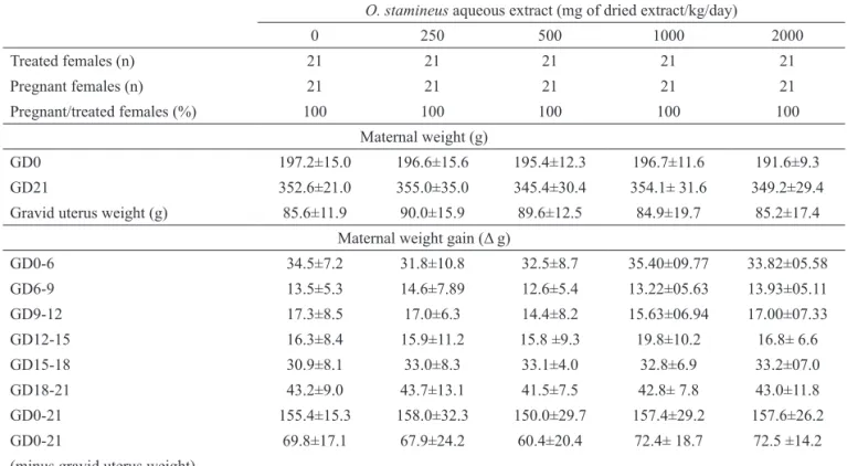
All control and treated dams survived to scheduled euthanasia. At the necropsy, no gross pathology alterations were found in OSAE treated females. Pregnancy weight gains - either with or without subtracting gravid uteri weights - were not altered at any dose level of OSAE (Table 1). Weight of
Table 1. Maternal weight gain of rats treated orally with Orthosiphon stamineus aqueous extract (0, 250, 500, 1000 and 2000 mg/kg/ day) on days 6-20 of gestation.
O. stamineus aqueous extract (mg of dried extract/kg/day)
0 250 500 1000 2000
Treated females (n) 21 21 21 21 21
Pregnant females (n) 21 21 21 21 21
Pregnant/treated females (%) 100 100 100 100 100
Maternal weight (g)
GD0 197.2±15.0 196.6±15.6 195.4±12.3 196.7±11.6 191.6±9.3
GD21 352.6±21.0 355.0±35.0 345.4±30.4 354.1± 31.6 349.2±29.4
Gravid uterus weight (g) 85.6±11.9 90.0±15.9 89.6±12.5 84.9±19.7 85.2±17.4
Maternal weight gain (Δ g)
GD0-6 34.5±7.2 31.8±10.8 32.5±8.7 35.40±09.77 33.82±05.58
GD6-9 13.5±5.3 14.6±7.89 12.6±5.4 13.22±05.63 13.93±05.11
GD9-12 17.3±8.5 17.0±6.3 14.4±8.2 15.63±06.94 17.00±07.33
GD12-15 16.3±8.4 15.9±11.2 15.8 ±9.3 19.8±10.2 16.8± 6.6
GD15-18 30.9±8.1 33.0±8.3 33.1±4.0 32.8±6.9 33.2±07.0
GD18-21 43.2±9.0 43.7±13.1 41.5±7.5 42.8± 7.8 43.0±11.8
GD0-21 155.4±15.3 158.0±32.3 150.0±29.7 157.4±29.2 157.6±26.2
GD0-21 69.8±17.1 67.9±24.2 60.4±20.4 72.4± 18.7 72.5 ±14.2
(minus gravid uterus weight)
and OSAE treated groups. Taken together, data on the resorption rate and mean number of live fetuses at term consistently showed that OSAE, given to dams in doses up to 2000 mg/kg/day on GD6-20, did not cause any increase of post-implantation losses over the incidence recorded in the control group. The mean number of males and females per litter also remained unaltered in control and OSAE treated groups.
Effects on placenta, fetal body weight and anogenital distance
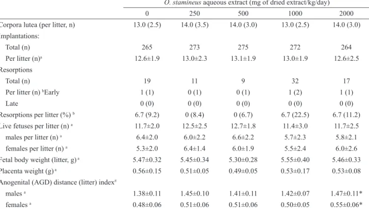
The mean weight of placentas in OSAE-treated groups was comparable to that of the control group (Table 2). The mean fetal body weight in OSAE treated groups did not differ from that in the control group either. The anogenital distance (AGD) index, however, was
signiicantly increased (p<0.05) over control AGD index measurements in male as well as in female offspring of dams treated with 2000 mg of OSAE/kg/day. Since AGD may vary with fetal body weight (Gallavan et al., 1999), AGD was normalized to body weight using the cube
root of the body weight (AGD/body weight⅓). The same
conclusion about the effect of OSAE on AGD was reached when statistical comparisons were made using direct measurements of AGD (not shown).
maternal organs (liver, kidneys, heart and lungs) did not differ between OSAE-treated and control rats (data not shown). Mild diarrhoea and softer faeces were noted in dams that received OSAE, i.e., in 1, 2, 3 and 1 dams from groups treated with 250, 500, 1000 and 2000 mg/kg/day, respectively. No behavioural alterations and no other clinical signs of toxicity were observed among treated dams. Food and water intakes of treated and control group dams were similar throughout pregnancy (data not shown).
Effects on the incidence of embryo-fetal death
Pregnancy was confirmed for all sperm positive females, i.e., all control and treated dams presented implantation sites in their uteri. The data obtained at the caesarean section are shown in Table 2. Mean numbers of corpora lutea graviditatis and implantation sites per litter did not differ between OSAE-treated and control groups. The foregoing findings indicated that exposure to OSAE from GD6 onwards did not induce peri-implantation losses. The occurrence of early and late resorptions was low in control and treated groups and the percentage of resorptions per litter (median) was not altered by OSAE administration. The average number of live fetuses per litter was similar in control
Table 2. Caesarean section data of rats treated orally with O. stamineus aqueous extract (0, 250, 500, 1000 and 2000 mg/kg /day) on days 6-20 of gestation.
O. stamineus aqueous extract (mg of dried extract/kg/day)
0 250 500 1000 2000
Corpora lutea (per litter, n) 13.0 (2.5) 14.0 (3.5) 14.0 (3.0) 13.0 (2.5) 14.0 (3.0) Implantations:
Total (n) 265 273 275 272 264
Per litter (n)a 12.6±1.9 13.0±2.3 13.1±1.9 13.0±1.9 12.6±2.5
Resorptions
Total (n) 19 11 9 32 17
Per litter (n) bEarly 1 (1) 0 (1) 0 (1) 1 (2) 1 (1)
Late 0 (0) 0 (0) 0 (0) 0 (0) 0 (0)
Resorptions per litter (%) b 6.7 (9.2) 0 (8.4) 0 (6.7) 6.7 (22.5) 6.7 (11.2)
Live fetuses per litter (n) a 11.7±2.0 12.5±2.5 12.7±1.8 11.4±3.0 11.7±2.5
males per litter (n) a 6.4±2.0 6.0±2.2 6.6±2.2 5.7±2.3 5.8±2.1
females per litter (n) a 5.3±2.0 6.4±1.4 6.0±1.9 5.5±2.4 6.0±2.6
Fetal body weight (litter, g) a 5.47±0.32 5.45±0.34 5.30±0.28 5.55±0.40 5.46±0.33
Placenta weight (g) a 0.56±0.15 0.51±0.05 0.49±0.05 0.53±0.17 0.53±0.08
Anogenital (AGD) distance (litter) index#
males a 1.38±0.11 1.45±0.10 1.41±0.11 1.42±0.07 1.47±0.11*
females a 0.48±0.06 0.51±0.06 0.51±0.06 0.50±0.05 0.55±0.06*
aData shown as means±SD were analyzed by ANOVA and Dunnett’s post hoc test and differences (p<0.05) are indicated by an asterisk (*). bData
shown as median and interquartile range (IQR) were analyzed by the Kruskal-Wallis test and no difference (p>0.05) among groups was detected.#
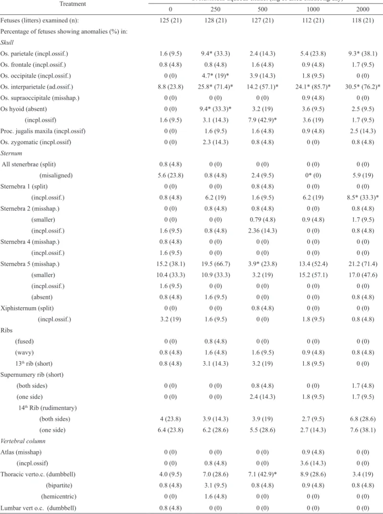
Effects on the occurrence of structural anomalies
OSAE administered on GD6-20 did not cause externally visible anomalies in the offspring of treated
dams. Similarly, examination of ixed fetuses for soft
tissue anomalies, including serial sections of brain, did not reveal any malformation (data not shown). The skeleton abnormalities found in control and OSAE-treated fetuses are shown in Table 3. The main OSAE treatment-related
skeletal abnormalities were incomplete ossiication of
some skull bones (os parietale, os occipitale and os hyoid), sternebra 1, and a forelimb long bone (os humerus), and a
poor ossiication of forelimb digits. Incidences of additional ossiication site in os interparietale and dumbbell shaped
thoracic vertebra centra were slightly enhanced as well.
Although being statistically signiicant at one or more
OSAE treated groups, enhanced occurrences of foregoing skeleton abnormalities were not dose dependent. The aforementioned skeleton abnormalities are generally
classiied as variations (Solecki et al., 2001). An increased
incidence of absent hyoid bones was noted in fetuses from dams treated with the lowest dose of OSAE (250 mg/kg body weight/day). Missing bones are a generally considered
as typical malformations and thus should be classiied the
absence of hyoid bone (Gallavan et al., 1999).
Discussion
Results from this study showed that, except for mild diarrhoea, OSAE administered orally to dams in doses up to 2000 mg/kg/day on GD6-20 did not cause overt maternal toxicity. The mild diarrheic symptoms noted in some rats treated with OSAE were not accompanied by reductions of body weight gain or alterations of food and water intakes and thus they were interpretedas being of minor toxicological significance. The absence of signs of maternal toxicity in this study was consistent with previous reports that O.stamineus
extracts and their major constituent (rosmarinic acid) present low acute and subacute toxicities (Abdullah et al., 2009; Yam et al., 2009). The oral LD50 of O. stamineus methanol extract in rats, for instance, was higher than 5000 mg/kg (Yam et al., 2009).
The fact that resorption rates (resorptions per litter) and litter sizes (number of live fetuses per litter) at term remained unaltered indicated that OSAE, did not cause embryo and fetal deaths. Likewise, unchanged ratios of treated (sperm positive) per pregnant females (i.e., with detected implantations sites) and numbers of corpora lutea graviditatis per litter suggested that OSAE did not cause peri-implantation losses either. Results, therefore, indicated that OSAE did not induce peri- or post-implantation gestational losses.
The absence of effects on placenta and fetal body weights showed that OSAE administered to dams
throughout embryogenesis (GD6-15) and fetal maturation (GD15-20) periods did not retard prenatal growth of exposed offspring. Furthermore, results from this study provided no evidence that administration of OSAE enhanced the incidence of externally visible and soft tissue anomalies in the exposed fetuses.
Skeletal abnormalities observed in
OSAE-exposed fetuses, such as incomplete ossiication of os
parietale, os hyoid, sternebra 1 and os humerus, poor
ossiication of forelimb digits and dumbbell shaped thoracic vertebra centra, are generally classiied as variations (Gallavan et al., 1999). Additional ossiication
in os interparietale occurs at a high background incidence in rats from our breeding stock (e.g., 8.8 % of fetuses and 23.8% of litters in the control group) and should also be considered as a variation. The absence of os hyoid,
on the other hand, is to be classiied as a malformation.
It should be borne in mind, however, that increased occurrences of the aforementioned skeletal variations and hyoid bone absence in OSAE exposed fetuses were not dose dependent effects. Owing to their low severity and or to the fact that observations were non-dose-related the foregoing skeletal abnormalities were not taken into account for setting the study-derived no- observed-adverse- effect-level (NOAEL) for OSAE.
A longer AGD in male and female offspring from mothers treated with the highest dose (2000 mg/kg/day) was the main developmental toxic effect of OSAE found in this study. AGD is a sexually dimorphic measure of genital development and a sensitive marker for endocrine disruption in rodent studies. Testosterone secretion by fetal testis increases the AGD in males relative to the
distance in females at term. The AGD is also inluenced by
intrauterine position of the conceptus, being a longer AGD associated with the presence of males on either side of the developing female fetus, and a shorter AGD associated with absence of males on either side of the developing female fetus (vom Saal et al., 1983; Hotchkiss et al., 2007; Han et al., 2008). Pre-natal exposure to hormonally-active compounds has also been shown to alter AGD. For instance, in utero (GD14-19) exposure of Sprague Dawley rats to the potent androgenic compound trenbolone was found to increase neonatal AGD, to delay puberty and to masculinize female offspring behavior (Han et al., 2008). The mode by which OSAE, at the highest dose tested, increased AGD in both male and female fetuses remains obscure. The presence of androgenic compounds in the extract is a plausible explanation for this effect and should be further investigated.
Treatment O. stamineus aqueous extract (mg of dried extract/kg/day)
0 250 500 1000 2000
Fetuses (litters) examined (n): 125 (21) 128 (21) 127 (21) 112 (21) 118 (21)
Percentage of fetuses showing anomalies (%) in: Skull
Os. parietale (incpl.ossif.) 1.6 (9.5) 9.4* (33.3) 2.4 (14.3) 5.4 (23.8) 9.3* (38.1)
Os. frontale (incpl.ossif.) 0.8 (4.8) 0.8 (4.8) 1.6 (4.8) 0.9 (4.8) 1.7 (9.5)
Os. occipitale (incpl.ossif.) 0 (0) 4.7* (19)* 3.9 (14.3) 1.8 (9.5) 0 (0)
Os. interparietale (ad.ossif.) 8.8 (23.8) 25.8* (71.4)* 14.2 (57.1)* 24.1* (85.7)* 30.5* (76.2)*
Os. supraoccipitale (misshap.) 0 (0) 0 (0) 0 (0) 0.9 (4.8) 0 (0)
Os hyoid (absent) 0 (0) 9.4* (33.3)* 3.2 (19) 3.6 (9.5) 2.5 (9.5)
(incpl.ossif) 1.6 (9.5) 3.1 (14.3) 7.9 (42.9)* 3.6 (19) 1.7 (9.5)
Proc. jugalis maxila (incpl.ossif) 0 (0) 1.6 (9.5) 1.6 (4.8) 0.9 (4.8) 2.5 (14.3)
Os. zygomatic (incpl.ossif) 0 (0) 2.3 (14.3) 0.8 (4.8) 0 (0) 0.8 (4.8)
Sternum
All stenerbrae (split) 0.8 (4.8) 0 (0) 0 (0) 0 (0) 0 (0)
(misaligned) 5.6 (23.8) 0.8 (4.8) 2.4 (9.5) 0* (0) 5.9 (19)
Sternebra 1 (split) 0 (0) 0 (0) 0.8 (4.8) 0 (0) 0 (0)
(incpl.ossif.) 0.8 (4.8) 6.2 (19) 1.6 (9.5) 6.2 (19) 8.5* (33.3)*
Sternebra 2 (misshap.) 0 (0) 0.8 (4.8) 0.8 (4.8) 0 (0) 0.8 (4.8)
(smaller) 0 (0) 0 (0) 0.79 (4.8) 0.9 (4.8) 1.7 (9.5)
(incpl.ossif.) 1.6 (9.5) 0.8 (4.8) 2.36 (14.3) 0 (0) 0.8 (4.8)
Sternebra 4 (misshap.) 0.8 (4.8) 0 (0) 0 (0) 0 (0) 0 (0)
(incpl.ossif.) 1.6 (9.5) 0 (0) 0 (0) 0 (0) 0 (0)
Sternebra 5 (misshap.) 15.2 (38.1) 19.5 (66.7) 3.9* (23.8) 13.4 (52.4) 21.2 (71.4)
(smaller) 10.4 (33.3) 10.9 (33.3) 3.2 (19) 15.2 (57.1) 17.0 (47.6)
(incpl.ossif.) 1.6 (9.5) 0 (0) 0 (0) 0 (0) 0 (0)
(absent) 0.8 (4.8) 1.6 (9.5) 0 (0) 0 (0) 0.8 (4.8)
Xiphisternum (split) 0 (0) 0 (0) 0.8 (4.8) 0 (0) 0 (0)
(incpl.ossif.) 3.2 (19) 1.6 (9.5) 0 (0) 1.8 (9.5) 0.8 (4.8)
Ribs
(fused) 0 (0) 0.8 (4.8) 0 (0) 0 (0) 0 (0)
(wavy) 0.8 (4.8) 1.6 (4.8) 1.6 (9.5) 0.9 (4.8) 0.8 (4.8)
13th rib (short) 0.8 (4.8) 3.1 (14.3) 3.2 (19) 1.8 (9.5) 0 (0)
Supernumery rib (short)
(both sides) 0 (0) 0 (0) 0.8 (4.8) 0 (0) 1.7 (4.8)
(one side) 0 (0) 0 (0) 2.4 (14.3) 1.8 (9.5) 1.7 (9.5)
14th Rib (rudimentary)
(both sides) 4 (23.8) 3.9 (14.3) 3.9 (19) 2.7 (9.5) 6.8 (28.6)
(one side) 6.4 (23.8) 6.2 (28.6) 5.5 (28.6) 2.7 (14.3) 7.6 (38.1)
Vertebral column
Atlas (misshap) 0 (0) 0 (0) 0 (0) 0.9 (4.8) 0 (0)
(incpl.ossif) 0 (0) 0.8 (4.8) 0 (0) 3.6 (14.3) 0 (0)
Thoracic verto.c. (dumbbell) 4.0 (9.5) 7.0 (28.6) 7.1 (42.9)* 8.9 (28.6) 3.4 (19)
(bipartite) 0.8 (4.8) 3.1 (9.5) 0.8 (4.8) 0.9 (4.8) 0.8 (4.8)
(hemicentric) 0 (0) 1.6 (4.8) 0 (0) 0 (0) 0 (0)
Lumbar vert o.c. (dumbbell) 0.8 (4.8) 0 (0) 0 (0) 0 (0) 0 (0)
as compared to that in control fetuses of the same gender was the most conspicuous developmental adverse effect of OSAE found in this study. The study-derived no-observed-adverse effect levels (NOAEL) for maternal and developmental toxicities were set at >2000 and 1000 mg of OSAE per kg body weight per day by the oral route, respectively. Taken into account that extraction yield was 4.8%, the foregoing NOAEL corresponds to 96 and 48 g of dried leaves of O. stamineus per kg body weight per day, respectively. It is of note that the study-derived NOAEL for developmental toxicity is far in excess of the estimated amount of O. stamineus currently consumed by women of childbearing age through the use of teas, medicinal potions and phytotherapeutic drugs.
Acknowledgment
This study was supported by the National Institute of Health, Malaysia, under project number JPP-IMR 05-007. The authors gratefully acknowledge the Director General of Health, Malaysia and Director of the Institute for Medical Research, Kuala Lumpur, for permission to publish this paper. FJRP is the recipient of a research fellowship from the National Research Council-Brazil (CNPq). The authors would also like to thank Rosangela De-Carvalho and Adlin Afzan for their technical assistance with the evaluation of soft tissue anomalies and phytochemistry analysis of the extract.
Authors contributions
HM ran the whole laboratory work as well as analyzing the data, drafting and reviewing the manuscript. SAS and ZI supervised the laboratory work and contributed to the review of the manuscript. FJRP supervised the data analyses and critically reviewed of the manuscript. All
authors have read the inal manuscript and approved its
submission.
References
Abdullah NR, Ismail Z, Ismail Z 2009. Acute toxicity of Orthosiphon stamineus Benth. standardized extract in
Sprague Dawley rats. Phytomedicine 16: 222-226. Adam Y, Somchit MN, Sulaiman MR, Nasaruddin AA, Zuraini
A, Bustamam AA, Zakaria Z 2009. Diuretic properties of Orthosiphon stamineus Benth. J Ethnopharmacol 124: 154-158.
Akowuah AG, Zhari I, Norhayati I, Sadikun A, Khamsah SM 2004. Sinensetin, eupatorin,
3’-hydroxy-5,6,7,4’-tetramethoxylavone and rosmarinic acid contents and
antioxidative effect of Orthosiphon stamineus from Malaysia. Food Chem 87: 569-666.
Ameer OZ, Salman IM, Asmawi MZ, Ibraheem ZO, Yam MF 2012. Orthosiphon stamineus: traditional uses, phytochemistry, pharmacology and toxicology: A review. J Med Food 15: 1-13.
Anon 2001.Orthosiphon. Medicinal and Poisonous Plants. Leidin: Buckhuys Publication. p. 368-371.
Arafat OM, Tham SY, Sadikun A, Zhari I, Houghton PJ, Asmawi MZ 2008. Studies of diuretic and hypouricemic effects of Orthosiphon stamineus methanol extract in rats. J Ethnopharmacol 118: 354-360.
Gallavan Jr RH, Holson JF, Stump DG, Knapp JF, Reynolds
VL 1999. Interpreting the toxicologic signiicance
of alterations in anogenital distance: potential for confounding effects of progeny body weights. Reprod Toxicol 13: 383-390.
Han CJ, Hussin AH, Ismail S 2008.Toxicity study of Orthosiphon stamineus Benth. (MisaiKucing) on Sprague-Dawley rats. Trop Med 25: 9-16.
Ho C-H, Noryati I, Sulaiman S-F, Rosma A 2010. In vitro antibacterial and antioxidant activities of Orthosiphon stamineus Benth. extracts against food-borne bacteria. Food Chem 122: 1168-1172.
Hotchkiss AK, Furr J, Makynen EA, Ankley GT, Gray LE Jr 2007. In utero exposure to the environmental androgen trenbolone masculinizes female Sprague-Dawley rats. Toxicol Lett 174: 31-41.
Muhammad H, Gomes-Carneiro MR, Poça KS, De-Oliveira AC, Afzan A, Sulaiman SA, Ismail Z, Paumgartten FJ 2011. Evaluation of the genotoxicity of Orthosiphon stamineus aqueous extract. J Ethnopharmacol 27: 647-653. Salewski E 1964.Farbemethoden zum makroskopischen
nachweiss von implantationsstellenan uterus der ratte. N-S Arch Ex Path Ph 247: 367.
(bipartite) 0.8 (4.8) 0 (0) 0 (0) 0.9 (4.8) 0 (0)
Forelimbs
Fingers (poorly ossiied) 14.4 (42.9) 29.7* (61.9) 35.4* (52.4) 11.6 (23.8) 33.9* (76.2)
Os humerus (incpl.ossif.) 0.8 (4.8) 6.2* (28.6) 10.2 (28.6) 5.4 (4.8) 15.2* (47.6)*
Hindlimbs
Os femur (misshap.) 4.8 (14.3) 3.1 (9.5) 3.2 (14.3) 8.9 (33.3) 4.2 (23.8)
(incpl.ossif.) 0 (0) 1.6 (9.5) 0.8 (4.8) 0 (0) 0 (0)
Values are % of fetuses (litters) showing the anomaly. Comparisons were made by the chi-square test or the Fisher’s exact test. Proportions different (p<0.05) from those of the control group are indicated by an asterisk (*). Ossif. Center: ossiication center; Incpl.ossif: incomplete ossiication.
Scheckel KA, Degner SC, Romagnolo DF 2008. Rosmarinic acid antagonizes activator protein-1-dependent activation of cyclooxygenase-2 expression in human cancer and non malignant cell lines. J Nutr 138: 2098-2105.
Solecki R, Bürgin H, Buschmann J, Clark R, Duverger M, Fialkowski O, Guittin P, Hazelden KP, Hellwig J, Hoffmann E, Hofmann T, Hübel U, Khalil S, Lingk W, Mantovani A, Moxon M, Müller S, Parkinson M, Paul M, Paumgartten F, Pfeil R, Platzek T, Rauch-Ernst M, Scheevelenbos A, Seed J, Talsness CE, Yasuda M, Younes M, Chahoud I 2001. Harmonisation of rat fetal
skeletal terminology and classiication. Report of the
Third Workshop on the Terminology in Developmental Toxicology. Berlin, 14-16 September 2000. Reprod Toxicol 15: 713-721.
Staples RE, Schnell UL 1964. Reinements in rapid clearing
techniques in the KOH-alizarin red S, methods for fetal bone. Stain Technol 39: 61-63.
vom Saal FS, Grant WM, McMullen CW, Laves KS 1983. High fetal estrogen concentrations: correlation with increased adult sexual activity and decreased aggression in male mice. Science 220: 1306-1309.
Yam MF, Ang LF, Basir R, Salman IM, Ameer OZ, Asmawi MZ 2009. Evaluation of the anti-pyretic potential of Orthosiphon stamineus Benth. standardized extract.
Inlammopharmacol 17: 50-54.
*Correspondence
Hussin Muhammad
Toxicology and Pharmacology Unit, Herbal Medicine Research Centre, Institute for Medical Research
Jln Pahang, 50588 Kuala Lumpur, Malaysia [email protected]