ABSTRACT
http://dx.doi.org/10.1590/1678-775720130488
Randomized controlled clinical trial of
long-term chemo-mechanical caries removal using
Papacarie
TM
gel
Lara Jansiski MOTTA1, Sandra Kalil BUSSADORI2, Ana Paula CAMPANELLI3, André Luis da SILVA3, Thays Almeida
ALFAYA4 2, Maria Fidela de Lima NAVARRO5
1- School of Dentistry, University Nove de Julho (UNINOVE), São Paulo, SP, Brazil.
2- Rehabilitation Sciences Post Graduation Program, University Nove de Julho (UNINOVE), São Paulo, SP, Brazil. 3- Department of Biological Sciences, Bauru School of Dentistry, University of São Paulo, Bauru, SP, Brazil. 4- Dental Clinic Post Graduation Program, Federal Fluminense University (UFF), Niterói, RJ, Brazil.
5- Department of Operative Dentistry, Endodontics and Dental Materials, Bauru School of Dentistry, University of São Paulo, Bauru, SP, Brazil.
Corresponding address: Sandra Kalil Bussadori - R. Vergueiro, 235/249 - Liberdade - São Paulo - SP - 01504-001 - Brazil - Phone: +55 11 3385 9222 - e-mail: [email protected]
!"#"$%&'()$&
A
have led to transformations in the restorative treatment of dental caries. Objectives: Compare the effectiveness of PapacarieTM gel for the chemo-mechanical removal of carious
lesions on primary teeth to conventional caries removal with a low-speed bur with regard randomized controlled clinical trial with a split-mouth design was carried out. The sample was composed of 20 children aged four to seven years, in whom 40 deciduous teeth were randomly divided into two groups: chemo-mechanical caries removal with PapacarieTM and
removal of carious dentin with a low-speed bur. Each child underwent both procedures and served as his/her own control. Restorations were performed with glass ionomer cement. The time required to perform the procedure was also analyzed. The patients underwent longitudinal clinical and radiographic follow-up of the restorations. Results: No statistically ! groups were found in the clinical evaluation at 6 and 18 months after treatment. Conclusion: PapacarieTM is as effective as the traditional method for the removal of carious dentin on
deciduous teeth, but offers the advantages of the preservation of sound dental tissue as well as the avoidance of sharp rotary instruments and local anesthesia.
Keywords: Dental caries. Papain. Dental atraumatic restorative treatment
INTRODUCTION
Advances in the field of cariology and the philosophy of minimally invasive intervention have led to transformations in the restorative treatment of dental caries. The most striking change involves the selective removal of carious tissue and maximal preservation of healthy dental tissue. Traditional methods involving a drill and a bur are incompatible with this philosophy1,4,19.
Chemo-mechanical caries removal (CMCR) is an alternative to the conventional method and consists
of the application of a proteolytic substance that softens carious dentin tissue and facilitates its removal using manual instruments17. This method
can be employed without the use of local anesthesia or burs, thereby preserving sound dental tissue4-6.
PapacarieTM is one of the products marketed for
CMCR. This gel contains papain and chloramine. Papain is an enzyme similar to human pepsin that acts as a debriding agent with no harm caused to healthy tissue. This substance accelerates the healing process and exhibits bactericidal,
Chloramine has properties related to disinfection11.
of PapacarieTM and report that its cost is lower than
similar products found on the market6,10,16,23.
There is a scarcity of well-standardized clinical trials comparing the traditional cavity preparation method to chemo-mechanical caries removal with the use of PapacarieTM gel. Thus, the aim of the
present study was to compare the effectiveness of PapacarieTM gel for the chemo-mechanical removal
of carious lesions on primary teeth to conventional caries removal with a low-speed bur with regard to execution time, clinical aspects and radiographic
MATERIAL AND METHODS
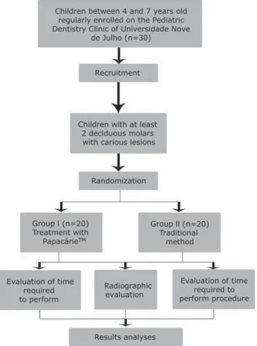
A randomized, controlled, clinical trial with a “split-mouth” design was carried out to assess
$TM gel. The investigation
was designed, analyzed and interpreted according to the Consolidated Standards of Reporting Trials (CONSORT) (Figure 1). Thirty children aged four to seven years who sought dental treatment at the Pediatric Dentistry Clinic of the University Nove de Julho (Brazil) were recruited. The control group was submitted to the traditional method (bur) for the removal of carious tissue. All parents/ guardians received information on the objectives
and procedures of the study and signed a statement of informed consent in compliance with Resolution 196/96 of the Brazilian National Health Board. This study received approval from the Human Research Ethics Committee of the University Nove de Julho (Brazil) under process nº 219047. The clinical trial registration number is NCT01811420.
Sample size calculation
The sample size was calculated using the Dinam 1.0 program with data from a pilot study. Calculations were performed considering time, discomfort, colony-forming bacteria and radiographic density. As the variable colony-forming bacteria required the largest number of teeth per
group, this variable was chosen as the reference (n=19 teeth per group).
Calibration exercise
The calibration exercise was carried out during the pilot study. An operator performed the treatment and a “gold standard” examiner performed the clinical evaluation of the removal of carious tissue. The examiner was blinded to the technique applied (chemo-mechanical caries removal with PapacarieTM
and removal of carious dentine with low-speed bur). The “gold standard” examiner evaluated all cavities following the respective interventions and was responsible for testing the hardness of the
remaining dentin. The clinical evaluation involved an inspection of the texture of the remaining dentin with a blunt exploratory probe, observing the vitreous aspect (cavity free of carious tissue). Caries removal was only considered complete when agreement was achieved between the operator and examiner. Intra-examiner agreement was determined using the Kappa statistic and was considered excellent (K=0.92).
Sample selection
Twenty children aged four to seven years participated in the study. The sample was made up of 40 primary teeth (two per child). The following were the inclusion criteria: good health, with no systemic conditions and good behavior. Clinically, the children need to have at least two primary molars with active, acute carious lesions not surpassing 2/3 of the dentin and involving only the occlusal facet, with no clinical or radiographic signs or symptoms of pulp involvement (spontaneous pain, pain upon tooth, periapical radiolucency, increase in the space of the periodontal ligament in the apical region, periapical radiopacity, lateral lesions and lesions in the furcation due to the impairment of accessory canals). The exclusion criteria were Class II, III or %& '* impossibility of restorations, carious lesion involving carious lesions in dentin (without access for manual excavators) and hidden caries.
Each child underwent both procedures and served as his/her own control. Randomization of the techniques was determined by lots using numbered tiles. For each individual, one tooth was randomly selected for one treatment (also randomly selected) and the other tooth automatically received the other form of treatment.
Group 1 (G1) – chemo-mechanical caries removal with PapacarieTM
Group 2 (G2) – removal of carious dentin with low-speed bur (traditional method - TM)
Treatments
Treatments were performed by a single dentist who had previously undergone the calibration exercise (Kappa statistic: 0.92). All procedures were initiated without the prior administration of local anesthesia, but the patients were informed that anesthesia was available if needed.
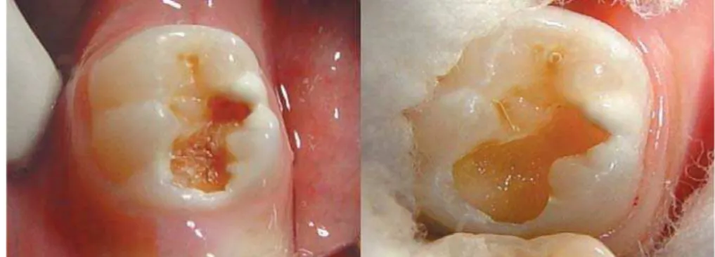
G1 – Chemo-mechanical caries removal with PapacarieTM
Initially, periapical radiographs were taken and prophylaxis was performed with a Robinson brush # (lip bumper, cotton roll and saliva aspirator) of the tooth. The starting time was then recorded. The PapacarieTM gel was applied. After 30 to 40 seconds,
the softened carious tissue was removed with the blunt end of a curette. The gel was reapplied, if necessary, until the complete removal of the carious tissue. Successful removal was determined by clinical examination involving the inspection of the texture of the remaining dentine with the use +5 ;< > time was then recorded. The restorative procedure was performed with glass ionomer cement (Ketac Molar Easy mix – 3M ESPETM, São Paulo, SP, Brazil).
The treated teeth were submitted to clinical and radiographic follow-up.
G2 – Removal of carious dentin tissue with low-speed bur (traditional method)
The initial protocol was the same as that used for G1. Once the starting time was recorded, caries removal was performed with low-speed burs, followed by a clinical evaluation and recording of the > same as that used in G1 and the treated teeth were submitted to clinical and radiographic follow-up.
Evaluations
Evaluation of time required to perform procedure
The time required to perform the procedure was measured in minutes and seconds using a digital chronometer (Kenko®).
Radiographic evaluation
$ * " criteria. Immediately after the clinical procedure, * > procedure was repeated in a standardized fashion on three different occasions. The radiographic evaluation was performed using the radiographic subtraction method and the assessment of the density of the remaining dentin immediately following the caries removal procedure as well as after one (T1), six (T2) and 18 (T3) months. Radiographic density was determined by gray-scale analysis.
A positioner for interproximal radiographs was used for standardization. A portion of self-curing acrylic resin was placed on the surface of the tooth analyzed and its antagonist for the impression of the anatomy of the surfaces and adapted to the positioner to allow the same positioning of the standardization of the same incidence of x-rays, the same vertical and horizontal angles and the same distance on all radiographs of the same patient. The interproximal radiographic images were scanned to allow the analysis of the difference in density between the different evaluation times using the Imagelab 2.3 program. Therefore, a greater degree of density on the image denoted greater success.
Clinical evaluation
The clinical evaluation of the restorations followed the criteria of atraumatic restorative treatment22, based on the retention of the material
in the cavity and the presence of secondary caries. The scoring system was as follows: 0=present, without defect; 1=present, small defects on the margin measuring less than 0.5 mm in depth, with no need for repair; 2=present, small defects on the margin measuring 0.5 to 1.0 mm in depth, with need for repair; 3=present, gross defects on the margin measuring 1.0 mm or more in depth, with need for repair; 4=absent, restoration completely lost, need for treatment; 5=absent, other treatment had been performed for some reason; 6=tooth absent for some reason; 7=present, wear on surface less than 0.5 mm, with no need for replacement; 8=present, wear on surface greater than 0.5 mm, with need for replacement; 9=diagnosis impossible12.
Statistical analysis
The SPSS 12.0 program for Windows was used for the statistical analysis. The following tests were performed: t-test complemented by the Mann-Whitney test for differences in the mean time required for treatment; Wilcoxon test for data related to the clinical evaluation and differences between evaluation times; and analysis of variance
(ANOVA) complemented by Tukey’s test for differences in mean radiographic density.
RESULTS
The sample was made of 20 children (10 girls and 10 boys) between four and seven years of age.
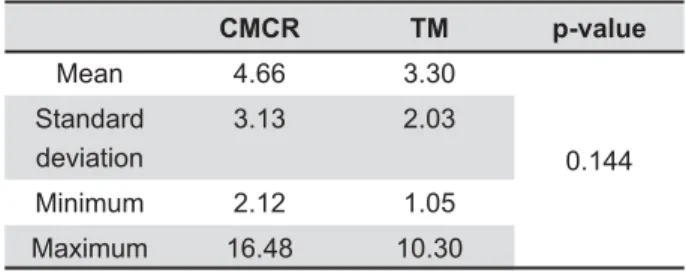
L between groups regarding the time required for the procedure (p=0.144) (Table 1). The use of anesthesia was only necessary in one case (G2). The administration of anesthesia was considered in the analysis of the time required for treatment, overall procedure.
Restorations having received a score of 0, 1 or 7 were considered successful and those having received a score of 2, 3, 4 or 8 were considered failures. Those having received a score of 5, 6 or 9 were excluded from the analysis. Evaluations were performed at one, six and 18 months after treatment. Table 2 displays the results of the evaluation of the restorations performed by an examiner who was blinded to the form of treatment employed prior to the restoration. Statistically in the clinical evaluation at the six-month and 18-month evaluations.
At the one-month evaluation, all restorations in both groups were considered successful. At the six-month evaluation, 5% (n=1) of the restorations in G1 and 10% (n=2) of the restorations in G2 had failed and were repaired. At the 18-month evaluation, 95% of the restorations in G1 and 80% in G2 were successful, with no occurrence of secondary caries (Table 3).
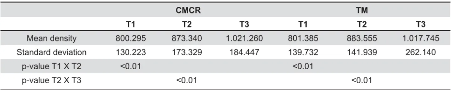
In the radiographic analysis, mean density of the affected dentin was 80.02 in G1 and 80.13 in X; ! gains in density in the radiolucent zones (affected dentin) were found in the entire sample (Table 4), with no differences between groups at the different evaluation times (T1: p=0.874; T2: p=0.661; T3: p=0.653).
CMCR TM p-value
Mean 4.66 3.30
0.144 Standard
deviation
3.13 2.03
Minimum 2.12 1.05
Maximum 16.48 10.30
CMCR=Chemo-mecanical caries removal TM=Traditional method
DISCUSSION
L groups was found regarding the time required to perform the different procedures. This is in Y al.16 (2009). However, other researchers have
investigated this issue and report that CMCR with PapacarieTM requires a shorter execution time in
comparison to conventional treatment8,13. While
execution time is essential in any dental procedure, especially in the treatment of children, anxious adults and individuals with disabilities7, CMCR
should be considered based on the fact that it causes less patient discomfort3,20 and is in line with
the philosophy of minimally invasive treatment4–6.
This was evidenced by Bohari, et al.3 (2012),
who compared four methods (burs, CarisolvTM,
PapacarieTM and Laser), as CMCR and laser are
minimally invasive methods and considered less
T1 (1 month) T2 (6 months) T3 (18 months)
Score CMCR
n (%)
Trad. Method
n (%)
p-value CMCR
n (%)
Trad. Method
n (%)
p- value CMCR n (%)
Trad. Method
n (%)
p- value
0 13 (65.0) 13 (65.0)
0.936
6 (30.0) 13 (65.0)
0.010
6 (30.0) 7 (35.0)
0.023
1 4 (20.0) 4 (20.0) 7 (35.0) 3 (15.0) 7 (35.0) 7 (35.0)
2 1 (5.0) 0 (0.0) 0 (0.0) 2 (10.0) 1 (5.0) 4 (20.0)
3 0 (0.0) 0 (0.0) 0 (0.0) 0 (0.0) 0 (0.0) 0 (0.0)
4 0 (0.0) 0 (0.0) 0 (0.0) 0 (0.0) 0 (0.0) 0 (0.0)
5 0 (0.0) 0 (0.0) 0 (0.0) 0 (0.0) 0 (0.0) 0 (0.0)
6 0 (0.0) 0 (0.0) 0 (0.0) 0 (0.0) 0 (0.0) 0 (0.0)
7 2 (10.0) 3 (15.0) 7 (35.0) 2 (10.0) 6 (30.0) 2 (10.0)
8 0 (0.0) 0 (0.0) 0 (0.0) 0 (0.0) 0 (0.0) 0 (0.0)
9 0 (0.0) 0 (0.0) 0 (0.0) 0 (0.0) 0 (0.0) 0 (0.0)
Total 20 (100.0) 20 (100.0) 20 (100.0) 20 (100.0) 20 (100.0) 20 (100.0)
CMCR=Chemo-mecanical caries removal
Table 2- Distribution of restoration scores at different clinical evaluations
T1 T2 T3
CMCR n (%)
Trad. Method n (%)
CMCR n (%)
Trad. Method n (%)
CMCR n (%)
Trad. Method n (%)
Success 20 (100.0) 20 (100.0) 19 (95.0) 18 (90.0) 19 (95.0) 16 (80.0)
Failure 0 (0.0) 0 (0.0) 1 (5.0) 2 (10.0) 1 (5.0) 4 (20.0)
Total 20 (100.0) 20 (100.0) 20 (100.0) 20 (100.0) 20 (100.0) 20 (100.0)
CMCR=Chemo-mechanical caries removal
Table 3-&OLQLFDOFODVVL¿FDWLRQDWGLIIHUHQWHYDOXDWLRQWLPHV
CMCR TM
T1 T2 T3 T1 T2 T3
Mean density 800.295 873.340 1.021.260 801.385 883.555 1.017.745
Standard deviation 130.223 173.329 184.447 139.732 141.939 262.140
p-value T1 X T2 <0.01 <0.01
p-value T2 X T3 <0.01 <0.01
CMCR=Chemo-mechanical caries removal TM=Traditional method
painful. Similar results are described in another study15.
In all the cases examined in the present study, the radiographic analysis revealed an increase in density in the affected dentin, demonstrating the success of treatment. The philosophy of minimally invasive treatment involves the maximum preservation of sound dental tissue that is capable of remineralization11,21,which is what
remains after the use of the gel. The outermost layer – denominated the infected dentin tissue – is irreversibly denatured, infected, incapable of remineralization and dead. The innermost layer – denominated the affected dentin tissue – is reversibly denatured, slightly infected or non-infected, capable of remineralization, sensitive and vital. In minimally invasive treatment, the former layer should be removed and the latter should be preserved14. Assessments of the mineral content
following the use of PapacarieTM on deciduous teeth
suggest that this gel only acts on carious tissue2.
Studies have also shown the lack of a smear layer after the use of PapacarieTM, along with
the deposition of minerals around the dentinal tubules10,13,which may have contributed to the
clinical success of the restorations following CMCR. The conventional method achieved an 80% success rate after 18 months. This may have occurred due to the fact that the use of a bur removes more enamel, which can affect the subsequent adhesion of the restoration and inherently leads to the formation of a smear layer. The cavity size is the same, but the support structure may be more compromised. It should be stressed that the manufacturer’s recommendations were followed for the restorative procedure in both groups, including the use of polyacrylic acid for 15 seconds in the pretreatment of the surface. This is a weak acid and there may have been residual remnants after its use, which would also affect the adhesion mechanism26. With the use of Papacarie™, there is
no formation of a smear layer. Moreover, studies addressing bond strength report satisfactory results with PapacarieTM12,17, whereas polyacrylic acid is a
weak acid and partially removes the smear layer, which implies lesser mechanical imbrication.
Clinical success has also been reported in a study carried out by Bussadori, et al.5 (2011) involving
young permanent molars treated with Papacarie™ and restored with glass ionomer cement, for which 13 of the 14 cases were successful throughout the 24-month follow-up period.
Survival rates of the restoration materials are related to factors such as the presence of secondary caries, fractures, type of tooth and pulp vitality26.
Despite the occurrence of failed restorations, no secondary carious lesions were found in either of the groups. This may be attributed to the inherent
properties of the restoration material, such as adhesiveness to dental tissue, biocompatibility
# 9. A previous study
involving restorations on the occlusal-proximal surface reports the susceptibility to the loss of restoration material in proximal areas. Defects in this region resemble carious lesions and plaque is thought to play an important role in this process24.
' $TM
and the traditional caries removal method are effective on deciduous teeth. However, PapacarieTM
offers the advantages of preserving sound dental tissue, as the disorganized tissue is removed with blunt manual instruments, thereby avoiding the use of a bur and local anesthesia.
ACKNOWLEDGMENTS
The authors would like to thank the volunteers for their participation. This study was funded by FAPESP – São Paulo Research Foundation (Grant no. 2008/08642-3).
REFERENCES
1- Balciuniene I, Sabalaite R, Juskiene I. Chemo-mechanical caries removal for children. Stomatologija. 2005;7:40-4.
2- Bittencourt ST, Pereira JR, Rosa AW, Oliveira KS, Ghizoni JS, Oliveira MT. Mineral content removal after Papacarie application in primary teeth: a quantitative analysis. J Clin Pediatr Dent. 2010;34:229-31.
3- Bohari MR, Chunawalla YK, Ahmed BM. Clinical evaluation of caries removal in primary teeth using conventional, chemo-mechanical and laser technique: an in vivo study. J Contemp Dent Pract. 2012;13:40-7
4- Bussadori SK, Castro LC, Galvão AC. Papain gel: a new chemo-mechanical caries removal agent. J Clin Pediatr Dent. 2005;30:115-9.
5- Bussadori SK, Guedes CC, Bachiega JC, Santis TO, Motta LJ. Clinical and radiographic study of chemical-mechanical removal of caries using Papacarie: 24-month follow up. J Clin Pediatr Dent. 2011;35:251-4.
6- Bussadori SK, Guedes CC, Hermida Bruno ML, Ram D. Chemo-mechanical removal of caries in an adolescent patient using a papain gel: case report. J Clin Pediatr Dent. 2008;32:177-80. 7- Caro TE, Aguilar AA, Saavedra JH, Alfaya TA, França CM, Fernandes KP, et al. Comparison of operative time, costs and self-reported pain in children treated with atraumatic restorative treatment and conventional restorative treatment. Med Sci Tech. 2012;53:159-63.
8- Carrillo CM, Tanaka MH, Cesar MF, Camargo MA, Juliano Y, Novo NF. Use of papain gel in disabled patients. J Dent Child (Chic). 2008;75:222-8.
9- Chadwick BL, Evans DJ. Restoration of class II cavities in ionomer cements: a systematic review of the literature. Eur Arch Paediatr Dent. 2007;8:14-21.
10- Corrêa FN, Rocha RO, Rodrigues Filho LE, Muench A, Rodrigues CR. Chemical versus conventional caries removal techniques in primary teeth: a microhardness study. J Clin Pediatr Dent. 2007;31:187-92.
12- Gianini RJ, Amaral FL, Florio FM, Basting RT. Microtensile bond strength of etch-and-rinse and self-etch adhesive systems to demineralized dentin after the use of a papain-based chemo-mechanical method. Am J Dent. 2010;23:23-8.
13- Jawa D, Singh S, Somani R, Jaidka S, Sirkar K, Jaidka R. \ " caries removal agent (Papacarie) and conventional method of caries removal: an in vitro study. J Indian Soc Pedod Prev Dent. 2010;28:73-7.
14- Kidd EA, Joyston-Bechal S, Beighton D. Microbiological validation of assessments of caries activity during cavity preparation. Caries Res. 1993;27:402-8.
15- Kochhar GK, Srivastava N, Pandit IK, Gugnani N, Gupta M. An evaluation of different caries removal techniques in primary teeth: a comparitive clinical study. J Clin Pediatr Dent. 2011;3:5-9 16- Kotb RM, Abdella AA, El Kateb MA, Ahmed AM. Clinical evaluation of Papacarie in primary teeth. J Clin Pediatr Dent. 2009;34:117-23.
17- Lopes MC, Mascarini RC, Silva BM, Flório FM, Basting RT. Effect of a papain-based gel for chemomechanical caries removal on dentin shear bond strength. J Dent Child (Chic). 2007;74:93-7. 18- Martins MD, Fernandes KP, Motta LJ, Santos EM, Pavesi VC, Bussadori SK. Biocompatibility analysis of chemo-mechanical $ subcutaneous tissue. J Dent Child (Chic). 2009;76:123-9.
19- Mathre S, Kumar S, Sinha S, Ahmed BM, Thanawala EA. Chemo-mechanical method of caries removal: a brief review. IJCDS. 2011;2:52-7.
20- Mickenautsch S, Yengopal V, Banerjee A. Atraumatic restorative treatment versus amalgam restoration longevity: a systematic review. Clin Oral Investig. 2010;14:233-40.
21- Murdoch-Kinch CA, McLean ME. Minimally invasive dentistry. J Am Dent Assoc. 2003;134:87-95.
22- Phantumvanit P, Songpaisan Y, Pilot T, Frencken JE. Atraumatic +]>< " Thailand - survival of one-surface restorations in the permanent dentition. J Public Health Dent. 1996;56:141-5; discussion 61-3. 23- Piva E, Ogliari FA, Moraes RR, Corá F, Henn S, Correr-Sobrinho L. Papain-based gel for biochemical caries removal: # '^ _ ] 2008;22:364-70.
24- Scholtanus JD, Huysmans MC. Clinical failure of class-II restorations of a highly viscous glass-ionomer material over a 6-year period: a retrospective study. J Dent. 2007;35:156-62. 25- Van de Sande FH, Opdam NJ, Rodolpho PA, Correa MB, ` 55 \ ! $ * { # of posterior composites. J Dent Res. 2013;92:78S-83S