Contents lists available atScienceDirect
Vaccine
j o u r n a l h o m e p a g e : w w w . e l s e v i e r . c o m / l o c a t e / v a c c i n e
Kinetics of cell migration to the dermis and hypodermis in dogs vaccinated
with antigenic compounds of
Leishmania braziliensis
plus saponin
Juliana Vitoriano-Souza
a, Alexandre B. Reis
a,b,c,∗, N ´adia D. Moreira
a, Rodolfo C. Giunchetti
a,c,
Rodrigo Correa-Oliveira
c, Cl ´audia M. Carneiro
a,baLaborat´orio de Imunopatologia, N´ucleo de Pesquisas em Ciˆencias Biol´ogicas (NUPEB), Universidade Federal de Ouro Preto, Ouro Preto, Minas Gerais, Brazil bDepartamento de An´alises Cl´ınicas, Escola de Farm´acia, Universidade Federal de Ouro Preto, Ouro Preto, Minas Gerais, Brazil
cLaborat´orio de Imunologia Celular e Molecular, Instituto de Pesquisas Ren´e Rachou, Fundac¸˜ao Oswaldo Cruz, Belo Horizonte, Minas Gerais, Brazil
a r t i c l e
i n f o
Article history:
Received 2 March 2008
Received in revised form 15 April 2008 Accepted 17 April 2008
Available online 22 May 2008
Keywords:
Canine visceral leishmaniasis Inflammation and cell migration Innate-immune response Vaccine
Saponin
a b s t r a c t
The search for new immunobiologicals against canine visceral leishmaniasis (CVL) has intensified in the last decade. However, it still remains to be elucidated that mechanisms of the innate immune response
in situafter immunization (a.i.). The aim of this study was to investigate the kinetics of cell migration in the skin dogs with distinct antigenic compounds of the LBSap vaccine. Our major findings indicated that saponin adjuvant alone or combined withLeishmania braziliensisantigen induced strong local acute inflammatory reaction. However, these reactions not progressed to ulcerated lesions. Overall, the cell profile found in Sap and LBSap was composed of neutrophils, lymphocytes and eosinophils. There was also increased production of iNOS in Sap and LBSap groups. Thus, we can conclude that dogs immunized by LBSap and the saponin adjuvant elicited a potential innate-immune activations status compatible with effective control of the resistance to infection byLeishmaniaand contributing to a better understanding of the innate-immunity events induced by the LBSap vaccine.
© 2008 Elsevier Ltd. All rights reserved.
1. Introduction
Visceral leishmaniasis (VL), a disease caused by Leishma-nia(Leishmania)chagasi(syn. Leishmania(Leishmania)infantum), constitutes a serious health problem in various regions of the Mediterranean and the Americas [1]. Since dogs are the most important domestic reservoirs of L.(L.)chagasi[2] within Latin America and Europe, the natural history of canine visceral leishma-niasis (CVL) has been studied extensively with respect to parasite load in different tissues and immunopathological changes relating to the progression of clinical forms[3–8]. Whilst such data have proven to be valuable in the development of tools employed in the evaluation of chemotherapies and vaccines against CVL, current treatment strategies have failed to achieve a consistent parasitolog-ical cure for the disease owing to the presence of latently infected cells[9,10]. In this respect, a canine vaccine may represent the most practical and effective method by which to reduce the incidence of
∗Corresponding author at: Laborat ´orio de Imunopatologia, N ´ucleo de Pesquisas em Ciˆencias Biol ´ogicas, ICEB II, Morro do Cruzeiro, Universidade Federal de Ouro Preto, 35400-000 Ouro Preto, Minas Gerais, Brazil. Tel.: +55 21 31 3559 1694; fax: +55 21 31 3559 1680.
E-mail address:[email protected](A.B. Reis).
human VL, and it could also provide a basis for the development of a similar vaccine for humans[11–13].
In the search for a potential vaccine against CVL, various approaches involving the dog model have been employed, and the use of purified fractions from parasite extracts (e.g. fucose mannose ligand [FML] antigen) [14,15] and from parasite cul-tures (excreted/secreted antigens)[16,17]have shown particular promise. Additionally, some remarkable results have been obtained following vaccination of dogs with killed parasite vaccines[18–23], thus demonstrating that vaccines prepared from antigenic extracts still remain a reliable proposition in consideration of their cost, safety and broad spectrum of antigenicity. In this context, we have recently demonstrated that killed Leishmania braziliensis, together with saponin (LBSap vaccine; Instituto Nacional da Pro-priedade Intelectual—patent PI 0601225-6, 17 February 2006), or with saponin and saliva ofLutzomyia longipalpis(LBSapSal vaccine), generates very high levels of immunogenicity in dogs. Vaccinated animals exhibited increased numbers of circulating T-cells (CD5+, CD4+ and CD8+), B-cells (CD21+) andL. chagasi-specific CD8+ T-cells[21,22], representing the key resistance profile against CVL[6]. Moreover, animals that had received saponin as adjuvant presented only minor local swelling as the major adverse reaction, indicating that in dogs, overall tolerance to the candidate vaccine appears to be adequate[21,22]. However, further studies are still required to
overcome potential problems in this area by searching for addi-tional safety biomarkers associated with the use of saponin as vaccine adjuvant.
On the basis of the above, we have investigated the kinetics of the inflammatory reaction and the expression of inducible nitric oxide synthase (iNOS) occurring in the skin of dogs following inoculation with different antigenic compounds considering initial time after each inoculum (1, 12, 24, 48 and 96 h). The approach presented herein represents an important tool through which it is possible to explore the cell profile and to identify additional safety biomarker parameters during the early events in the dermis of dogs that have been immunized with LBSap vaccine and its separate components.
2. Materials and methods
2.1. Animals
The details of the proposed study were presented to, and approved by, the Ethical Committee for the Use of Experimental Animals of the Universidade Federal de Minas Gerais, Belo Hori-zonte, MG, Brazil. The animal population consisted of 12 mongrel dogs (males and females), 8–12 months old, which were born and raised in the kennels of the Institute of Biological Sciences, Uni-versidade Federal de Ouro Preto, Ouro Preto, MG, Brazil. The dogs were treated with an anthelmintic and vaccinated against rabies (Tecpar, Curitiba, PR, Brazil), canine distemper, type 2 adenovirus, coronavirus, parainfluenza, parvovirus and leptospira (Vanguard® HTLP 5/CV-L; Pfizer Animal Health, New York, NY, USA). Two days prior to the experiments, the dorsal area of all animals was shaved and no local reactions were observed.
2.2. Immunization protocol
The study population was divided into four groups of three dogs per group. The Sap group was inoculated with 1 mg of saponin (Sigma Chemical Co., St. Louis, MO, USA), the LB group was inocu-lated with 600g ofL. braziliensispromastigote protein, the LBSap
group was inoculated with a 600g ofL. braziliensis
promastig-ote prpromastig-otein and 1 mg of saponin, and the control (S) group was inoculated with an equivalent volume of sterile 0.9% saline solu-tion. Adjuvants and antigens were diluted with sterile 0.9% saline solution to a total volume of 250L. Following application of a
gen-eral anaesthetic (Thiopentax®
; Crist ´alia, Itapira, SP, Brazil; 7 mg/kg of body weight), the antigenic components of the LBSap vaccine and the placebo were inoculated into the shaved dorsal area of the animals via intradermic injection. Skin biopsies in the inoculation areas were performed at 1, 12, 24, 48 and 96 h in order to evaluate the kinetics of cell migration, and alterations (i.e. nodules or ulcer-ous lesions) produced by the different substances in these areas were recorded. Skin fragments were fixed with 10% buffered for-malin (pH 7.2), embedded in paraffin and cut into 5m sections
for histochemical and histopathological analyses.
2.3. Immunohistochemical analysis and assessment of iNOS expression
Endogenous peroxide was blocked by incubating skin sections with 3% hydrogen peroxide (H2O2) in methanol for 30 min. Sec-tions were then de-waxed by heating in a microwave oven (700 W) for 10 min to retrieve the antigens and cooled to room temper-ature. After washing with phosphate buffered saline (PBS), the sections were further blocked with normal horse serum (Vector Laboratories Burlingame, CA, USA) to reduce non-specific anti-body binding, and incubated with the primary antianti-body against iNOS diluted 1:200 (Cat. No. sc-651; Santa Cruz Biotechnology Inc.,
Santa Cruz, CA, USA) at 4◦C overnight. After washing with PBS for 3× 5 min, the sections were incubated with the secondary
anti-body conjugated with biotin (Elite ABC Kit, Vector Laboratories) for 30 min at 37◦C, washed again with PBS, and incubated with streptavidin–peroxidase complex for 30 min at 37◦C. The reac-tion products of peroxidase were visualized by incubareac-tion with PBS containing 50 mg 3,3′-diaminobenzidine (DAB) and 500L of H2O2. Finally the sections were stained for nuclei with Harris haematoxylin solution. Negative control slides were prepared in the absence of the primary antibody.
2.4. Histopathological analysis
For standard histological examination (morphometric analysis and leukocyte differential counting) sections were stained with haematoxylin and eosin (HE). The kinetics of cell migration was evaluated within three skin layers (outer dermis, inner dermis and hypodermis) by submitting the stained histological preparations to immunohistochemical analysis for iNOS and subsequent examina-tion under an Olympus Optical Co. (Tokyo, Japan—model CH3RF100 optical microscope). The intensity and predominance of cells in the inflammatory infiltrate and their distribution within the skin layers were assessed, together with the distribution of iNOS-positive cells and the intensity of expression as registered semi-quantitatively (i.e. absent, discreet, moderate or intense).
A quantitative (morphometric) analysis of the inflammatory cells present in the skin layers was performed by acquiring digital images of pre-marked areas that had been found to be asso-ciated with iNOS expression. Images were captured at 40 and 100×magnification using a Leica DM5000B micro-camera (Leica
Microsystems-Switzerland Ltd., Heerbrugg, Switzerland) and Leica Application Suite software (version 2.4.0 R1), and analysed with the aid of Leica QWin (V3) software. Twenty random images (total area = 1.5×10⁶m2) were determined to be sufficient for the
rep-resentative analysis of a slide. In order to identify which types of cells were recruited to the sites of different inoculations sites, the inflammatory cells (neutrophils, eosinophils, macrophages and lymphocytes) were counted and the results were expressed in per-centages.
2.5. Statistical analyses
Statistical analyses were performed with the aid of the software package Prism 4.0 (Prism Software, Irvine, CA, USA). Normality of the data was established using the Kolmogorov–Smirnoff test. One-way analysis of variance (ANOVA) and Tukey post hoc tests were used to investigate differences between groups with respect to cel-lular infiltrates in the dermal and hypodermal layers. Associations between leukocytes (%) or between iNOS and cell infiltrates in the dermis and hypodermis were investigated using Pearson’s rank cor-relation. In all cases, differences were considered significant when Pvalues were <0.05.
3. Results
3.1. Effects of the saponin adjuvant
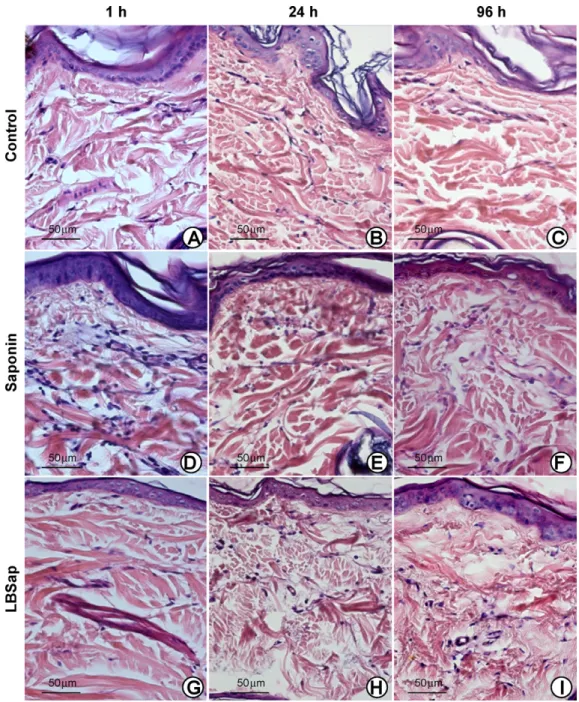
inoculated areas, together with signs of significant focal inflam-mation. Such alterations were observed particularly in the early period (1 h) following inoculation (Fig. 1D and G) and continued until the late period (up to 96 h) (Fig. 1F and I). Sap and LBSap inoculums induced a pronounced inflammatory reaction in the dermis and hypodermis of the study animals, and this was posi-tively correlated with iNOS expression (Fig. 2, upper and lower right panels).
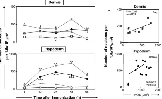
Significant increases in inflammatory infiltrate were observed in the dermis of animals of the Sap group after 1, 12, 24 and 96 h com-pared with control animals (S group). At 24 h, LB group animals also exhibited a significant increase in inflammatory infiltrate compared with the S group (Fig. 2, upper left panel). Longitudinal evalua-tion of the results showed that there were no differences within the groups concerning inflammatory infiltrate during the period of investigation. A positive correlation (P= 0.0265/r= 0.6924) between
the total number of cell nuclei and the marked areas of iNOS expres-sion could be established only in the Sap group (Fig. 2, upper right panel).
In the hypodermis, the inflammatory infiltrate increased sig-nificantly within the Sap and LBSap groups at 12, 24 and 48 h in comparison with the S and LB groups (Fig. 2, lower left panel and
Fig. 3). It is important to emphasise that the increases observed within the Sap and LBSap groups occurred at similar times. At 1 and 96 h, the inflammatory infiltrate within the Sap group was signifi-cantly enhanced compared with the S group. Longitudinal analysis revealed that in the Sap and LBSap groups, inflammatory infiltrate was significantly increased within the period 12–48 h in compar-ison with the level at 1 h after inoculation. There was a positive correlation (P= 0.0427/r= 0.5683) between the total number of cell nuclei and the marked areas of iNOS expression in the hypodermis of animals within the LBSap group (Fig. 2, lower right panel).
Fig. 2.Left panels: analysis of the inflammatory infiltrates in the dermis and hypodermis of dogs at various times after inoculation with saline (control group S;), saponin (Sap group; ), antigen ofL. braziliensis(LB group; ) or antigen ofL. braziliensisplus saponin (LBSap group;䊉). Significant differences (P< 0.05) between the values associated with the S, Sap and LB groups are indicated by the letters “a”, “b” and “c”, respectively. Right panels: correlations between the number of cell nuclei and iNOS activity within skin layers of the Sap and LBSap groups (r= Pearson correlation coefficient).
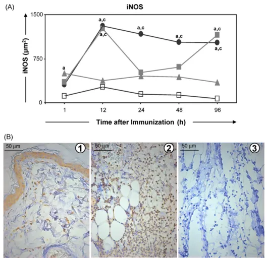
3.2. Assessment of iNOS expression in the different skin layers
Anti-iNOS immunohistochemical reactions were observed in all three skin layers of Sap group as well as in the epidermis (ker-atinocytes) and annexes (sebaceous and sudoriparous glands). Such reactions varied on a semi-quantitative basis from moderate to intense in the keratinocytes, from discreet to intense in the der-mis, and from absent to intense in the hypodermis. Similar levels of iNOS expression were also observed in the LB group. Within the LBSap group, iNOS expression was intense in the keratinocytes and dermis during the whole study period, whilst in the hypodermis, expression varied from moderate to intense. iNOS expression was also observed in the glands, fibroblasts and endothelial cells of all studied groups (Fig. 4B).
On a quantitative basis, iNOS expression increased significantly within the Sap and LBSap groups at 12 h compared with the level determined at 1 h after inoculation (Fig. 4). Moreover, the area of iNOS expression was significantly increased within the LB group compared with the S group at 1 h after inoculation, whilst at 12 h both the Sap and LBSap groups presented significantly larger areas of iNOS expression than the S and LB groups. Increased intensities of iNOS were also observed between 24 and 96 h within the LBSap group and at 96 h within the Sap group, and these differences were statistically significant in comparison with the S and LB groups.
3.3. Mobilisation of inflammatory cells to the outer dermis
Longitudinal evaluation of cell migration revealed that the per-centage of neutrophils increased significantly within the Sap group at 12 h compared with 1 h after inoculation (Fig. 5, upper left panel), and this was followed by significant reductions at 48 and 96 h. The number of neutrophils in the outer dermis of animals of the Sap group was significantly increased at 12 h compared with the levels determined in the S, LB and LBSap groups.
The percentage of eosinophils within the LBSap group (Fig. 5, lower left panel) increased significantly at 96 h compared with val-ues recorded at earlier times (12, 24 and 48 h), although differences between groups were not statistically significant. Concerning the
macrophages, a reduction was observed within the Sap group at 12 h compared with 1 h, the former value being significantly lower than that of the S group (Fig. 5, upper middle panel). The LBSap group presented a significantly reduced number of macrophages at 48 h compared with the S and LB groups. There was a positive cor-relation (P= 0.0445/r= 0.5876) between iNOS expression and the percentage of macrophages in the outer dermis of LB groups (Fig. 5, right panel).
In contrast to the above, the number of lymphocytes increased significantly in the outer dermis of members of the LBSap group at 48 h compared with the value determined at 1 h following inoc-ulation, and this increase was statistically different from those determined in the S and LB groups at 48 h (Fig. 5, middle left panel). Additionally, the number of lymphocytes within the Sap and LBSap groups increased significantly at 24 h compared with the S and LB groups.
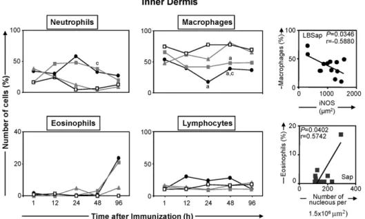
3.4. Mobilisation of inflammatory cells to the inner dermis
Longitudinal analysis showed that the number of neutrophils, macrophages and lymphocytes present in the inner dermis of animals within each group did not vary significantly during the study period. In contrast, significant variations in inflammatory cell profiles were recorded between the groups of animals. Thus, the number of neutrophils increased significantly at 48 h after inoculation within the Sap group compared with the LB group (Fig. 6, upper left panel). Furthermore, the numbers of macrophages were reduced significantly at 24 and 48 h within the LBSap group compared with the S group, and at 48 h compared with the LB group (Fig. 6, upper middle panel). Additionally, the percentage of macrophages was significantly reduced at 48 h within the Sap group compared with the S group, Regarding the eosinophils, there was a significant increase within the Sap group at 96 h after inocu-lation compared with the levels at 1, 12, 24 and 48 h (Fig. 6, lower left panel), and also within the LBSap group at 96 h after inoculation compared with the values at 12 and 24 h.
Fig. 3.Photomicrographs of the hypodermis of dogs immunized with saline (upper panels; control), saponin (middle panels) or LBSap (antigen ofL. braziliensisplus saponin; lower panels) and acquired at 1, 24 and 96 h after inoculation (slides shown at 40×magnification; bar = 50m).
eosinophils and the total number of cell nuclei (Fig. 6, lower right panel), whilst in members of the LBSap group there was a negative correlation (P= 0.0346/r=−0.5880) between the
per-centage of macrophages and iNOS expression (Fig. 6, upper right panel).
3.5. Mobilisation of inflammatory cells to the hypodermis
Longitudinal analysis showed that the percentage of neutrophils in the hypodermis was significantly reduced within the LBSap group at 96 h compared with the levels at 1 and 24 h after inocula-tion (Fig. 7, upper left panel). Moreover, compared with the S group, the numbers of neutrophils within the Sap and LBSap groups were significantly increased at 1 and 24 h after inoculation, whilst in the LB group the neutrophil number was significantly increased at 24 h compared with the S group. Additionally, the LB group exhibited a significant reduction in the number of neutrophils at 48 and 96 h
Fig. 4.Upper panel: kinetics of expression of iNOS in the skin of dogs at various times after inoculation with saline (control group S;), saponin (Sap group; ), antigen ofL. braziliensis(LB group; ) or antigen ofL. braziliensisplus saponin (LBSap group;䊉). Significant differences (P< 0.05) between the values associated with the S and LB groups are indicated by the letters “a” and “c”, respectively. Lower panel: photomicrographs showing the immunohistochemical detection of iNOS expression in the dermis (plate 1) and the hypodermis (plate 2) of dogs and recorded 12 h after immunization with LBSap: plate 3 shows the negative control of the reaction (slides shown at 40×
magnification; bar = 50m). Anti-iNOS immunohistochemical reactions were observed in all three skin layers as well as in the epidermis (keratinocytes), fibroblasts and inflammation cells.
Fig. 6.Left and middle panels: comparative analyses of the selective migration of cells to the inner dermis of dogs at various times after inoculation with saline (control group S;), saponin (Sap group; ), antigen ofL. braziliensis(LB group; ) or antigen ofL. braziliensisplus saponin (LBSap group;䊉). Significant differences (P< 0.05) between the values associated with the S and LB groups are indicated by the letters “a” and “c”, respectively. Right panels: correlation between the percentage of macrophages and iNOS activity in the inner dermis of dogs in the LBSap group, and correlation between the percentage of eosinophils and the number of cell nuclei in the inner dermis of dogs in the Sap group (r= Pearson correlation coefficient).
within the Sap group at 48 h compared with 24 h (Fig. 7, lower right panel).
4. Discussion
The emergent and re-emergent character of VL results from the failure of authorities fully to control reservoirs and vectors, and also from opportunistic infection by the parasite of vulnerable
individuals, particularly those affected by AIDS [24–26]. More-over, the escalation of the disease has been aggravated by the development of drug-resistant strains ofLeishmania[27]. Numer-ous anti-CVL vaccines containing diverse antigens and adjuvants have been tested in Brazil, and some have shown promising results
[14,18,21–23,28–32]. In our continuing effort to develop a vaccine against CVL that is both efficient and safe, we have conducted stud-ies on two new preparations, namely, LBSap and LBSapSal. The
results obtained so far have revealed that these vaccines possess strong immunogenic capacities and can induce high levels of anti-LeishmaniaIgG (IgG1 and IgG2) as well as lymphocytes, particularly T CD8+(circulating andin vitro L. chagasi-specific) cells. Further-more, we have demonstrated that these vaccines induce an immune response that is compatible with the control of the etiological agent of CVL, i.e. intense proliferative activity ofL. chagasi-specific lym-phocytes and increased production of NO duringin vitrostimulation by solubleL. chagasiantigens[21,22].
In the present study, the inflammatory processes induced in the inoculation area by the antigen and adjuvant present in LBSap vac-cine were evaluated during the initial 96 h period. On the basis that immunization against infectious agents requires the participation of innate and adaptive immune responses, the determination of the kinetics of cell migration to the inoculation area is extremely relevant since the number and types of cells recruited immedi-ately after inoculation will stimulate the innate-immune system and will influence the development of acquired immunity[33]. Although most studies tend to focus on the cytokine profile of the inflammatory reaction[34–36], knowledge of the cell profile of the local inflammatory infiltrate can provide information concerning the immune response in the microenvironment of the inoculation
[37,38].
The local oedema observed following inoculation with Sap or LBSap[21]suggests an acute inflammatory response to the saponin adjuvant, and this is important for inducing a specific immune reaction[39]. However, the oedemas did not evolve into ulcerated lesions, thus demonstrating that saponin (in isolation or in combi-nation with theL. braziliensisantigen) is safe and can be employed as an adjuvant. Giunchetti et al.[21]observed a similar reaction in dogs vaccinated with LBSap and demonstrated that the nodules that emerged after vaccination disappeared during the later stages, thus indicating that LBSap vaccine was well tolerated despite the pres-ence of the saponin adjuvant. Following inoculation with an FML antigen vaccine (Leishmune®), dogs presented moderate adverse reactions, including pain, anorexia and local puffiness, which spon-taneously disappeared before the second immunization[40].
The adverse events following immunizations to be defined are fever, local reactions, intussusceptions, inconsolable crying, seizure, hypotonic hyporesponsive episode, allergic reaction, rash, asthenia, paresthesia, myalgia and idiopathic thrombocytopenia in humans[60]. Biomarkers in local reactions associated with safety are formation of induration and swelling (information about onset, duration and size of nodules), presence of granuloma (subcategory of nodules at injection site, which can present as persistent nodules many months post-immunization). Other biomarkers associated with local reactions: firmness, tenderness or pain and pruritus[61]. In present study, vaccination was not associated with hyperther-mia, pain, fever, lymphadenopathy or any other general adverse reactions.
Herein, histopathological alterations (oedema, haemorrhage and congestion) were observed chiefly within the Sap and LBSap groups. The intensity of such symptoms may well depend on the purity of the saponin employed[41]. In this context, moderate to intense inflammatory reactions, with oedemas lasting from 2 to 3 days, have been reported in sheep following subcutaneous inocula-tion with 1 mg of saponin[42]. Additionally, we observed discreet alterations in the skin of the LB animals at 12 and 24 h after inocu-lation, and these could have been induced by the inoculation itself together with increased vascular permeability. It is important to emphasise, however, that these alterations were much less intense in the absence of the saponin adjuvant.
Morphometric analysis revealed that saponin plays an impor-tant role in the transfer of cells to the dermis judging by the number of cells in the inflammatory infiltrate at 1, 12, 24 and 96 h following
inoculation with Sap. This finding reinforces the assumption that saponin modulates the immune response by stimulating antibody production and non-specific reactions, such as inflammation, and cell traffic[41,43]. The numbers of cells observed in the hypodermis of Sap and LBSap animals were larger than in the dermis, probably by virtue of the more extensive vascularisation in the former layer. As demonstrated in the present study, the inflammatory infiltrate increased within the Sap and LBSap groups at 12, 24 and 48 h com-pared with the S and LB groups, and also in the Sap group at 1 and 96 h compared with the S group. Since, throughout the whole experimental period, there was greater cell recruitment within the Sap group than in the LBSap group, the initial assumptions regard-ing the role of saponin in the transfer of inflammatory cells to the inoculation area are strengthened.
Within the Sap and LBSap groups, increases in the numbers of neutrophils and lymphocytes and decreases in the num-bers of macrophages were observed at various times during the study period. In the late period following inoculation, there were increases in the numbers of eosinophils in the inner dermis and hypodermis of Sap animals, and in the inner and outer dermis of LBSap animals. Based on these results it is possible to hypothesise that neutrophils function at the front line of the immune system, responding immediately upon request, and direct the migration of other cells to the inoculation area some hours afterwards. It appears, therefore, that neutrophils participate effectively in the adaptive immune response from the very beginning of the process. This assumption is supported by several studies in which the role of neutrophils has been considered[44,45]. According to Appel-berg[44], neutrophils not only act as phagocytes but also determine the inflammatory immune response and cooperate with other cells in the amplification of such response. Moreover, neutrophils play a role in acquired (or adaptive) immunity since they are antigen-presenting cells (APCs) that can activate na¨ıve T lymphocytes[45]. In this context, neutrophils have been shown to migrate to the infec-tion site before macrophages and dendritic cells, and are the first cells to interact with CD8+T lymphocytes[44]. Moreover, together with saponin[41], the neutrophils stimulate the activation of T CD8+lymphocytes, hence contributing to the entire immunogenic process. Some reports have suggested that neutrophils operate not only in an indirect manner as APCs, but are also instrumen-tal in the recruitment of other cells (i.e. T lymphocytes, monocytes, macrophages and immature dendritic cells) through the production of chemokines[46].
The increase in the number of eosinophils within the Sap and LBSap groups at the end of the experimental period (48 and 96 h) suggests that immunization with the LB antigen and saponin gener-ated a mixed cell medigener-ated immune response. Based on this finding, it is possible to hypothesise that eosinophils, together with neu-trophils, participate effectively in the innate-immune response at the inoculation area.
With respect to macrophages, the reduction in the numbers of these cells was possibly due to the intense migration of neutrophils to the inoculation area, which occurs in any acute process[47], and not because they were inhibited by the antigenic components of the vaccine. Indeed, the macrophages are known to play an important role in the immunogenic response induced by saponin-containing vaccines[41].
mechanism since the cells produce IFN-␥via the interaction with
IL-12 and IL-18 cytokines, independent of the T-cell receptor-major histocompatability complex (TCR-MHC) mechanism, and destroy infected cells hence controlling the multiplication of the infectious agent and mediating the initial stages of the acquired immune response[48]. The presence of CD4+T lymphocytes is also impor-tant since these cells produce IFN-␥ and activate macrophages,
thus increasing the microbicide capability and the production of cytokines that take part in the adaptive response and control of Leishmaniainfection[49].
Based on such evidence[48,49], it is possible to infer that the selective recruitment of lymphocytes, particularly IFN-␥-producing
CD8+T-cells, contribute to the protective immune response against CVL. Another feature that demonstrates the importance of the saponin adjuvant in prompting the immunogenicity of the LB antigen is the fact that within the LB group the recruitment of inflammatory cells to the inoculation area was rather weak.
The contribution of inflammatory cells to the creation of a microenvironment suitable for immune reaction against Leishma-niainfection has also been investigated by examining the activity of iNOS in the inoculation area. It is known that iNOS expression is a key factor in the immune adaptive response to external stimuli and virulent pathogens[50,51], and that NO is particularly important in the control ofLeishmaniaparasites[52]. Semi-quantitative anal-ysis demonstrated that iNOS was expressed in the skin layers of all animal groups including those of the control group S. This finding leads us to believe that all stimuli have the capacity to increase iNOS expression, contributing to the production of NO and participating in a broad range of processes ranging from non-specific reactions to the modulation of the immune system[53].
Quantitative analysis of iNOS expression showed that at 1 h after inoculation the area of expression of the enzyme was enlarged within the LB group. This observation can be explained by the pres-ence of a higher number of macrophages, cells that are considered to be important NO producers, recruited to the outer dermis dur-ing this period in response to the LB inoculum. In contrast, the Sap and LBSap groups showed larger areas of iNOS expression at 12 h onwards in comparison with the other groups. The increased iNOS expression in these groups may be explained by the great number of NO-producing cells recruited to the inoculation area, activation of which would contribute to the eradication of the parasite. The correlation between the number of nuclei and iNOS expression in the dermis and hypodermis of animals of the Sap and LBSap groups confirms these assumptions.
It is important to emphasise that iNOS is expressed in many types of cells (macrophages, neutrophils and fibroblasts) in response to diverse stimuli including cytokines and lipopolysaccha-rides[54]. Indeed, numerous reports have demonstrated that type 1 cytokines (IFN-␥, TNF-␣and IL-18) induce iNOS expression[55,56],
whilst type 2 cytokines (IL-4, IL-13 and IL-10) diminish leishmani-cidal activity in murine models and in humans by down regulating iNOS expression[57]. In the present study it was shown that in the outer dermis of LB animals, the percentage of macrophages was positively correlated with the area of iNOS expression sug-gesting that the LB inoculum activated NO-producing macrophages. In addition, within the Sap group there was a positive correlation between the number of cell nuclei and eosinophils present in the inner dermis, suggesting that these cells contribute to the innate response induced by the strong adjuvant power of saponin.
The role of NO in inflammatory reactions of the skin is very com-plex since at low levels it induces the dilation of blood vessels and the migration of neutrophils, whilst at high levels it down regulates the production of cell adhesion molecules, suppresses the activa-tion of inflammatory cells and induces their apoptosis[58]. Hence, NO influences the balance between type 1 and type 2 responses. The
high levels of NO observed in the groups inoculated with saponin may be related to the high levels of type 1 cytokines (IFN-␥and
TNF-␣)[59]that are essential for the activation of effector
mech-anisms such as the production of NO for the control ofLeishmania parasites.
The results presented in this paper demonstrate that the LBSap vaccine and the isolated saponin adjuvant were able to induce intense cell migration in the skin of inoculated dogs, thus trigger-ing the initial immunogenic events. Moreover, the components of the vaccine were shown to be safe since no ulcerated lesions were observed at the inoculation sites during the study period. The selec-tive recruitment of neutrophils to the skin, and the intense iNOS expression exhibited by animals in the Sap and LBSap groups, were similar to the reactions observed in animals resistant to infection byLeishmania.
Acknowledgements
The study was supported by the Fundac¸˜ao de Amparo `a Pesquisa do Estado de Minas Gerais, Brazil (PRONEX 2007). RCO and P-SCB thank CNPq for fellowships. The authors wish to extend their grat-itude to the staff of the kennels at the Federal University of Ouro Preto for their help and dedication throughout the execution of this project. The authors are also grateful for the use of facilities at CEBIO, Universidade Federal de Minas Gerais and Rede Mineira de Bioterismo (FAPEMIG), and for support with the provision of experimental animals.
References
[1] Desjeux P. Leishmaniasis: current situation and new perspectives. Comp Immunol Microbiol Infect Dis 2004;27:305–18.
[2] Deane LM, Deane MP. Leishmaniose visceral urbana (no c˜ao e no homem) em Sobral, Cear ´a. Hospital 1955;47:75–87.
[3] Chamizo C, Moreno J, Alvar J. Semi-quantitative analysis of cytokine expres-sion in asymptomatic canine leishmaniasis. Vet Immunol Immunopathol 2005;103:67–75.
[4] Reis AB, Martins-Filho OA, Teixeira-Carvalho A, Carvalho MG, Mayrink W, Franca-Silva JC, et al. Parasite density and impaired biochemical/hematological status are associated with severe clinical aspects of canine visceral leishmani-asis. Res Vet Sci 2006;81:68–75.
[5] Reis AB, Teixeira-Carvalho A, Vale AM, Marques MJ, Giunchetti RC, Mayrink W, et al. Isotype patterns of immunoglobulins: hall-marks for clinical status and tissue parasite density in Brazilian dogs naturally infected byLeishmania
(Leishmania)chagasi. Vet Immunol Immunopathol 2006;112:102–16. [6] Reis AB, Teixeira-Carvalho A, Giunchetti RC, Guerra LL, Carvalho MG, Mayrink W,
et al. Phenotypic features of circulating leucocytes as immunological markers for clinical status and bone marrow parasite density in dogs naturally infected byLeishmania chagasi. Clin Exp Immunol 2006;146:303–11.
[7] Giunchetti RC, Mayrink W, Genaro O, Carneiro CM, Correia-Oliveira R, Martins-Filho AO, et al. Relationship between canine visceral leishmaniosis and the
Leishmania(Leishmania)chagasiburden in dermal inflammatory foci. J Comp Pathol 2006;135:100–7.
[8] Lage RS, Oliveira GC, Busek SU, Guerra LL, Giunchetti RC, Correa-Oliveira R. Analysis of the cytokine profile in spleen cells from dogs naturally infected by
Leishmania chagasi. Vet Immunol Immunopathol 2007;115:135–45. [9] Baneth G, Shaw SE. Chemotherapy of canine leishmaniosis. Vet Parasitol
2002;106:315–24.
[10] Noli C, Auxilia ST. Treatment of canine Old World visceral leishmaniasis: a systematic review. Vet Dermatol 2005;16:213–32.
[11] Hommel M, Jaffe CL, Travi B, Milon G. Experimental models for leishma-niasis and for testing anti-leishmanial vaccines. Ann Trop Med Parasitol 1995;89(Suppl 1):55–73.
[12] Gradoni L. An update on anti-leishmanial vaccine candidates and prospects for a canineLeishmaniavaccine. Vet Parasitol 2001;100:87–103.
[13] Mauel J. Vaccination againstLeishmaniainfections. Curr Drug Targets Immune Endocr Metabol Disord 2002;2:201–26.
[14] Da Silva V, Borja-Cabrera GP, Correia Pontes NN, de Souza EP, Luz KG, Palatnik M. A phase III trial of efficacy of the FML-vaccine against canine Kala-azar in an endemic area of Brazil (S˜ao Gonc¸alo do Amaranto, RN). Vaccine 2000;19:1082–92.
[16] Lemesre JL, Holzmuller P, Cavaleyra M, Goncalves RB, Hottin G, Papierok G. Protection against experimental visceral leishmaniasis infection in dogs immunized with purified excreted secreted antigens ofLeishmania infantum
promastigotes. Vaccine 2005;23:2825–40.
[17] Lemesre JL, Holzmuller P, Goncalves RB, Bourdoiseau G, Hugnet C, Cavaleyra M, et al. Long-lasting protection against canine visceral leishmaniasis using the LiESAp-MDP vaccine in endemic areas of France: double-blind randomised efficacy field trial. Vaccine 2007;25:4223–34.
[18] Mayrink W, Genaro O, Silva JCF, da Costa RT, Tafuri WL, Peixoto Toledo VPC, et al. Phases I and II open clinical trials of a vaccine againstLeishmania chagasi
infections in dogs. Mem Inst Oswaldo Cruz 1996;91:695–7.
[19] Lasri S, Sahibi H, Sadak A, Jaffe CL, Rhalem A. Immune responses in vaccinated dogs with autoclavedLeishmania majorpromastigotes. Vet Res 1999;30:441–9. [20] Panaro MA, Acquafredda A, Lisi S, Lofrumento DD, Mitolo V, Sisto M. Nitric oxide production by macrophages of dogs vaccinated with killedLeishmania infantum
promastigotes. Comp Immunol Microbiol Infect Dis 2001;24:187–95. [21] Giunchetti RC, Correa-Oliveira R, Martins-Filho O, Teixeira-Carvalho A, Roatt
BM, Aguiar-Soares RDO, et al. Immunogenicity of a killedLeishmaniavaccine with saponin adjuvant in dogs. Vaccine 2007;25:7674–86.
[22] Giunchetti RC, Corrˆea-Oliveira R, Martins-Filho OA, Teixeira-Carvalho A, Roatt BM, Aguiar-Soares RDO, et al. A killed Leishmaniavaccine with sand fly saliva extract and saponin adjuvant displays immunogenicity in dogs. Vaccine 2008;26:623–38.
[23] Giunchetti RC, Reis AB, Da Silveira-Lemos D, Martins-Filho OA, Corrˆea-Oliveira R, Bethony J, et al. Antigenicity of a whole parasite vaccine as promising candi-date against canine leishmaniasis. Res Vet Sci 2008;85:106–12.
[24] Badar ´o R, Rocha H, Carvalho EM, Queiroz AC, Jones TC.Leishmania donovani: an opportunistic infection associated with progressive disease in three immuno-compromised patients. Lancet 1986;8482:647–9.
[25] Altes J, Salas A, Riera M, Udina M, Galmes A, Balanzat J, et al. Visceral leishma-niasis: another HIV-associated opportunistic infection? Report of eight cases and review of literature. AIDS 1991;5:201–20.
[26] Gradoni L, Scalone A, Gramicia M. HIV-Leishmaniaco-infections in Italy: sero-logical data as an indication of the sequence of acquisition of two infections. Trans R Soc Trop Med Hyg 1993;87:94–6.
[27] World Heath Organization. Magnitude of the problem; 2008, available at http://www.who.int/leishmaniasis/burden/magnitude/burden magnitude/en/ index.html[last accessed March 1, 2008].
[28] Genaro O, de Toledo VP, Da Costa CA, Hermeto MV, Afonso LC, Mayrink W. Vaccine for prophylaxis and immunotherapy, Brazil. Clin Dermatol 1996;14:503–12.
[29] Palatnik de Sousa CB, Previato JO, Mendonc¸a-Previato L, Borojevic R. A new approach to phylogeny ofLeishmania: species-specificity of glycoconjugate lig-ands for promastigote internalization into murine macrophages. Parasitol Res 1990;76:289–93.
[30] Palatnik de Sousa CB, Moreno MB, Paraguai de Souza E, Borojevic R. The FML vaccine (fucose-mannose ligand) protects hamsters from experimental Kala-azar. Cienc Cult (J Braz Assoc Adv Sci) 1994;46:290–6.
[31] Palatnik de Sousa CB, Paraguai de Souza E, Gomes EM, Borojevic R. Experimental murineLeishmania donovaniinfection immunoprotection by fucose mannose ligand (FML). Braz J Med Biol Res 1994;27:547–51.
[32] Borja-Cabrera GP, Correia Pontes NN, Da Silva VO, Paraguai de Souza E, Santos WR, Gomes EM, et al. Long lasting protection against canine Kala-azar using the FML-QuilA saponin vaccine in an endemic area of Brazil (S˜ao Gonzalo do Amarante, RN). Vaccine 2002;20:3277–84.
[33] Teixeira CR, Teixeira MJ, Gomes RBB, Santos CS, Andrade BB, Raffaele-Neto I, et al. Saliva fromLutzomyia longipalpisinduces CC chemokine ligand 2/monocyte chemoattractant protein-1 expression and macrophage recruitment. J Immunol 2005;175:8346–53.
[34] Cohen S. Cell mediated immunity and the inflammatory system. Hum Pathol 1976;7:249–64.
[35] Dunn CJ, Hardee MM, Staite ND. Acute and chronic inflammatory responses to local administration of recombinant IL-1 alpha, IL-beta, TNF alpha, IL-2 and IFN gamma in mice. Agents Actions 1989;27:290–3.
[36] Berg DJ, Leach MW, K ¨uhn R, Rajewsky K, M ¨uller W, Davidson NJ, et al. Interleukin 10 but not interleukin 4 is a natural suppressant of cutaneous inflammatory responses. J Exp Med 1995;182:99–108.
[37] Stewart RJ, Holloway LJ, Isbister WH. Peritoneal neutrophilia: a poten-tial indicator of the surgical acute abdomen. Aust NZ J Surg 1984;54: 565–8.
[38] De Souza, RCA. Estudo da cin ´etica celular da resposta inflamat ´oria causada pela crioterapia experimental em pele normal de camundongos utilizando o m ´etodo de bolsas de ar no subcutˆaneo. MSc dissertation. Florian ´opolis: Universidade Federal de Santa Catarina; 2001.
[39] Jancar S. Imunidade natural e inflamac¸˜ao. In: Calish V, Vaz C, editors. Imunolo-gia. Rio de Janeiro: Editora Revinter; 2001. p. 11–29.
[40] Parra LE, Borja-Cabrera GP, Santos FN, Souza LO, Palatnik-de-Sousa CB, Menz I. Safety trial using the Leishmune vaccine against canine visceral leishmaniasis in Brazil. Vaccine 2007;25:2180–6.
[41] Kensil CR. Saponins as vaccine adjuvants. Crit Rev Ther Drug 1996;13:1–55. [42] Houdayer M, Rouze P, Dalsgaard K, Metzger JJ. Adjuvant effect of Quil A in
porcine humoral immune response. Ann Immunol Cell Biol (Institute Pauster) 1978;129(1):107–12.
[43] De Oliveira CAC, Perez AC, Merino G, Prieto JG, Alvarez AI. Protective effects ofPanax ginsengon muscle injury and inflammation after eccentric exercise. Comp Biochem 2001;130:369–77.
[44] Appelberg R. Neutrophils and intracellular pathogens: beyond phagocytosis and killing. Trends Microbiol 2007;15:87–92.
[45] Beauvillain C, Delneste Y, Scotet M, Peres A, Gascan H, Guermonprez P, et al. Neutrophils efficiently cross-prime naive T cellsin vivo. Blood 2007;110: 2965–73.
[46] Scapini P, Lapinet-Vera JA, Gasperini S, Calzetti F, Bazzoni F, Cassatella MA. The neutrophil as a cellular source of chemokines. Immunol Rev 2000;177: 195–203.
[47] Terui T, Ozawa M, Tagami H. Role of neutrophils in induction of acute inflamma-tion in T-cell-mediated immune dermatosis, psoriasis: a neutrophil-associated inflammation-boosting loop. Exp Dermatol 2000;9:1–10.
[48] Berg RE, Forman J. The role of CD8 T cells in innate immune and in antigen non-specific protection. Curr Opin Immunol 2006;18:338–43.
[49] Bittar RC, Nogueira RS, Vieira-Gonc¸alves R, Pinho-Ribeiro V, Mattos MS, Oliveira-Neto MP, et al. T-cell responses associated with resistance to leish-mania infection in individuals from endemic areas forLeishmania(Viannia)
braziliensis. Mem Inst Oswaldo Cruz 2007;102:625–30.
[50] Nathan C, Xie Q. Nitric oxide synthases: roles, tolls, and controls. Cell 1994;78:915–8.
[51] Nathan C, Xie Q. Regulation of biosynthesis of nitric oxide. J Biol Chem 1994;269:13725–8.
[52] Hall LR, Titus RG. Sandfly vector saliva selectively modulates macrophage func-tions that inhibit killing ofL. majorand nitric oxide production. J Immunol 1995;155:3501–6.
[53] Bogdan C. Nitric oxide and the immune response. Nat Immunol 2001;2:907–16. [54] Liew FY, Wei XQ, Proudfoot L. Cytokines and nitric oxide as effector molecules
against parasitic infections. Philos Trans Roy Soc B 1997;352:1311–5. [55] Liew FY, O’Donnell CA. Immunology of leishmaniasis. Adv Parasitol
1993;32:161–259.
[56] Bogdan C, Vodovotz Y, Paik J, Xie QW, Nathan C. Traces of bacterial lipopolysac-charide suppress IFN-gamma-induced nitric oxide synthase gene expression in primary mouse macrophages. J Immunol 1993;151:301–9.
[57] Vouldoukis I, Becherel PA, Riveros-Moreno V, Arock M, Da Silva O, Debre P, et al. Interleukin-10 and interleukin-4 inhibit intracellular killing ofLeishmania infantumandLeishmania majorby human macrophages by decreasing nitric oxide generation. Eur J Immunol 1997;27:860–5.
[58] Ross R, Reske-Kunz AB. The role of NO in contact hypersensitivity. Int Immunopharmacol 2001;1:1469–78.
[59] Kaim U, Moritz A, Failing K, Baumg ¨artner W. The regression of a canine Langer-hans cell tumour is associated with increased expression of IL-2, TNF-alpha, IFN-gamma and iNOS mRNA. Immunology 2006;118:472–82.
[60] Bonhoeffer J, Kohl K, Chen R, Duclos P, Heijbel H, Heininger U, et al. The Brighton Collaboration: addressing the need for standardized case definitions of adverse events following immunization (AEFI). Vaccine 2002;13:298–302.