Abundant
NDRG2
Expression Is Associated
with Aggressiveness and Unfavorable
Patients
’
Outcome in Basal-Like Breast
Cancer
Vera Kloten*☯, Martin Schlensog☯, Julian Eschenbruch☯, Janina Gasthaus,
Janina Tiedemann, Jolein Mijnes, Timon Heide, Till Braunschweig, Ruth Knüchel, Edgar Dahl
Molecular Oncology Group, Institute of Pathology, Medical Faculty of the RWTH Aachen University, Aachen, Germany
☯These authors contributed equally to this work. *vkloten@ukaachen.de
Abstract
NDRG2, a member of the N-myc downstream-regulated gene family, is thought to be a puta-tive tumor suppressor gene with promising clinical impact in breast cancer. Since breast cancer comprises heterogeneous intrinsic subtypes with distinct clinical outcomes we investigated the pivotal role of NDRG2 in basal-type breast cancers. Based on subtype classified tumor (n = 45) and adjacent normal tissues (n = 17) we examinedNDRG2mRNA expression and CpG-hypermethylation, whose significance was further validated by inde-pendent data sets from The Cancer Genome Atlas (TCGA). In addition, NDRG2 protein expression was evaluated immunohistochemically using a tissue micro array (TMA, n = 211).In vitro, we investigated phenotypic effects caused by NDRG2 silencing in the basal A-like HCC1806 as well as NDRG2 over-expression in basal A-like BT20 compared to lumi-nal-type MCF7 breast cancer cells. Our tissue collections demonstrated an overall low NDRG2mRNA expression in breast cancer subtypes compared to normal breast tissue in line with an increased CpG-hypermethylation in breast cancer tissue. Independent TCGA data sets verified a significant (P<0.001) expression loss ofNDRG2in breast tumors. Of interest, basal-like tumors more frequently retained abundantNDRG2expression concor-dant with a lower CpG-hypermethylation. Unexpectedly, basal-like breast cancer revealed an association ofNDRG2expression with unfavorable patients’outcome. In line with this observation,in vitroexperiments demonstrated reduced proliferation and migration rates (~20%) in HCC1806 cells followingNDRG2silencing. In contrast, NDRG2 over-expressing luminal-type MCF7 cells demonstrated a 26% decreased proliferation rate. Until now, this is the first study investigating the putative role of NDRG2 in depth in basal-type breast cancer. Our data indicate that the described putative tumor suppressive function of NDRG2 may be confined to luminal- and basal B-type breast cancers.
a11111
OPEN ACCESS
Citation:Kloten V, Schlensog M, Eschenbruch J, Gasthaus J, Tiedemann J, Mijnes J, et al. (2016) AbundantNDRG2Expression Is Associated with Aggressiveness and Unfavorable Patients’Outcome in Basal-Like Breast Cancer. PLoS ONE 11(7): e0159073. doi:10.1371/journal.pone.0159073
Editor:Javier S Castresana, University of Navarra, SPAIN
Received:February 1, 2016
Accepted:June 27, 2016
Published:July 11, 2016
Copyright:© 2016 Kloten et al. This is an open access article distributed under the terms of the Creative Commons Attribution License, which permits unrestricted use, distribution, and reproduction in any medium, provided the original author and source are credited.
Data Availability Statement:All relevant data are within the paper and its Supporting Information files.
Funding:The authors received no specific funding for this work.
Introduction
Breast cancer remains the most frequently diagnosed cancer and the leading cause of cancer deaths in European women [1]. Based on the high breast cancer-related mortality rate, the understanding of tumor biological and molecular consequences is mandatory, enabling an individual and targeted development of breast cancer therapy. However, adapting current diag-nostic and therapeutic strategies to each patient is a challenging task due to the heterogeneous molecular aspects of breast tumors. Breast cancer can be classified into four main intrinsic sub-types, i.e. luminal A, luminal B, HER2-enriched and basal-like, based upon global gene expres-sion profiles demonstrated for the first time in 2000 by Perou and colleagues [2]. While particularly patients with luminal A tumors benefit from systemic endocrine therapy, thera-peutic targets for the basal-like class of breast cancer is still insufficient due to the lack of under-standing of the driving oncogenic mechanisms [3], resulting in chemotherapy treatment. In addition, breast cancer patients affected with basal-type cancer show worse survival outcome compared to patients with e.g. luminal A-type breast cancer [3].
A putative tumor suppressor gene implicated in cancer development [4] and progression [5–7] is NDRG2, a member of the N-myc downstream-regulated gene family. NDRG2 has been widely implicated in carcinogenesis including breast cancer invasion [8], angiogenesis [9] and metastasis [10–12]. Recent studies indicated decreasedNDRG2expression due to promoter DNA-hypermethylation [13,14], underlining a possible tumor suppressive role of NDRG2 in breast carcinogenesis. So far, NDRG2 was shown to inhibit invasive and meta-static capacity of breast cancer cells by reducing the production of active TGF-β[12] or suppression of MMP-9 activity [15]. Recently, Kim et al. [11] demonstrated a retarded STAT3 signaling by NDRG2 resulting in an inhibition of EMT progression due to the down-regulation of SNAIL expression. Nevertheless, studies evaluating the putative tumor suppressive biological and clinical impact of NDRG2 were irrespective of intrinsic breast cancer subtypes or mainly focused on luminal or basal B breast cancer cell modelsin vitro
andin vivo.
This is the first study giving evidence that the described putative tumor suppressive function of NDRG2 may be confined to luminal- or basal B-type breast tumors: A more frequently retainedNDRG2mRNA expression associated with unfavorable clinical outcome in basal-type breast cancer patients. Moreover, NDRG2 knockdown in the basal A breast cancer cell line HCC1806 caused a reduced proliferation and migration rate while NDRG2 over-expression in basal A-like BT20 breast cancer cells, lacking endogenous NDRG2 expression, resulted in an increased cell proliferation. In contrast, over-expression of NDRG2in luminal MCF7 cells showed a 26% decreased proliferation rate compared to control cells while significance slightly missed (P = 0.064).
Methods
TCGA patients
’
data set and breast cancer-related online tools
Subtype-specific patients
’
tissue collective
A total of 62 tissue samples, including breast tumors (n = 45) and adjacent normal tissues (n = 17) were obtained through the RWTH centralized biomaterial bank (RWTH cBMB). All patients gave written informed consent for retention and analysis of their tissue for research purposes (local ethical review board of the medical faculty of the RWTH Aachen, ref no. EK-206/09). Tumor material was snap-frozen in liquid nitrogen directly after surgery. Haematoxy-lin and eosin-stained sections were prepared for assessment of the percentage of tumor cells. The median percentage of vital tumor cells was 100% in the selected samples. Breast cancer molecular subtypes were defined according to St. Gallen criteria [18]. Ki67 staining was per-formed to make a distinction between luminal A and luminal B breast tumors. The Ki67 stain-ing was performed at the Uniklinik RWTH Aachen pathology department, accordstain-ing to standard protocols. The percentage of cells stained positive for Ki67 was determined using the free available ImmunoRatio software [19]. An overview of the clinical characteristics of breast cancer patients of this study is summarized inS2 Table.
Cell lines
Basal A-type HCC1806 and BT20 as well as luminal-type MCF7 breast cancer cell lines were obtained from the American Type Culture Collection (ATCC, Manassas, VA), which assures the molecular authentication of cell lines (ATCC Bulletin 2010:Maintaining high standards in cell culture,https://www.atcc.org/~/media/PDFs/CellBiologyStandards.ashx). MCF7, HCC1806 and BT20 cells were cultured in RPMI media (Gibco Life Science) supple-mented with 10% fetal calf cerum (FCS), 1% L-Glutamin and penicillin/streptomycin. MCF7 cell medium additionally complemented with 1% sodium pyruvat, 1% non-essential amino acids and 1% insulin. All cell lines were incubated in a humidified incubator at 37°C supplied with 5% carbon dioxide. Cells were regularly tested for Mycoplasma infection using the PCR-based Venor1GeM Mycoplasma Detection Kit (Minerva Biolabs, Berlin, Germany).
Nucleic acid extraction and reverse transcription PCR
Total cellular RNA from breast cancer and normal breast tissues as well as cultured breast can-cer cells was prepared by using TRIzol reagent (Invitrogen-Life Technologies). cDNA was syn-thesized using the reverse transcription system (Promega, Madison, WI) as previously
described [20].
Real-time PCR
cDNAs were amplified by semi-quantitative real-time PCR using SYBR-Green PCR mix (Bio-Rad Laboratories, Munich, Germany) performed in an iCycler IQ5 (Bio-(Bio-Rad Laboratories) and quantified as previously described [21]. All used primers spanned at least one intron, and are listed inS3 Table.
Pyrosequencing
Bisulfite modification
The extracted tissue DNA was bisulfite-converted using the EZ DNA methylation kit (Zymo Research, Orange, CA) as previously described [20].
Western blot analysis
For Western blot analysis, cultured cells were washed in ice-cold PBS solution and prepared under reducing (50 mM DTT) conditions by using NuPAGE LDS electrophoresis sample buffer (Invitro-gen-Life Technologies). Samples were separated on a 4–12% polyacrylamide gel (Invitrogen-Life Technologies) using MOPS-SDS running buffer. Proteins were electroblotted to nitrocellulose membranes and unspecific binding sites were blocked in TBS-T [10 mM Tris-HCl, 150 mM NaCl, 0.1% (v/v) Tween 20, pH 7.6] containing 5% (w/v) non-fat milk powder. The membranes were then probed overnight with a polyclonal rabbit anti-NDRG2 (Atlas Antibodies, HPA002896, 1:500) (4°C). Membranes were washed three times with TBS-T and incubated with horseradish peroxidase-conjugated secondary antibodies (Dako, Glostrup, Denmark), and the signal was detected by chemiluminescence (Pierce ECL, Thermo Scientific, Rockford, IL). Equal protein load-ing was monitored by probload-ing with a ß-actin specific antibody (Sigma-Aldrich (A5316), 1:2000).
Immunohistochemistry
NDRG2 protein expression was assessed using a TMA with 161 breast cancer and 50 normal tissue cases that have been described previously [20,23]. Paraffin-embedded tissue sections (2μm) were subjected to immunostaining using the UltraVision Quanto Detection System
HRP (Thermo Scientific) following the manufacturer’s instructions. Antigen retrieval was per-formed by pre-treatment in citrate buffer (pH 6) in a microwave oven (30 min). The sections were incubated for 1 h at room temperature with anti-NDRG2 (1:150). Slides were incubated for 10 min with secondary antibody (HRP Polymer Quanto; Thermo Scientific). DAB Quanto chromogensubstrate (Thermo Scientific) was used for antibody detection. An experienced breast cancer pathologist scored the immunohistochemical staining intensity according to the scoring system suggested by Remmele and Stegner (1987).
Transient transfection
Transient transfection of human BT20 and MCF7 breast cancer cells with NDRG2-pT-Rex-DEST 30 (Invitrogen-Life Technologies) expression vector, containing the full-length human
NDRG2cDNA, was performed as recently described [20]. In brief, 6 x 10⁵cells per 6-well were transfected using the FugeneHD reagent (Invitrogen-Life Technologies) at a ratio of 2:6 (DNA: reagent) for 48 h. Thereafter cells were cultured for one week in complete medium under posi-tive selection using geneticin (G418, 100μg/ml).
RNA Interference
Cell proliferation assay
The XTT proliferation assay (Roche) for BT20 and MCF7 (gain-of-function) as well as HCC1806 (loss-of-function) cell models was used and performed as previously described [20].
Cell growth assay
Increase of cell number was recorded for BT20 and MCF7 NDRG2-positive and negative cells over 96 h. 2 x 10⁴cells were seeded in 6-well culture plates and cell number was determined with theCASY1Cell Counter and Analyzer(OLS OMNI Life Science, Bremen, Germany) after 24 h, 48 h, 72 h and 96 h. Experiments were performed in triplicate.
Wound healing (
“
Scratch
”
) assay
Thein vitromotility was assessed by performing a monolayer scratch wound assay in BT20 and HCC1806 cell models as previously described [24].
Statistical analysis
Statistical analyses were performed using SPSS 22.0 (SPSS, Chicago, IL) and GraphPad Prism 5.0 (GraphPad Software Inc., La Jolla, CA). Box Plot graphs are shown as follows:Horizontal lines: grouped medians.Boxes: 25–75% quartiles.Vertical lines: range, peak and minimum. The non-parametric Mann-Whitney U-test was used in order to comparein vitroresults of the con-trol and NDRG2 set, respectively. Correlation analysis was performed by calculating a Spear-mancorrelation coefficient. Differences were considered statistically significant if the two sided p-values were equal or below 5% (0.05).
Results
NDRG2
revealed a divergent expression and methylation pattern in
basal- compared to luminal-type breast cancer
Although NDRG2 is thought to be a potential tumor suppressor in breast cancer, tumorigene-sis studies investigating the tumor suppressive role of NDRG2 were irrespective of intrinsic breast cancer subtypes or mainly focused on luminal or basal B breast cancer cell modelsin vitro[8,9,15,25] andin vivo[12]. To take a deeper look in the biological relevance of NDRG2 regarding the heterogeneous nature of breast cancer, we initially analysed mRNA expression in 45 subtype-classified breast tumors, including triple negative (n = 28), HER2-enriched (n = 4), luminal A (n = 7), luminal B (n = 4) breast cancer specimens, and 15 normal breast tissues by real-time PCR. We showed a significant (P<0.001) loss ofNDRG2mRNA expression
consider-ing all breast cancer subtypes when compared to normal breast tissues (median expression level: 1.3) (Fig 1A). In more detail, we revealed a pronounced downregulation ofNDRG2
mRNA expression (median FC: 2.7-fold downregulation) (Fig 1B and 1C). In addition to the PAM50 intrinsic subtype classification we showed an association of abundantNDRG2 expres-sion to ER-, PR-, and HER2-negative breast cancer specimen of the TCGA data cohort
(Table 1). Furthermore, we demonstrated a reducedNDRG2mRNA expression in invasive
ductal carcinoma (IDC) with regard to invasive lobular breast cancer (ILC) (Table 1).
Recent studies investigated promoter hypermethylation as the molecular cause forNDRG2
expression loss in different cancer types including breast cancer [13]. As expected, by analyzing TCGA data we identified an inverse correlation (Pearson r = -0.548, P<0.001) ofNDRG2
pro-moter hypermethylation andNDRG2mRNA expression in primary breast tumors supporting CpG-hypermethylation as the molecular cause of its gene silencing (Fig 1D). Concerning our own subtype-stratified breast tissue collective we showed a significant (P<0.001) higher
CpG-hypermethylation in luminal A (median methylation: 16.3%), luminal B (median methylation: 20.7%) and HER2-enriched (median methylation: 22.7%) tumors with respect to methylation frequency in normal breast tissue. In line with abundantNDRG2mRNA expression, CpG-hypermethylation in triple negative tumors (median methylation: 6.2%) was similar to normal breast tissue (median methylation: 3%) (Fig 1E).
NDRG2 protein is downregulated in the course of breast tumor
progression
DifferentialNDRG2mRNA expression and CpG-hypermethylation levels between luminal-and basal-like or triple negative breast cancers animated us to investigate NDRG2 protein expression in normal and malignant breast tissue using a TMA containing 161 invasive breast carcinomas and 50 normal breast tissue samples. In general, NDRG2 protein was localized in epithelial cells of the normal breast with absence in fibroblasts, adipocytes and endothelial cells
(Fig 2A) with a strong expression (median IRS9) in 70% (35/50) of normal breast tissue
samples (Fig 2B). However, invasive breast carcinomas showed a significant (P<0.001)
reduc-tion or complete loss (median IRS<6) of NDRG2 expression in 80% (128/161) of cases (Fig 2B). In more detail, stratification of breast cancer specimen in triple negative (n = 24), luminal-type (n = 87) and HER2-enriched (n = 27) breast cancers showed a pronounced downregula-tion of NDRG2 in luminal-type and HER2-enriched tumors (P<0.001) compared to triple
neg-ative cancers (P = 0.025) (Fig 2C). In line, a significant higher intensity of NDRG2
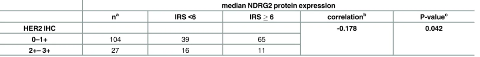
immunohistochemical staining was achieved in triple negative cancers in contrast to luminal-type and HER2-enriched specimens (Fig 2D). Next, clinicopathological characteristics were correlated with NDRG2 immunohistochemistry results for descriptive data analysis. Of inter-est, strong (IRS6) NDRG2 immunohistochemical staining was significantly associated with low HER2-receptor expression (0–1+) concordant to abundant transcriptionalNDRG2mRNA expression in breast carcinomas with low HER2-expression level (Table 2).
Fig 1.NDRG2revealed a divergent expression and methylation pattern in basal- compared to luminal-type breast cancer. (A)Box plot analysis (based on our own subtype classified tissue collective) illustrates a decreasedNDRG2mRNA expression in breast cancer compared with normal breast tissue. An increasedNDRG2mRNA expression (median expression level: 0.334) in triple negative tumors is observed comparing to HER2-enriched (median expression level: 0.275) and luminal-type (median expression level: 0.229) carcinomas.(B)Heatmap ofNDRG2expression is shown.Red: high-,black: mean-,green: low-expression. Left panel: breast cancer subtypes.Middle panel:NDRG2mRNA expression.Right panel: sample type (dark grey: primary tumor; light grey: solid normal tissues). Breast tumor samples are stratified by subtypes [3].(C)Box plot demonstrating a significant loss of NDRG2mRNA expression in luminal-type and HER2-enriched tumors with respect to basal-type cancer specimens.(D)Statistical association ofNDRG2mRNA expression andNDRG2hypermethylation in breast cancer. Pearson correlation coefficient: r = -0.5478, P<0.001.(E)Box plot analysis (based on our own subtype classified tissue collective) showing significant increased CpG-hypermethylation of theNDRG2promoter in luminal-type and HER2-enriched tumors compared to normal tissue samples. Horizontal lines: grouped medians. Boxes: 25–75% quartiles. Vertical lines: range, peak, and minimum.***P<0.001,**P<0.01, ns: not significant.
Abundant
NDRG2
mRNA expression in invasive ductal carcinoma is
associated with unfavorable survival gene signatures
Based on hormone-receptor and HER2-negative cancer specimen showing abundant NDRG2 expression, we hypothesized that the described NDRG2 expression loss and thus a putative tumor suppressive function of NDRG2 may be confined to luminal-type breast cancers. To address a possible subtype-associated prognostic significance ofNDRG2mRNA expression we performed a subtype stratified univariate survival analysis on Kaplan Meier-Plotter (KMP) and TCGA data. Analyzing KMP data showed a favorable RFS (P<0.001) and OS (P = 0.014) in
luminal A breast cancer patients with abundant NDRG2 mRNA expression underlining the known tumor suppressive function of NDRG2 (S1A and S1B Fig). In contrast, basal-type breast cancer patients showing abundantNDRG2expression revealed an unfavorable OS (P = 0.038) and tend to have a worse RFS while significance was barely missed (P = 0.093) (S1C and S1D Fig). Moreover, analyzing distinct gene signatures predicting breast cancer patients’outcome
Table 1. Clinicopathological parameters in relation toNDRG2mRNA expression of the TCGA data portal.
Variable NDRG2mRNA expression
na 1067b >1067b P-valuec correlationd
Clinicopathological factors
Age at diagnosis (median: 58 years; range: 26–90 years)
58 years 512 248 264 0.311 -0.032
>58 years 484 250 234
Histological type
IDC 727 401 326 0.001 0.102
ILC 169 43 126
Tumor size (pT)
pT 1–2 836 430 406 0.061 0.060
pT 3–4 155 67 88
Lymph node status (pN)
pN negative 473 231 242 0.583 -0.018
pN positive 506 256 250
Distant metastasis status (pM)
pM negative 845 434 411 0.413 -0.028
pM positive 18 11 7
ER status
negative 225 82 143 <0.001 -0.144
positive 722 385 337
PR status
negative 315 134 181 0.003 -0.095
positive 629 331 298
HER2 status
negative 618 322 296 <0.001 -0.146
positive 103 75 28
aOnly female patients with primary and unilateral invasive breast cancer were included bMedianNDRG2mRNA expression values
cFisher
’s exact test at a two-sided significance level of 0.05 dPearson correlation; IDC, invasive ductal carcinoma
ILC, invasive lobular carcinoma;NDRG2, N-myc downstream regulated gene 2; ER, Estrogen receptor; PR, Progesterone receptor; HER2, Human epidermal growth factor receptor 2; Significant P-values marked in bold face.
based on gene expression score values (high vs. low score) [26,27] indicated a clear clinical sig-nificance concerningNDRG2mRNA expression level (Fig 3). In patients with IDC abundant
NDRG2mRNA expression showed a significant association (Pearson r: 0.2274, P<0.001) with
a high breast cancer 21-gene recurrence scorepredicting poor prognosis in tamoxifen-treated, node-negative breast cancer [26] (Fig 3A and 3B). Contrary abundantNDRG2mRNA expres-sion in patients with ILC is associated with a low recurrence score indicating favorable progno-sis (Pearson r: -0.3100, P<0.001) (Fig 3A and 3C). Further, ILC with abundantNDRG2
expression showed an inverse correlation (Pearson r: -0.3795, P<0.001) with highbreast cancer HRneg/Tneg survival scoreassociated with poor metastatic outcome in early stage TNBC [27] while correlation in IDC showing abundant NDRG2 was low (Pearson r: -0.0736, P = 0.0474)
Fig 2. Loss of NDRG2 protein expression in human breast cancer. (A)Strong NDRG2 expression in epithelial cells of normal breast tissue (aandb). Moderate to low NDRG2 immunoreactivity in luminal-type (candd) and triple-negative (eandf) breast carcinoma.(B)Box plot analysis showing significant loss of NDRG2 expression in tumor tissue.(CtoD)Box plot analysis illustrating pronounced loss of NDRG2 expression in luminal-type and HER2-enriched tumors (P<0.001) compared to triple negative cancers (P = 0.025)(C)and in line with a significant higher median NDRG2 staining intensity in triple negative cancer(D). Horizontal lines: group medians. Boxes: 25–75% quartiles. Vertical lines: range, peak and minimum.***P<0.001.*P<0.05.
(S2 Fig). With respect to hormone-receptor positive breast cancer (i.e. particularly histological invasive lobular and intrinsic luminal breast carcinoma) these data underline a tumor suppres-sive role ofNDRG2probably depending on positive hormone-receptor expression.
NDRG2 loss-of-function and gain-of-function
in vitro
tumor models in
basal B and luminal-type breast cancer cells
To provide for the first time insight into NDRG2 biology beyond the assumed tumor suppres-sive role in luminal- and basal B-type breast carcinoma cell models, we established two differ-ential basal A-typein vitrotumor models: (I) A small-interfering RNA (siRNA)-mediated NDRG2 knockdown loss-of-function model in HCC1806 breast cancer cells showing abundant wild type (WT) NDRG2 expression and (II) NDRG2 over-expression in a gain-of-function model using a full-length NDRG2-pT-Rex-DEST 30 expression vector (+NDRG2) or the empty vector alone (-NDRG2) in WT BT20 tumor cells showing no endogenous NDRG2 expression. To address published tumor suppressive function of NDRG2 in luminal-type breast cancer we performed NDRG2 over-expression in luminal MCF7 cells showing low endogenous NDRG2 expression.
Two high quality and independent commercial siRNAs (#4 and #6) and a combination of both were used for all transient knockdown experiments. UsingNDRG2-specific siRNAs a complete loss of NDRG2 protein was achieved after a 144 h treatment (Fig 4A), a knockdown of the respective mRNA of 76% (#4), 73% (#6) and 80% (combination of #4 and #6) was dem-onstrated (Fig 4A). For all following experiments, cells were treated with siRNAs for 144 h to assure an efficient NDRG2 protein knockdown. In contrast, the negative control, i.e. a non-coding (nc)-siRNA, exhibitedNDRG2expression similar to the HCC1806 WT, which indi-cated the absence of unspecific side effects potentially caused by transfection procedures. On the other hand, BT20 and MCF7 cells transient transfected with full-length NDRG2-pT-Rex-DEST 30 expression vector showed a median expression fold change (FC) of 161 and 147 com-pared to NDRG2-negative cells, respectively (S3A and S3B Fig).
Abundant NDRG2 expression forces proliferation and migration in basal
A-type HCC1806 and BT20 cells
Addressing the potentially biological role of NDRG2 in basal-type breast cancer carcinogenesis, the impact of NDRG2 expression on tumor cell proliferation and migration was studied based on ourin vitrotumor models. In HCC1806 cells proliferation was significantly (P = 0.007) reduced by siRNA knockdown of NDRG2 compared to cells treated with control siRNA (Fig
4B and 4C). Of interest, in BT20 cells, we observed that in a bulk of transiently transfected
Table 2. Correlation of the NDRG2 protein expression with HER2-receptor expression.
median NDRG2 protein expression
na IRS<6 IRS6 correlationb P-valuec
HER2 IHC -0.178 0.042
0–1+ 104 39 65
2+–3+ 27 16 11
a
Only female patients with primary and unilateral invasive breast cancer were included b
Pearson product-moment correlation coefficient cFisher
’s exact test at a two-sided significance level of 0.05. IHC, immunohistochemistry; IRS, immunoreactivity score.
cells, i.e., a part of non-transfected BT20 cells may be present, NDRG2 re-expression led to a significant (P = 0.042) increased cell growth by 38%, compared with the mock-transfected cells 96 h after plating (S3C Fig). However, measurement of cell proliferation by using XTT reagent revealed no significant influence of NDRG2 over-expression on BT20 cell proliferation (S3E Fig). Underlining the published functional consequences of NDRG2 re-expression in luminal type breast cancer we demonstrated a 26% decreased proliferation rate in NDRG2 over-expressing MCF7 compared to control cells while significance was barely missed (P = 0.064)
(S3F Fig).
Next, we focused on cell migration performing a wound healing assay. While the motility of BT20 cells was not clearly altered by NDRG2 overexpression (data not shown), loss of NDRG2 expression inhibits cell migration of basal-type HCC1806 cells, i.e. HCC1806 NDRG2 positive
Fig 3. AbundantNDRG2mRNA expression in invasive ductal carcinoma is associated with unfavorable patients’recurrence score. (A)Heatmap of NDRG2expression and breast cancer 21-gene recurrence score is shown.Red: high-,black: mean-,green: low-expression respectively score values.Left panel: breast cancer histological subtypes (light orange: invasive ductal carcinoma (IDC);dark orange: invasive lobular carcinoma (ILC);light grey: solid normal tissues).Middle panel:NDRG2mRNA expression.Right panel: 21-gene signature score values.(BtoC)Statistical association ofNDRG2mRNA expression and 21-gene recurrence score in(B)IDC samples (Pearson correlation coefficient: r = 0.2274, P<0.0001) and(C)ILC cases (Pearson correlation coefficient: r = -0.310, P<0.0001).
cells repopulated the wounded area notably faster than corresponding NDRG2 negative cells
(Fig 4D–4F): After 48 h HCC1806-control siRNA cells repopulated the wounded area
completely compared to corresponding HCC1806 cells treated with NDRG2 siRNA (Fig 4D
and 4E). In more detail, 30 h after wounding, cell movement of HCC1806-NDRG2 siRNA cells
was maximal reduced (Fig 4E). At this time point HCC1806-control cells had repopulated 88.3% of the wound, whereas HCC1806-NDRG2 siRNA cells covered on average 70% of the scratch area. A detailed comparison of wound closure between different NDRG2 and negative siRNAs 30 h after wounding is shown inFig 4F.
Discussion
Today, several lines of evidence suggest a potential suppressive role of NDRG2 in tumorigene-sis. Current studies in colon [6] and breast carcinoma [12] revealed that NDRG2 antagonizes transforming growth factorβ(TGF-β)–mediated cell invasion. In addition, it has been demon-strated that NDRG2 inhibits tumor cell proliferation and increases p53- or hypoxia-mediated apoptosis and its expression is correlated with patient survival and prognosis [4,8,9,28–30]. In a recent study, Maet al. [31] demonstrated an association of abundant NDRG2 expression with glucose transport in breast carcinoma cells associated with a favorable patients’outcome. However, studies evaluating the putative tumor suppressive biological and clinical impact of NDRG2 in breast cancer were irrespective of intrinsic breast cancer subtypes or as mentioned in this study before, mainly based on luminal- and basal B-type cell models. The current study is the first to analyze in depth NDRG2 expression, as well as its potential clinical and functional impact toward intrinsic breast cancer subtypes.
Initially, we verified by both real-time PCR and immunohistochemistry that NDRG2 was downregulated in human breast tumor tissue, underlining recent studies showing NDRG2 expression loss in the course of tumor progression. We correlated NDRG2 loss to the breast cancer subtypes as defined by St. Gallen criteria [18] and PAM50 [3] in two independent tissue collectives. The St. Gallen criteria characterised breast tumors mainly on the expression level of ER, PR and HER2, defining triple negative breast cancer (TNBC) by the lack of these receptors. In contrast, the PAM50 array investigates expression of distinct gene signatures (including a basal-like gene expression signature) for classification of breast tumors. Therewith, the major-ity of TNBC (*70%) falls into the classification of the basal-like subtype characterized e.g. by
the expression of cytokeratins 5, 6, 14, and 17 [32,33]. Interestingly,NDRG2downregulation was abundantly found in luminal A, luminal B and HER2-enriched breast cancer while TNBC (own tissue collective, St. Gallen criteria) and basal-like tumors (TCGA data, PAM50) more frequently retainedNDRG2mRNA expression. Next, to analyze the molecular cause of down-regulation, we investigated the epigenetic configuration of theNDRG2gene promoter in our own subtype-stratified breast tissue collective, as it is known that theNDRG2promoter
Fig 4. NDRG2 expression loss leads to reduced cell proliferation and migration in basal type HCC1806 cells. (A)NDRG2 knockdown in basal-type HCC1806 cells.Upper graph:NDRG2mRNA expression loss after 48 h, 96 h and 144 h RNA interference treatment.Vertical lines: standard deviation of three independent analyses. House-keeping geneGAPDHwas used for
normalization.Lower graph: Representative western blot illustrates the NDRG2 protein expression loss after 48 h, 96 h and 144 h RNA interference treatment.β-Actin served as loading control.(BtoC)Cell proliferation due to transient NDRG2 knockdown:(B) Cell proliferation rate is decreased due to NDRG2 expression loss (green line; median proliferation rate of NDRG2-specific siRNA #4, #6 as well as a combination of #4 and #6) compared to HCC1806 cells treated with control siRNA (red line).Vertical lines: standard error of mean(SEM) of triplicates(C)Box plot represents averages of triplicate experiments. (DtoF) Cell migration was analyzed by performing a scratch wound healing assay.(D)Representative images of the wound size are shown for
HCC1806-siRNA control and HCC1806-NDRG2 siRNA for 0 h, 30 h and 48 h is shown.(E)Mean migration rate of a control cell set (HCC1806-siRNA control) and independent HCC1806-NDRG2 siRNA cells over 48 h is shown. Cell-free area on day 0 was set as 100% and used for standardization.ΔFC 30 hours: differences of cell-free areas after 30 h.(F)Detailed comparison of wound closure after 30 h.
sequence contains distinct CpG islands. In fact,NDRG2gene promoter was methylated in 66% of the analyzed breast tumor tissues, while in TNBC median methylation was similar to normal breast tissue methylation. Again, TCGA data analyses confirmed our results by indicating a fre-quent hypermethylation in primary tumor tissue and, accordingly, demonstrated an inverse correlation (r =–0.548, P<0.001) ofNDRG2methylation and mRNA expression indicating
promoter hypermethylation as the molecular cause of theNDRG2loss particularly in luminal and HER2-enriched breast cancer.
Previous studies indicated that the expression of NDRG2 is regulated by many hormones, including dexamethasone, insulin, androgens, and aldosterone [34–36]. In addition, analysis of the promoter region flanking 5’of theNDRG2gene revealed a putative estrogen-response ele-ment (ERE), which suggests that estrogen may also play a regulative role of NDRG2 expression [37]. Furthermore, a recent study by Maet al. [38] showed a co-localization of NDRG2 with the estrogen receptorβ(ERβ) in astrocytes and up-regulation of NDRG2 expression by estro-gen. In agreement with a possible hormone-depending regulation of NDRG2 expression, we demonstrated a positive association of abundantNDRG2mRNA expression with ILC which is almost always hormonally regulated (90–95% of cases express ERαand ERβ) [39]. Owing to that we further revealed a significant correlation of abundantNDRG2mRNA expression in ILC concerning a defined low recurrence [26] and metastasis prediction [27] score. Unexpect-edly, abundantNDRG2expression in IDC significantly associated with a high recurrence and metastasis prediction score, i.e. unfavorable patients’outcome. Moreover, the described poten-tial clinical tumor suppressive impact of abundantNDRG2mRNA expression is impaired in hormone-receptor negative breast cancer: While in luminal A breast cancer patients, therewith ER-positive tumors, abundantNDRG2expression significantly predicted both a favorable RFS and OS, patients with basal-like breast cancer showed worse prognosis upon increasedNDRG2
mRNA expression. Of clinical importance, in breast cancer that may be or is hormone-receptor negative, like invasive ductal and basal-like cancer, transcriptional regulation of theNDRG2
gene seems to be irrespective of hormone expression thus the putative tumor suppressive func-tion of NDRG2 may be confined to luminal-type breast cancers. Besides the putative estrogen-dependent regulation of NDRG2 in luminal type breast cancer, divergent expression profile of NDRG2 may be due to the metastatic behaviour of basal-like tumors. In this context, Smid et al. [40] found a 648-gene signature (includingNDRG2)up-regulated in basal type breast cancer. Underlining the reversal prognostic impact ofNDRG2in basal- compared to luminal-type breast cancer, the tumor suppressor geneSFRP1, a key antagonist of the WNT/β-catenin signaling pathway, also tends to be associated with unfavorable patients’outcome (Kaplan Meier-Plotter data) in basal-like breast cancer. Since basal-type tumors frequently metastasize to the brain, one may speculate that high expression of NDRG2 as well as WNT/β-catenin sig-naling molecules, known to have an important putative role in the development and mainte-nance of normal brain tissue [41–44], facilitate metastasis of basal tumors. Thus NDRG2 could support the thesis that the seed grows better in the soil it resemble [45] as mentioned by Smid et al. Since biological evidence supporting this hypothesis was lacking, the present study aimed to proof the relevance of NDRG2 in basal-type breast cancer in two independent transient basal A-typein vitrocell models: BT20 and HCC1806 belong to the basal A subtype while pub-lished MDA-MB-231 cell models, showing a tumor suppressive function of NDRG2, belongs to the basal B subtype. Stratification of basal type breast cancer cell lines into two subgroups was firstly demonstrated 2006 by Neve et al. [46] and could be validated by further studies [47–
MDA-MB-231 are more accurately classified as a mesenchymal or mesenchymal stem like tri-ple negative breast cancer (TNBC) cell line reflecting the clinical“triple-negative”tumor type rather than basal-like tumors [46,48,49]. In contrast, basal A cells were characterized by the expression of basal cytokeratins 5, 6, 14, and 17 and lack of vimentin expression as clear evi-dence for basal origin [46,48,49]. Of interest, comparison of expression patterns between sub-type classified cell lines and 86 breast tumors by Kao et al. [48] showed all basal-like tumors most resembled basal A cancer cell lines. Since the basal A lines cluster matches closely the PAM50 gene expression signature [46] we performed functional NDRG2-analysis in basal A lines to shed light on the basal-like subtype beyond the impact of NDRG2 mainly shown in mesenchymal basal B cell lines. In parallel to the abundant NDRG2 expression in basal-type primary breast cancer, we observed a clear tumor suppressive impact mediated by NDRG2 knockdown in metastatic, basal A-like HCC1806. In fact, cell proliferation in HCC1806 cells was effectively suppressed by NDRG2 knockdown. Consistent with that, a wound healing assay confirmed an inhibition of cell motility of metastatic HCC1806 cells upon NDRG2 expression loss. In addition to that, we revealed an increased cell growth upon NDRG2 over-expression in BT20 cells lacking endogenous NDRG2 and demonstrated a 26% decreased pro-liferation rate in NDRG2 over-expressing luminal-type MCF7.
In summary, we provide for the first time clinical and functional evidence that the described putative tumor suppressive function of NDRG2 may be confined to luminal-type and basal B-type (more reflecting mesenchymal TNBC) breast cancers. Our data propose a fundamental clinical tumor suppressive role of NDRG2 in hormone-receptor positive breast cancer while in basal-like breast cancer patient’s abundant NDRG2 expression is associated with unfavorable patients’outcome and a more aggressive phenotypein vitro. Further investigations considering transcriptionalNDRG2regulation and clinical significance in basal-type cancer are needed that may help to understand underlying pathways in more detail, finally helping to improve disease management.
Supporting Information
S1 Fig. NDRG2 expression in human basal-type breast tumors predicts unfavorable overall
(OS) and recurrence-free (RFS) survival in an independent data set.(AtoB) Kaplan-Meier
analyses illustrating RFS(A)and OS(B)of luminal A-type breast cancer patients with high
NDRG2(red curve) compared to reducedNDRG2expression (black curve).(CtoD)Survival curves display RFS (C) and OS (D) of basal-type breast cancer patients with highNDRG2(red curve) compared to reducedNDRG2expression (black curve).
(TIF)
S2 Fig. AbundantNDRG2mRNA expression in invasive ductal carcinoma is associated
with unfavorable patients’recurrence score. (A)Heatmap ofNDRG2expression and breast
cancer triple negative (TNBC) score is shown.Red: high-,black: mean-,green: low-expression respectively score values.Left panel: breast cancer histological subtypes (light orange: invasive ductal carcinoma (IDC);dark orange: invasive lobular carcinoma (ILC);light grey: solid normal tissues).Middle panel:NDRG2mRNA expression.Right panel: TNBC-gene signature score val-ues.(BtoC)Statistical association ofNDRG2mRNA expression and TNBC-gene signature score in(B)IDC samples (Pearson correlation coefficient: r = 0.2274, P<0.0001) and(C)ILC
cases (Pearson correlation coefficient: r = -0.310, P<0.0001).
(TIF)
S3 Fig. Forced NDRG2 expression promotes cell proliferation in basal-type BT20 cells and
BT20(A)and luminal-type MCF7(B).Upper graph:NDRG2mRNA expression after tran-siently transfection.Vertical lines: standard deviation of three independent analyses.GAPDH
expression was used for normalization.Lower graph: Representative western blot illustrating NDRG2 protein expression after transient transfection.β-Actin served as loading control.(C
toF)Cell number is increased in BT20 cells following NDRG2 over-expression(CandE)or decreased in MCF7 cells(DandF).Vertical lines: standard error of mean (SEM) of three inde-pendent experiments.
(TIF)
S1 Table. Clinicopathological breast cancer patients’data of the TCGA portal.
(DOCX)
S2 Table. Clinicopathological data of the subtype-specific patients’tissue collective.
(DOCX)
S3 Table. Sequences for the real-time PCR and pyrosequencing primer and performing conditions.
(DOCX)
Author Contributions
Conceived and designed the experiments: VK MS JE RK ED. Performed the experiments: VK MS JE JG JT JM. Analyzed the data: VK MS JE JG JT JM TH TB. Contributed reagents/materi-als/analysis tools: VK MS JE JT JM TB RK ED. Wrote the paper: VK.
References
1. Jemal A, Bray F, Center MM, Ferlay J, Ward E, Forman D. Global cancer statistics. CA Cancer J Clin 2011 Mar; 61(2):69–90. doi:10.3322/caac.20107PMID:21296855
2. Perou CM, Sorlie T, Eisen MB, van de Rijn M, Jeffrey SS, Rees CA, et al. Molecular portraits of human breast tumours. Nature 2000 Aug 17; 406(6797):747–52. PMID:10963602
3. Nielsen TO, Parker JS, Leung S, Voduc D, Ebbert M, Vickery T, et al. A comparison of PAM50 intrinsic subtyping with immunohistochemistry and clinical prognostic factors in tamoxifen-treated estrogen receptor-positive breast cancer. Clin Cancer Res 2010 Nov 1; 16(21):5222–32. doi: 10.1158/1078-0432.CCR-10-1282PMID:20837693
4. Nakahata S, Ichikawa T, Maneesaay P, Saito Y, Nagai K, Tamura T, et al. Loss of NDRG2 expression activates PI3K-AKT signalling via PTEN phosphorylation in ATLL and other cancers. Nat Commun 2014; 5:3393. doi:10.1038/ncomms4393PMID:24569712
5. Lee DC, Kang YK, Kim WH, Jang YJ, Kim DJ, Park IY, et al. Functional and clinical evidence for NDRG2 as a candidate suppressor of liver cancer metastasis. Cancer Res 2008 Jun 1; 68(11):4210–
20. doi:10.1158/0008-5472.CAN-07-5040PMID:18519680
6. Shen L, Qu X, Ma Y, Zheng J, Chu D, Liu B, et al. Tumor suppressor NDRG2 tips the balance of onco-genic TGF-beta via EMT inhibition in colorectal cancer. Oncogenesis 2014; 3:e86. doi:10.1038/oncsis. 2013.48PMID:24492480
7. Zheng J, Li Y, Yang J, Liu Q, Shi M, Zhang R, et al. NDRG2 inhibits hepatocellular carcinoma adhesion, migration and invasion by regulating CD24 expression. BMC Cancer 2011; 11:251–9. doi:10.1186/ 1471-2407-11-251PMID:21676268
8. Kim MJ, Kim HS, Lee SH, Yang Y, Lee MS, Lim JS. NDRG2 controls COX-2/PGE(2)-mediated breast cancer cell migration and invasion. Mol Cells 2014 Oct 31; 37(10):759–65. doi:10.14348/molcells. 2014.0232PMID:25256221
9. Ma J, Liu W, Yan X, Wang Q, Zhao Q, Xue Y, et al. Inhibition of endothelial cell proliferation and tumor angiogenesis by up-regulating NDRG2 expression in breast cancer cells. PLoS One 2012; 7(2): e32368. doi:10.1371/journal.pone.0032368PMID:22393400
11. Kim MJ, Lim J, Yang Y, Lee MS, Lim JS. N-myc downstream-regulated gene 2 (NDRG2) suppresses the epithelial-mesenchymal transition (EMT) in breast cancer cells via STAT3/Snail signaling. Cancer Lett 2014 Nov 1; 354(1):33–42. doi:10.1016/j.canlet.2014.06.023PMID:25153349
12. Oh SS, Kim D, Kim DH, Chang HH, Sohn KC, Kim KH, et al. NDRG2 correlated with favorable recur-rence-free survival inhibits metastasis of mouse breast cancer cells via attenuation of active TGF-beta production. Carcinogenesis 2012 Oct; 33(10):1882–8. doi:10.1093/carcin/bgs211PMID:22696597 13. Jeschke J, Van NL, Glockner SC, Dhir M, Calmon MF, Deregowski V, et al. Biomarkers for detection and prognosis of breast cancer identified by a functional hypermethylome screen. Epigenetics 2012 Jul; 7(7):701–9. doi:10.4161/epi.20445PMID:22647880
14. Liu N, Wang L, Liu X, Yang Q, Zhang J, Zhang W, et al. Promoter methylation, mutation, and genomic deletion are involved in the decreased NDRG2 expression levels in several cancer cell lines. Biochem Biophys Res Commun 2007 Jun 22; 358(1):164–9. PMID:17470364
15. Shon SK, Kim A, Kim JY, Kim KI, Yang Y, Lim JS. Bone morphogenetic protein-4 induced by NDRG2 expression inhibits MMP-9 activity in breast cancer cells. Biochem Biophys Res Commun 2009 Jul 24; 385(2):198–203. doi:10.1016/j.bbrc.2009.05.038PMID:19450561
16. The Cancer Genome Atlas Network. Comprehensive molecular portraits of human breast tumours. Nature 2012 Oct 4; 490(7418):61–70. doi:10.1038/nature11412PMID:23000897
17. Gyorffy B, Surowiak P, Budczies J, Lanczky A. Online survival analysis software to assess the prognos-tic value of biomarkers using transcriptomic data in non-small-cell lung cancer. PLoS One 2013; 8(12): e82241. doi:10.1371/journal.pone.0082241PMID:24367507
18. Goldhirsch A, Winer EP, Coates AS, Gelber RD, Piccart-Gebhart M, Thurlimann B, et al. Personalizing the treatment of women with early breast cancer: highlights of the St Gallen International Expert Con-sensus on the Primary Therapy of Early Breast Cancer 2013. Ann Oncol 2013 Sep; 24(9):2206–23. doi:10.1093/annonc/mdt303PMID:23917950
19. Tuominen VJ, Ruotoistenmaki S, Viitanen A, Jumppanen M, Isola J. ImmunoRatio: a publicly available web application for quantitative image analysis of estrogen receptor (ER), progesterone receptor (PR), and Ki-67. Breast Cancer Res 2010; 12(4):R56. doi:10.1186/bcr2615PMID:20663194
20. Veeck J, Chorovicer M, Naami A, Breuer E, Zafrakas M, Bektas N, et al. The extracellular matrix protein ITIH5 is a novel prognostic marker in invasive node-negative breast cancer and its aberrant expression is caused by promoter hypermethylation. Oncogene 2008 Jan 31; 27(6):865–76. PMID:17653090 21. Noetzel E, Rose M, Bornemann J, Gajewski M, Knuchel R, Dahl E. Nuclear transport receptor
karyo-pherin-alpha2 promotes malignant breast cancer phenotypes in vitro. Oncogene 2012 Apr 19; 31 (16):2101–14. doi:10.1038/onc.2011.403PMID:21909132
22. Noetzel E, Rose M, Sevinc E, Hilgers RD, Hartmann A, Naami A, et al. Intermediate filament dynamics and breast cancer: aberrant promoter methylation of the Synemin gene is associated with early tumor relapse. Oncogene 2010 Aug 26; 29(34):4814–25. doi:10.1038/onc.2010.229PMID:20543860 23. Dahl E, Kristiansen G, Gottlob K, Klaman I, Ebner E, Hinzmann B, et al. Molecular profiling of
laser-microdissected matched tumor and normal breast tissue identifies karyopherin alpha2 as a potential novel prognostic marker in breast cancer. Clin Cancer Res 2006 Jul 1; 12(13):3950–60. PMID: 16818692
24. Kristiansen G, Hu J, Wichmann D, Stiehl DP, Rose M, Gerhardt J, et al. Endogenous myoglobin in breast cancer is hypoxia-inducible by alternative transcription and functions to impair mitochondrial activity: a role in tumor suppression? J Biol Chem 2011 Sep 19.
25. Kim MJ, Lim J, Yang Y, Lee MS, Lim JS. N-myc downstream-regulated gene 2 (NDRG2) suppresses the epithelial-mesenchymal transition (EMT) in breast cancer cells via STAT3/Snail signaling. Cancer Lett 2014 Nov 1; 354(1):33–42. doi:10.1016/j.canlet.2014.06.023PMID:25153349
26. Paik S, Shak S, Tang G, Kim C, Baker J, Cronin M, et al. A multigene assay to predict recurrence of tamoxifen-treated, node-negative breast cancer. N Engl J Med 2004 Dec 30; 351(27):2817–26. PMID: 15591335
27. Yau C, Esserman L, Moore DH, Waldman F, Sninsky J, Benz CC. A multigene predictor of metastatic outcome in early stage hormone receptor-negative and triple-negative breast cancer. Breast Cancer Res 2010; 12(5):R85. doi:10.1186/bcr2753PMID:20946665
28. Liu N, Wang L, Li X, Yang Q, Liu X, Zhang J, et al. N-Myc downstream-regulated gene 2 is involved in p53-mediated apoptosis. Nucleic Acids Res 2008 Sep; 36(16):5335–49. doi:10.1093/nar/gkn504 PMID:18689861
30. Yamamura A, Miura K, Karasawa H, Morishita K, Abe K, Mizuguchi Y, et al. Suppressed expression of NDRG2 correlates with poor prognosis in pancreatic cancer. Biochem Biophys Res Commun 2013 Nov 8; 441(1):102–7. doi:10.1016/j.bbrc.2013.10.010PMID:24134849
31. Ma J, Liu W, Guo H, Li S, Cao W, Du X, et al. N-myc downstream-regulated gene 2 expression is asso-ciated with glucose transport and correlated with prognosis in breast carcinoma. Breast Cancer Res 2014; 16(2):R27. doi:10.1186/bcr3628PMID:24636131
32. Bertucci F, Finetti P, Cervera N, Esterni B, Hermitte F, Viens P, et al. How basal are triple-negative breast cancers? Int J Cancer 2008 Jul 1; 123(1):236–40. doi:10.1002/ijc.23518PMID:18398844 33. Rakha EA, Elsheikh SE, Aleskandarany MA, Habashi HO, Green AR, Powe DG, et al. Triple-negative
breast cancer: distinguishing between basal and nonbasal subtypes. Clin Cancer Res 2009 Apr 1; 15 (7):2302–10. doi:10.1158/1078-0432.CCR-08-2132PMID:19318481
34. Boulkroun S, Fay M, Zennaro MC, Escoubet B, Jaisser F, Blot-Chabaud M, et al. Characterization of rat NDRG2 (N-Myc downstream regulated gene 2), a novel early mineralocorticoid-specific induced gene. J Biol Chem 2002 Aug 30; 277(35):31506–15. PMID:12072429
35. Foletta VC, Prior MJ, Stupka N, Carey K, Segal DH, Jones S, et al. NDRG2, a novel regulator of myo-blast proliferation, is regulated by anabolic and catabolic factors. J Physiol 2009 Apr 1; 587(Pt 7):1619–
34. doi:10.1113/jphysiol.2008.167882PMID:19204049
36. Boulkroun S, Le MC, Blot-Chabaud M, Farman N, Courtois-Coutry N. Expression of androgen receptor and androgen regulation of NDRG2 in the rat renal collecting duct. Pflugers Arch 2005 Nov; 451 (2):388–94. PMID:16142456
37. Li Y, Yang J, Li S, Zhang J, Zheng J, Hou W, et al. N-myc downstream-regulated gene 2, a novel estro-gen-targeted gene, is involved in the regulation of Na+/K+-ATPase. J Biol Chem 2011 Sep 16; 286 (37):32289–99. doi:10.1074/jbc.M111.247825PMID:21771789
38. Ma YL, Qin P, Feng DY, Li Y, Zhang LX, Liu ZY, et al. Estrogen regulates the expression of Ndrg2 in astrocytes. Brain Res 2014 Jun 20; 1569:1–8. doi:10.1016/j.brainres.2014.04.036PMID:24796879 39. McCart Reed AE, Kutasovic JR, Lakhani SR, Simpson PT. Invasive lobular carcinoma of the breast:
morphology, biomarkers and 'omics. Breast Cancer Res 2015; 17:12. doi:10.1186/s13058-015-0519-x PMID:25849106
40. Smid M, Wang Y, Zhang Y, Sieuwerts AM, Yu J, Klijn JG, et al. Subtypes of breast cancer show prefer-ential site of relapse. Cancer Res 2008 May 1; 68(9):3108–14. doi:10.1158/0008-5472.CAN-07-5644 PMID:18451135
41. Liebner S, Corada M, Bangsow T, Babbage J, Taddei A, Czupalla CJ, et al. Wnt/beta-catenin signaling controls development of the blood-brain barrier. J Cell Biol 2008 Nov 3; 183(3):409–17. doi:10.1083/ jcb.200806024PMID:18955553
42. Malaterre J, Ramsay RG, Mantamadiotis T. Wnt-Frizzled signalling and the many paths to neural devel-opment and adult brain homeostasis. Front Biosci 2007; 12:492–506. PMID:17127312
43. Okuda T, Kokame K, Miyata T. Differential expression patterns of NDRG family proteins in the central nervous system. J Histochem Cytochem 2008 Feb; 56(2):175–82. PMID:17998568
44. Takeichi T, Takarada-Iemata M, Hashida K, Sudo H, Okuda T, Kokame K, et al. The effect of Ndrg2 expression on astroglial activation. Neurochem Int 2011 Aug; 59(1):21–7. doi:10.1016/j.neuint.2011. 03.019PMID:21672576
45. Paget S. The distribution of secondary growths in cancer of the breast. 1889. Cancer Metastasis Rev 1989 Aug; 8(2):98–101. PMID:2673568
46. Neve RM, Chin K, Fridlyand J, Yeh J, Baehner FL, Fevr T, et al. A collection of breast cancer cell lines for the study of functionally distinct cancer subtypes. Cancer Cell 2006 Dec; 10(6):515–27. PMID: 17157791
47. Charafe-Jauffret E, Ginestier C, Monville F, Finetti P, Adelaide J, Cervera N, et al. Gene expression profiling of breast cell lines identifies potential new basal markers. Oncogene 2006 Apr 6; 25(15):2273–
84. PMID:16288205
48. Kao J, Salari K, Bocanegra M, Choi YL, Girard L, Gandhi J, et al. Molecular profiling of breast cancer cell lines defines relevant tumor models and provides a resource for cancer gene discovery. PLoS One 2009; 4(7):e6146. doi:10.1371/journal.pone.0006146PMID:19582160