1 Gümüşhane State Hospital, Department of Cardiovascular Surgery, Gümüşhane, Turkey 2 Akdeniz University School of Medicine, Department of Cardiovascular Surgery, Antalya, Turkey
Yazışma Adresi /Correspondence: Dr. Mehmet Erdem Memetoğlu,
Gümüşhane State Hospital, Cardiovascular Department, Gümüşhane, Turkey Email: [email protected]
Geliş Tarihi / Received: 14.03.2012, Kabul Tarihi / Accepted: 29.06.2012 ORIGINAL ARTICLE / ÖZGÜN ARAŞTIRMA
Laser saphenous ablation results with at least one year of follow-up
En az bir yıl takipleriyle birlikte lazer safen ablasyon sonuçlarımız
Mehmet Erdem Memetoğlu1, Ozan Erbasan2
ÖZET
Amaç: Bu retrospektif çalışma, 940 nanometre dalga boyu ile endovenöz lazer ablasyonun etkinlik ve kalıcılı -ğını, en az 1 yıllık takibiyle birlikte değerlendirmeyi amaç -lamıştır.
Gereç ve yöntem: Aralık 2009 ve Şubat 2012 arasında, inkompetan 68 büyük safen ven ve 4 küçük safen ven, 940 nanometre dalga boyu kullanarak, endovenöz lazer ablasyonla tedavi edildi. Hastaların, endovenöz lazer ab -lasyonu sonrası ortalama 18 ay (aralığı 12 ile 26 ay) ile standart klinik ve dupleks muayeneleri yapıldı. Prosedür ile ilgili hasta memnuniyeti, görsel analog skala kullanımı (aralığı 1 ila 100) ile değerlendirildi.
Bulgular: İşlem sonrası dupleks taramalarda, büyük sa -fen veninin 56 (%97) hastada total okluzyonu ve 2 (%3) hastada sub-total okluzyonu tespit edildi. İşlem sonrası dupleks taramalarla, küçük safen ven için 4 (100%) has -tada total okluzyon tespit edildi. İşlem öncesi ortalama modiiye klinik tablo, etyoloji, anatomik dağılım ve patoiz -yoloji klinik skor, 12 ay sonra önemli ölçüde düzeldi. Se -rimizin komplikasyonları olarak, 3 (%5) hastada şişme ve endurasyon; 3 (%5) hastada cilt pigmentasyonu görüldü. Cerrahi sonuçla ilgili hasta memnuniyeti 83,17 % (±11,79, n=58) bulundu.
Sonuç: Sonuçlarımız tatmin edicidir ve bu çalışma, bü -yük safen ven yetmezliği tedavisinde 940 dalga boyu ile endovenöz lazer ablasyon etkinliğini ve kalıcılığını teyit etmiştir.
Anahtar kelimeler: Dupleks ultrason, endovenöz teknik, safen ven, venöz yetmezlik
ABSTRACT
Objectives: This retrospective study aimed to evaluate the eficacy and durability of endovenous laser ablation with 940 nanometer wavelength with at least one-year follow-up.
Materials and methods: Between December 2009 and February 2012, a total of 68 incompetent great saphe -nous veins and 4 small saphe-nous veins were treated by endovenous laser ablation, using 940 nanometer wave -lengths.
Patients underwent standard clinical and duplex follow-up examinations with a mean of 18 months (range 12 to 26 months) after endovenous laser ablation. Patient satisfac -tion regarding the procedure was assessed with the use of a visual analog scale (range 1 to 100).
Results: Post-procedural duplex scans showed total oc -clusion of the treated great saphenous veins in 56 pa -tients (97%) and sub-total occlusion in 2 (3%) pa-tients. For small saphenous veins, post-procedural duplex scans showed total occlusion in 4 (100%) patients.
The average pre-procedure modiied clinical picture, eti -ology, anatomic distribution and pathophysiology clinical score improved signiicantly after 12 months. Complica -tions from our series included swelling and induration in 3 patients (5%), skin pigmentation in 3 patients (5%). Pa -tient satisfaction with the surgical outcome was 83.17 % (±11.79, n=58).
Conclusions: Our results have been satisfying, and this study has reafirmed the effectiveness and durability of endovenous laser ablation with 940 wavelength in the treatment of great saphenous vein insuficiency.
INTRODUCTION
Varicose vein disease is an important cause of mor
-bidity and a substantial public health burden. The disease affects up to 20% of the population in the developed countries and the occurrence increases with age to exceed 65% in women and 50% in men over the age of 45. Common symptoms include leg pain, swelling and skin changes.1
The traditional, most common treatment for varicose vein disease is surgical vein stripping and removal of affected veins.2 However, research has shown that the clinical results are not always as ex
-pected and that severe side effects, such as infection or nerve damage, are not uncommon.3
Recurrence occurs in approximately one-third to two-thirds of patients after ive years. Other dis
-advantages of surgery are the necessity for general anesthesia and the development of scars and post
-operative pain.4
Endovenous laser ablation (EVLA) of the great saphenous vein (GSV) and small saphenous vein (SSV) is an alternative, minimally invasive tech
-nique for the treatment of the venous insuficiency. EVLA avoids the need for surgical incisions, and the complications of surgical exploration of the groin or popliteal fossa, and stripping. The proce
-dure is commonly performed under local anesthe
-sia, with immediate mobilization and rapid return to normal activity.5 EVLA produces a transmural vein wall injury, typically associated with perforations and carbonization. The pattern of injury is eccen
-trically distributed, with maximum injury occurring along the path of laser contact.6
Our purpose is to report 940 nm laser saphe
-nous ablation results from the safety and effective
-ness point of view with at least 1 year of duplex follow-up.
MATERIALS AND METHODS
Study population
There were 58 patients treated for varicose veins with saphenous relux consisting of 38 (65%) fe
-males and 20 (35%) -males. The patients’ mean age was 43.71± 16.53. Mean age for the males was 45.8 ± 16.07 and for the females was 42.61 ± 16.88. The mean body mass index for the patients was 24,6 kg/ m2. Premorbid conditions in our patients included
hypertension in 3 (5%) patients and diabetes mel
-litus in 3 (3%) patients (Table 1).
The most common symptoms were cramping and pain in the lower limbs in 28 (48%) of the pa
-tients. Other symptoms included lower limb swell
-ing in 12 (21%) of the patients, skin pigmentation in 3 (5%) of the patients (Table 1). Fifteen (26%) of our patients chose to undergo to surgery for cos
-metic reasons.
Table 1. Preoperative clinical presentations and charac-teristics of the patients
Presentation/ Characteristics Number of patients (%) Total number of patients 58
Unilateral limbs 48 (83%)
Bilateral limbs 10 (17%)
Total number of limbs 68 Age (range) (years) 48 (17-70) Gender
Female 38 (65%)
Male 20 (35%)
Premorbid conditions
Hypertension 3 (5%)
Diabetes mellitus 3 (5%)
CEAPa clinical class
II 48 (83%)
III 7 (12%)
IV 3 (5%)
V/VI 0
Varicose vein
Few 12 (21%)
Calf 24 (41%)
Calf and thigh 12 (21%)
Pain/ Cramping
Occasional 38 (65%)
Daily 20 (35%)
Oedema 7 (12%)
Pigmentation
Small area 2 (3%)
Large area 1 (2%)
Patients with documented saphenous vein in
-suficiency through duplex venous examination, and in modiied clinical picture, etiology, anatomic distribution and pathophysiology (CEAP) clinical class II or above were studied (Table 1). Duplex scanning was performed by a radiologist using an Acuson 120XP10 (Aspen, California, USA) device to document the patency of the deep veins and to evaluate the extent and severity of the relux in the supericial venous system (GSV, SSV and perfora
-tors) of patients in the standing position. The com
-petence of the leg perforators was also assessed during the examination. Venous relux is deined as a reverse low of more than 0.5 seconds, while per
-forators are considered incompetent if the diameter is 4 mm or more and/or have an outward directional low exceeding 0.5 seconds.7
Great saphenous vein diameter was measured at a location that was 3 cm below the sapheno-fem
-oral junction, and SSV diameter was measured at a location that was 1.5 cm below the sapheno-poplite
-al junction while the patient was standing.
Patients were excluded if there was any evi
-dence of deep venous thrombosis (DVT), superi
-cial thrombophlebitis, healing ulcers or non-palpable pedal pulses. Patients with very super
-icial or tortuous GSV, and patients with ancillary procedures included (phlebectomy/sclerotherapy) after EVLA were also excluded. A written consent was obtained from all patients, and our local ethical committee approved the study.
Endovenous laser ablation procedure and postoperative course
The anaesthetic solution for tumescent anaesthesia included 500 mL saline, 5 mL 10% lidocaine, 10 mL 8.4% sodium bicarbonate, and 1 mL adrenaline. After the sapheneous veins were punctured and the laser ibers were inserted to proper location, 250 to 500 mL of tumescent anaesthesia solution was administered under duplex ultrasonography (US) guidelines. We used a 300-600-μm bare-tip laser iber for each procedure, and the ibers were not used again. During the EVLA, we preferred 10-12 W power, 1 s duration, and 1 s interval using the pulse mode. After administering the tumescent an
-aesthesia, we performed EVLA (940 nm/ delivering 70-100 joules/cm energy).
After each vein was ablated, the iber and the sheath/catheter were removed, and the puncture area was covered with sterile tape. Tinzaparin 100 anti-Xa IU/kg administered subcutaneously. An elastic bandage was then wrapped around the leg and patients were immediately requested to walk for 20-30 minutes.
Patients were given a non-steroidal anti-inlam
-matory drug (diclofenac 100 mg/daily) for three days postoperatively. They wore elastic bandages for three days and class II (30-40 mmHg) stock
-ings or eccentric compression bandages for at least one month. They were also advised to walk at least one hour/day, warned to avoid intense exercise and standing for a long period of time.
Patients were followed up with duplex US by the same radiologist and clinically assessed for at least one-year postoperatively. Tibial and popliteal veins of treated legs were also checked for duplex evidence of DVT. All the patients attended post-procedural duplex examination and clinical follow-ups. To assess treatment satisfaction after 1-year, patients were asked to express their overall appre
-ciation by using of a visual analog scale between 0 and 100, 0 (the extreme left side) indicated not satisied to 100 (the extreme right side) indicated entirely satisied.
Statistical analysis
For statistical analysis, non-normally distributed data were analyzed with Mann-Whitney U-test (for two-group comparison). Mann Whitney U Test was used in the analysis of age and satisfaction scores between males and females. Spearman correlation test was applied for the correlation between satis
-faction score and age variable. P values smaller than 0,05 were accepted signiicant statistically. Analy
-ses were done by using SPSS 18.00 packet program.
RESULTS
Between December 2009 and February 2012, we performed EVLA in 58 patients. 10 patients had EVLA for both of their legs. GSVs and SSVs of the same limb were treated in 4 (7%) patients by EVLA. In total, 68 GSVs were treated with EVLA.
The lengths of GSVs and SSVs treated ranged from 24 to 50 cm (mean, 35 cm) for GSVs and 12 to 20 cm (mean, 15.2 cm) for SSVs. The diameters of the GSVs and SSVs shrunk from 18 to 8 mm (mean, 12 mm) for GSV, and 16 to 6 mm (mean, 8.25 mm) for SSV. The mean tumescent anesthesia solution was 402 mL (range 250-500). The mean operating time, and mean energy delivered per unit of length were 38 minutes (range 25-60), and 80 joules/cm (range 70-100) respectively.
After 1-year, post-procedural duplex scans showed total occlusion of the treated GSVs for 56 patients (97%) and sub-total occlusion for 2 (3%) patients, respectively. For SSVs, postprocedural du
-plex scans showed total occlusion in 4 (100%) pa
-tients. At the duplex examination, the patients treat
-ed with EVLA but still having visible GSVs, there was a diameter reduction of about 50% and these were the patients in whom sub-total occlusion of the GSV was determined. At the duplex examination of GSVs treated with EVLA, in 56 of patients (97%), there were no longer GSVs detectable by US. The commonest duplex inding in the groin was an open, competent, SFJ with a < or =4-cm patent terminal GSV segment (97%). Two GSV trunks had sub-to
-tal occlusion, but only one reluxed. Neovascular
-ity was not identiied in any groin. Recurrent vari
-cosities including telangiectasias and isolated small tributary branches were observed in 3 patients, and 2 patients of recurrent varicosities group had sub-total occluded GSVs post-procedurally.
For the two GSVs where sub-total occlusion was observed, the diameter was greater than 12 mm. During follow-up, all patients had resolution of their varicosities and improvement in their symp
-toms postoperatively. The modiied CEAP clinical score improved from 3 to 0.8 (mean value). The complications of EVLA experienced by our patients after one year postprocedurally included swelling and induration in 3 patients (5%) and skin pigmen
-tation in 3 patients (5%) (Table 2). The mean body mass index in patients with skin pigmentation fol
-lowing EVLA was 18. The mean body mass index in patients without skin pigmentation was 25 kg/ m2. Patient satisfaction with the surgical outcome was 83.17 % (±11.79, n=58). In statistical analyses, no signiicant difference was observed between the ages of males and females (p=0.461) and between the satisfaction scored (p=0.993). No patient under
-went a secondary surgical procedure. None devel
-oped pulmonary embolism.
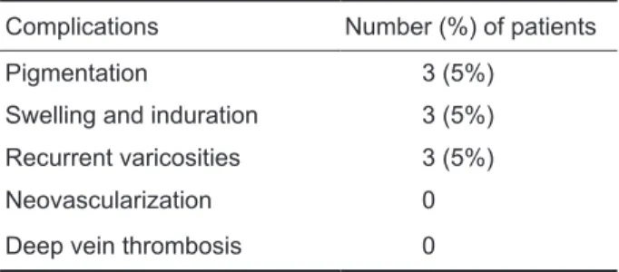
Table 2. List of complications of all endovenous laser ab-lation performed after 1 year of postprocedural
Complications Number (%) of patients
Pigmentation 3 (5%)
Swelling and induration 3 (5%) Recurrent varicosities 3 (5%)
Neovascularization 0
Deep vein thrombosis 0
DISCUSSION
Recent studies show that EVLA has a high success rate of over 90% after several years of follow-up studies and a minimal complication rate compared with traditional ligation plus stripping.8 EVLA for saphenous vein insuficiency treatment is proven to be successful for over 10 years.
The prospective randomised study of endo
-venous radiofrequency obliteration (closure) ver
-sus ligation and vein stripping ( EVOLVeS) study showed a better quality-of-life score for the endove
-nous group compared with conventional treatment for varicose veins at the one- and two-year assess
-ments.9
Endovenous ablation had advantages over con
-ventional surgery in terms of less postoperative pain, shorter periods of sick leave, earlier return to normal activities, and reduced overall costs to soci
-ety.10 In a number of large case series the technical success rate was close to 100%, and the long-term success rate (up to 5 years) ranged from 90% to 100%.10-12
Laser energies of various wavelengths includ
-ing 810 nm,13 940 nm,7 980 nm,14 1320 nm 15 and 1470 nm 16 have been applied to obliterate the GSV. A prospective randomised study 17 comparing the use of 980 nm and 810 nm laser for endovenous obliteration procedures and another one 18 compar -ing the use of 810, 940, and 980 nm diode lasers showed no signiicant difference in their effective
pulmonary embolism seldom occur. An exceptional complication is a material or device that by acci
-dent remains inside the body after the procedure. Ecchymosis, pain, induration, skin burns, dyses
-thesia, supericial thrombophlebitis, and hematoma were classiied as minor complications. Deep vein thrombosis and nerve injury were classiied as ma
-jor complications.19
Incidence of hypoaesthesia, swelling, bruising and discomfort following EVLA are not rare, but these impairments are usually self-limited and of
-ten improve within months.10,20 Hyperpigmentation along the course of the treated vein can also be seen at times, especially if the vein is above the fascial level and in thin individuals, but this complication also gradually fades over time.10 The mean body mass index for our patients with hyperpigmenta
-tion following EVLA was signiicantly low and they were thin individuals. We think that this factor might contribute to the skin discoloration following EVLA.
Our results are comparable with those of other studies.12,20 However, only a few studies have re -ported late results, especially late recanalization.11,12 Ravi and et al.12 have reported recanalization with a ratio of 0.007 as the most frequent complication in the long term follow up of a 3000-leg study. We haven’t come across with a recanalization in our study. After one year post-procedurally, most of the our postprocedure complications were transient and self limiting; the most common complications were swelling, induration, and hyperpigmentation in our study. No major complications, such as deep vein thrombosis, occurred. In conclusion, our EVLA study is satisfactory from the point of 1-year results and major complications observed. However, for the enlightenment of possible major complications, long term results of large scale patient studies are important.
We think that EVLA with 940 nm wavelength is safe and effective in all suitable patients especial
-ly from the point of satisfaction factor regardless of age and gender differences.
REFERENCES
1. Robertson L, Evans C, Fowkes FG. Epidemiology of chronic venous disease. Phlebology 2008;23(3):103-11.
2. Somers P, Knaapen M. The histopathology of varicose vein dis -ease. Angiology 2006;57(5): 546-55.
3. Rigby KA, Palfreyman SJ, Beverley C et al. Surgery versus sclerotherapy for the treatment of varicose veins. Cochrane Database Syst Rev 2004;18(4):CD004980.
4. van Rij AM, Jiang P, Solomon C, Christie RA, Hill GB. Re -currence after varicose vein surgery: a prospective long-term clinical study with duplex ultrasound scanning and air plethys -mography. J Vasc Surg 2003;38(5):935-43.
5. Durai R, Srodon PD, Kyriakides C. Endovenous laser abla -tion for supericial venous insuficiency. Int J Clin Pract 2010;64(1):61-6.
6. Fan CM, Rox-Anderson R. Endovenous laser ablation: mecha -nism of action. Phlebology 2008;23(5):206-13.
7. Tan KK, Nalachandran S, Chia KH. Endovenous laser treatment for varicose veins in Singapore: a single centre experience of 169 patients over two years. Singapore Med J 2009;50(6):591-4. 8. Bos R, Arends L, Kockaert M et al. Endovenous therapies of
lower extremity varicosities: a meta-analysis. J Vasc Surg 2009;49(1):230-9.
9. Lurie F, Creton D, Eklof B, et al. Prospective randomised study of endovenous radiofrequency obliteration (closure) versus li -gation and vein stripping (EVOLVeS): two-year follow-up. Eur J Vasc Endovasc Surg 2005;29(1):67-73.
10. van Groenendael L, Flinkenlögel L, van der Vliet JA, et al. Conventional surgery and endovenous laser ablation of recur -rent varicose veins of the small saphenous vein: a retrospec -tive clinical comparison and assessment of patient satisfaction. Phlebology 2010;25(3):151-7.
11. Spreaico G, Kabnick L, Berland TL, et al. Laser saphenous ablations in more than 1,000 limbs with long-term duplex ex -amination follow-up. Ann Vasc Surg 2011;25(1):71-8. 12. Ravi R, Trayler EA, Barrett DA, et al. Endovenous thermal
ablation of supericial venous insuficiency of the lower ex -tremity: single-center experience with 3000 limbs treated in a 7-year period. J Endovasc Ther 2009;16(4):500-5.
13. Goode SD, Chowdhury A, Crockett M, et al. Laser and radio -frequency ablation study (LARA study): a randomised study comparing radiofrequency ablation and endovenous laser abla -tion (810 nm). Eur J Vasc Endovasc Surg 2010;40(2):246-53. 14. Ergenoglu MU, Sayin M, Kucukaksu S. Fate of vena saphe
-na mag-na stump after endovenous laser ablation with 980-nm diode laser: 12-month follow-up. Photomed Laser Surg 2010;28(5):659-62.
15. Proebstle TM, Herdemann S. Early results and feasibility of in -competent perforator vein ablation by endovenous laser treat -ment. Dermatol Surg 2007;33(2):162-8.
16. Rathod J, Taori K, Joshi M, et al. Outcomes using a 1470-nm laser for symptomatic varicose veins. J Vasc Interv Radiol 2010;21(12):1835-40.
17. Kabnick LS. Outcome of different endovenous laser wave -lengths for great saphenous vein ablation. J Vasc Surg 2006;43(1):88-93.
18. Proebstle TM, Sandhofer M, Kargl A, et al. Thermal damage of the inner vein wall during endovenous laser treatment: key role of energy absorption by intravascular blood. Dermatol Surg 2002;28(7):596-600.
19. Van Den Bos RR, Neumann M, De Roos KP, Nijsten T. En -dovenous laser ablation-induced complications: review of the literature and new cases. Dermatol Surg 2009;35(8):1206-14. 20. Theivacumar NS, Dellagrammaticas D, Mavor AI, Gough MJ.