CLINICAL SCIENCE
Genomic instability at the 13q31 locus and somatic
mtDNA mutation in the D-loop site correlate with
tumor aggressiveness in sporadic Brazilian breast
cancer cases
Gilson Costa dos Santos-Jr,IAndre´a Carla de Souza Go´es,IHumberto de Vitto,IICarla Cristina Moreira,I Elizabeth Avvad,IIIFranklin David Rumjanek,II Claudia Vitoria de Moura GalloI
IUniversidade do Estado do Rio de Janeiro, Instituto de Biologia Roberto Alcantara Gomes, Departamento de Gene´tica, Rio de Janeiro/RJ, Brazil.
IIUniversidade Federal do Rio de Janeiro, Instituto de Bioquı´mica Me´dica, Rio de Janeiro/RJ, Brazil. IIIInstituto Fernandes Figueira, FIOCRUZ,
Departamento de Patologia, Rio de Janeiro/RJ, Brazil.
OBJECTIVE:Genomic instability is a hallmark of malignant tissues. In this work, we aimed to characterize nuclear and mitochondrial instabilities by determining short tandem repeats and somatic mitochondrial mutations, respectively, in a cohort of Brazilian sporadic breast cancer cases. Furthermore, we performed an association analysis of the molecular findings and the clinical pathological data.
METHODS: We analyzed 64 matched pairs of breast cancer and adjacent non-cancerous breast samples by genotyping 13 nuclear short tandem repeat loci (namely, D2S123, TPOX, D3S1358, D3S1611, FGA, D7S820, TH01, D13S317, D13S790, D16S539, D17S796, intron 12 BRCA1 and intron 1 TP53) that were amplified with the fluorescent AmpFlSTR Identifiler Genotyping system (Applied Biosystems, USA) and by silver nitrate staining following 6% denaturing polyacrylamide gel electrophoresis. Somatic mtDNA mutations in the D-loop site were assessed with direct sequencing of the hypervariable HVI and HVII mitochondrial regions.
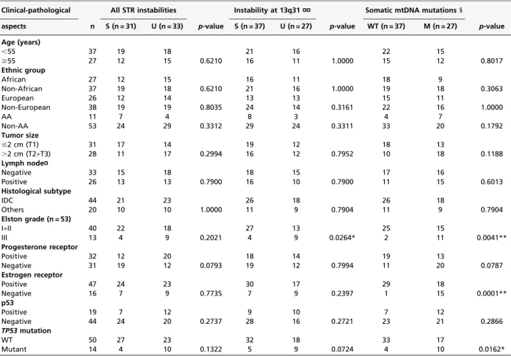
RESULTS:Half of the cancer tissues presented some nuclear instability. Interestingly, the D13S790 locus was the most frequently affected (36%), while the D2S123 locus presented no alterations. Forty-two percent of the cases showed somatic mitochondrial mutations, the majority at region 303-315 poly-C. We identified associations between Elston grade III, instabilities at 13q31 region (p= 0.0264) and mtDNA mutations (p= 0.0041). Furthermore, instabilities at 13q31 region were also associated with TP53 mutations in the invasive ductal carcinoma cases (p= 0.0207).
CONCLUSION:Instabilities at 13q31 region and the presence of somatic mtDNA mutations in a D-loop site correlated with tumor aggressiveness.
KEYWORDS: Breast Cancer; STRs; Allelic Imbalance; LOH; Somatic mtDNA Mutation.
Santos GC-Jr, Goes AC, Vitto H, Moreira CC, Avvad E, Rumjanek FD, et al. Genomic instability at the 13q31 locus and somatic mtDNA mutation in the D-loop site correlate with tumor aggressiveness in sporadic Brazilian breast cancer cases. Clinics. 2012;67(10):1181-1190.
Received for publication onApril 12, 2012;First review completed onMay 28, 2012;Accepted for publicationJune 19, 2012
E-mail: [email protected]
Tel.: 55 21 2334-0858
INTRODUCTION
Breast cancer is the most prevalent cancer that affects women worldwide. One of the most striking characteristics of this disease is the heterogeneity of its genetic and patholo-gical aspects (1). Genomic instability is one of the hallmarks of cancerous tissues, and it increases in advanced and more aggressive tumors (2,3). This instability may involve large
chromosomal alterations, such as chromosomal deletions or duplications, and lead to allelic loss or amplification. In addition to the epigenetic mechanisms, the loss of hetero-zygosity (LOH), which results in allelic imbalance, is a common method of hampering tumor suppressor gene activities during carcinogenesis. TP53 and RB are good examples of tumor suppressor genes that are frequently altered by allelic imbalance (3). Short tandem repeats (STRs) or microsatellites are polymorphic regions that are widely used to analyze allelic imbalance in tumors. In breast cancer, LOH has been detected at several loci in both familial and sporadic breast cancers, with frequencies ranging between 20% and 79% (4,5). Recently, Tokunaga et al. (6) studied the microsatellite instability of five randomly selected loci in Japanese primary breast cancer samples. They observed that
Copyrightß2012CLINICS– This is an Open Access article distributed under the terms of the Creative Commons Attribution Non-Commercial License (http:// creativecommons.org/licenses/by-nc/3.0/) which permits unrestricted non-commercial use, distribution, and reproduction in any medium, provided the original work is properly cited.
a high frequency of LOH was associated with triple-negative and high-grade HER2 breast cancers. When the same research group specifically evaluated microsatellite instabil-ity at the BRCA1 locus, they demonstrated that LOH at this region was independently associated with disease-free survival (7). In addition to nuclear genomic instabilities, researchers have also considered mitochondrial genomic alterations as indicators of cell commitment to carcinogen-esis. Although their involvement is currently not well understood, somatic mitochondrial DNA (mtDNA) muta-tions seem to participate in cancer development in different ways (8,9). Lim et al. (10) demonstrated that mtDNA mutations in colorectal cancer might be implicated in risk factors that induce poor outcomes and tumorigenesis. Tseng et al. (11) suggested that somatic mtDNA mutations may play a critical role in breast cancer progression.
The aim of this study was to characterize nuclear instabilities and mitochondrial genomic mutations in a cohort of Brazilian sporadic breast cancer cases. We analyzed matched pairs of breast cancer and adjacent non-cancerous breast samples by genotyping 13 nuclear STR loci [namely, D2S123, TPOX, D3S1358, D3S1611, FGA, D7S820, TH01, D13S317, D13S790, D16S539, D17S796, intron 12 BRCA1 and intron 1 TP53] and by directly sequencing HVI and HVII mitochondrial regions. Fur-thermore, we performed an association analysis of the molecular findings and clinical pathological data from the cases.
PATIENTS AND METHODS
Tumor samples
Tissue specimens from sporadic primary breast cancer tumors and the corresponding adjacent tumor-free areas were obtained between 2005 and 2009 from the biopsies of 64 women at the Fernandes Figueira Institute, FIOCRUZ, Rio de Janeiro, Brazil. After excision, the tissues were snap-frozen in liquid nitrogen and stored at -70oC. Cancer diagnosis was confirmed by histopathology. Sixty-four percent of cases were diagnosed as invasive ductal carcinoma, and 36% were classified as invasive lobular carcinoma, mucinous, or micropapillary. DNA was extracted from the tissue samples using a salting-out method (12). The DNA was quantified using ethidium bromide staining in agarose gels and UV spectrophotometry at 260 nm. The P53 and estrogen/progesterone receptor levels, which were assessed by immunohistochemistry, and the clinical-pathological data were obtained from records of the department of pathology, IFF-FIOCRUZ. The study protocol was approved by the local ethics committee.
mtDNA sequencing
Hypervariable mitochondrial DNA regions I and II (D-loop region) were sequenced using the dideoxy chain termination method (BigDyeHTerminator v3.1 Cycle Sequencing Kit) and analyzed in an automated ABI310 Sequencer (Applied Biosystems, USA). All of the sequences were aligned to the Revised Cambridge Reference Sequence, accession number NC_012920. The primer pairs designed for the PCR and direct sequencing of mtDNAs are provided in Supple-mentary Table 1. The mitochondrial somatic mutation data were assessed by comparing cancerous and adjacent non-cancerous breast samples.
STR typing of nuclear DNA andTP53mutation detection
Nuclear genomic instability was assessed by PCR analysis of 13 STR markers. The TPOX, D3S1358, FGA, D7S820, TH01, D13S317 and D16S539 loci were amplified with the fluor-escent AmpFlSTR Identifiler Genotyping system according to the manufacturer’s recommendations (Applied Biosystems, USA) and then analyzed using the automated ABI3100 Genetic Analyzer platform and GeneMapper Software (Applied Biosystem, USA). The D13S790 locus was amplified with an independent FAM-fluorescent system and analyzed using the ABI3100 Genetic Analyzer platform (Applied Biosystems, USA). The D2S123, D3S1611, D17S796, intron 12 BRCA1 and intron 1 TP53 loci were analyzed using silver nitrate staining following a 6% denaturing polyacrylamide gel electrophoresis. Nuclear genome instability was assessed by observing the allelic imbalances, which are usually identified as LOH. Supplementary Table 1 shows the STR loci localizations and the primer sequences. When the allelic patterns differed between the matched normal and tumor DNAs, the PCRs and electrophoresis were performed twice. Eventually, the lymphocyte DNAs of patients were also genotyped and compared to normal and tumor DNAs to confirm results. In a previous study,TP53mutation detection was performed for exons 4-9 (13). The association analyses were performed with Fisher’s exact test with a significance level of 95% using GraphPadHsoftware.
RESULTS
Clinical-pathological aspects of cases
To obtain all the possible noteworthy clinical-pathological data from the studied cases, the 64 patients were evaluated for age, ethnicity, histological classification, TNM, Elston grade, p53 and estrogen and progesterone receptor expres-sion levels (Table 1 and Supplementary Tables 2 and 3). The average age of the studied patients was 53, and the ages ranged from 27 to 76 years. The ethnic classification was based on mitochondrial haplogroups. The patients were classified into three ethnic groups: African (42%), European (40%) and Asian-Amerindians (18%). Most of the cases (69%) were diagnosed as invasive ductal carcinomas (IDCs). The other histological subtypes, which represented a total of 18 cases (31%), included the following subtypes: invasive papillary carcinoma, comedocarcinoma, mucinous and medullar intraductal carcinoma. Most of the cases (75%) were classified at low or intermediate grades, although 25% were Elston grade III (high aggressiveness). Fifty percent of the cases were progesterone-positive, and 74% were estro-gen-positive. In relation to the p53 tumor suppressor protein, 70% of the cases were protein-negative, and 22% were mutant (13).
Nuclear and mitochondrial genome instability
of the cases displayed microsatellite instability to some extent; this instability was characterized by allelic imbal-ances and 41% of cases exhibited alterations in three or more loci. Among the 13 analyzed STR loci, only the D2S123 locus was stable and the D7S820 locus had the lowest frequency of
instability (1%). The intron 1 TP53 and D13S317 loci were each unstable in 16% of cases. Interestingly, the D13S790 locus had the highest frequency of instability among the STR loci (36%). Figure 2 displays the distribution of the number of instabilities in the STR loci. Supplementary Table
Table 1- Clinical-pathological aspects of the cases and an association analysis of STR instabilities and mtDNA mutations (n = 64).
Clinical-pathological All STR instabilities Instability at 13q31 ¤¤ Somatic mtDNA mutations1
aspects n S (n = 31) U (n = 33) p-value S (n = 37) U (n = 27) p-value WT (n = 37) M (n = 27) p-value
Age (years)
,55 37 19 18 21 16 22 15
$55 27 12 15 0.6210 16 11 1.0000 15 12 0.8017
Ethnic group
African 27 12 15 16 11 18 9
Non-African 37 19 18 0.6210 21 16 1.0000 19 18 0.3063
European 26 12 14 13 13 15 11
Non-European 38 19 19 0.8035 24 14 0.3161 22 16 1.0000
AA 11 7 4 8 3 4 7
Non-AA 53 24 29 0.3312 29 24 0.3311 33 20 0.1792
Tumor size
#2 cm (T1) 31 17 14 19 12 18 13
.2 cm (T2+T3) 28 11 17 0.2994 16 12 0.7952 10 18 0.1188
Lymph node¤
Negative 33 15 18 18 15 17 16
Positive 26 13 13 0.7900 16 10 0.7900 11 15 0.6013
Histological subtype
IDC 44 21 23 26 18 26 18
Others 20 10 10 1.0000 11 9 0.7904 11 9 0.7904
Elston grade (n = 53)
I+II 40 22 18 27 13 25 15
III 13 4 9 0.2021 4 9 0.0264* 2 11 0.0041**
Progesterone receptor
Positive 32 12 20 18 14 19 13
Negative 31 19 12 0.0793 19 12 0.7994 11 20 0.0787
Estrogen receptor
Positive 47 24 23 30 17 29 18
Negative 16 7 9 0.7735 7 9 0.2397 1 15 0.0001**
p53
Positive 19 7 12 9 10 7 12
Negative 44 24 20 0.2737 28 16 0.2721 23 21 0.2866
TP53mutation
WT 50 27 23 32 18 33 17
Mutant 14 4 10 0.1322 5 9 0.0724 4 10 0.0162*
¤¤13q31 region: D13S317 and D13S790 STR loci.
n - Total number of samples; S - Number of stable samples; U - Number of unstable samples; AA - Asian-Amerindian; mtDNA – Mitochondrial DNA. WT - Wild type; M – Mutation; IDC - Invasive Ductal Carcinoma.
1
Mitochondrial alteration within the D-loop region.
¤Lymph node metastasis: Negative (N0); Positive (N1+N2+N3).
*Fisher’s exact test (p
#0.05 statistically significant).
**Fisher’s exact test (p
#0.05 highly statistically significant).
4 summarizes the data that was obtained from each of the 64 cases. Regarding the mitochondrial genome analysis, 42.18% of cases had somatic mutations, most of which were at the 303-315 poly-C region (Supplementary Table 4). Figure 3 illustrates an example of mtDNA mutation assessed by direct sequencing.
Association with clinical-pathological aspects
Following the determination of nuclear instabilities and mitochondrial genomic alterations, an association study with clinical-pathological aspects was performed. Interestingly, when the most frequent unstable genome region (13q31, assessed here through the microsatellite markers D13S317 and D13S790) was analyzed separately, it was statistically associated with Elston grade III (p= 0.0264) (Table 1). Furthermore, a positive association was also observed with the presence ofTP53mutations in IDCs (p= 0.0207) (Table 2). A highly positive association with Elston grade III was also observed with the presence of somatic mtDNA mutations (p= 0.0041). Moreover, reinforcing their correlation with parameters of tumor aggressiveness, the mtDNA mutations were statistically associated with negative estrogen receptor expression (p= 0.0001) andTP53mutations (p= 0.0162). There was no correlation between the STR instabilities and the somatic mtDNA mutations.
DISCUSSION
Several molecular mechanisms are involved in the forma-tion and progression of breast carcinomas, particularly sporadic breast cancers. An important feature of breast tumor
development is the characteristic but highly heterogeneous genomic instability (14). Recently, the advantageous utiliza-tion of genome-scale analysis and microarray-based gene expression profiling has stressed the complexity of breast cancer progression (15,16). This study was designed and executed to provide further understanding of genomic instability in Brazilian breast cancer cases. We performed nuclear STR loci genotyping and direct sequencing of HVI and HVII mitochondrial regions of 64 matched pairs of cancerous and adjacent non-cancerous breast samples. Our main aims were to detect genomic instabilities in well-known DNA regions using selected STR loci and the mitochondrial D-loop region and to analyze their association with clinical aspects. With the results, we could expect to have a clearer understanding of local and defined genomic changes, both nuclear and mitochondrial, and their clinical consequences. Surprisingly, through the microsatellite markers D13S317 and D13S790, we found that 13q31 was the most frequent unstable genomic region. It was most apparent at the D13S790 locus, with more than 20 cases presenting LOH. When analyzed separately from the other chromosomal loci, 13q31 was shown to be statistically associated with Elston grade III in all breast tumors and withTP53 mutations in invasive ductal carcinomas, both of which are clinical parameters of tumor aggressiveness (17,18). The 13q31 locus has been described as a chromosome region that shows different genetic alterations depending on the cancer type. Genetic gains have been observed in sarcoma (19) and colorectal cancer (20). Genetic losses have also been verified in breast cancer (21,22). Eiriksdottir et al. (23) analyzed chromosome 13q in detail in 139 sporadic breast tumors with 18 polymorphic microsatel-lite markers and identified 3 LOH target regions: 13q12-q13, 13q14 and 13q31-q34. In another study, correlations were
Figure 2 -Distribution of STR instabilities among the loci. The D2S123 locus presented no alterations. N: number of genetic instabilities at each STR locus.
Figure 3 -Detection of the mtDNA somatic mutation (16192 CC/T) in a case of breast cancer. The arrow indicates the mutation. N: normal tissue; T: tumor tissue.
Table 2 -Association analysis ofTP53and mtDNA mutations with STR instabilities in invasive ductal carcinoma cases (n = 44).
Clinical-pathological All STR instabilities Instability at 13q31¤
aspects n S (n = 23) U (n = 21) p-value S (n = 26) U (n = 18) p-value
TP53mutation 14
WT 35 21 24 11
Mutant 9 2 7 0.0642 2 7 0.0207*
mtDNA mutations1
WT 26 15 11 17 9
Mutant 18 8 10 0.5406 9 9 0.3613
¤13q31region: D13S317 and D13S790 STR loci.
n – Total number of samples; S - Number of stable samples; U - Number of unstable samples; WT - Wild Type. 1
Somatic mtDNA mutations within the D-loop region.
*Fisher’s exact test (P
detected between the allelic loss of the D13S1694 marker (telomeric to BRCA2) and both larger tumor sizes and negative estrogen receptors (24). More recently, Schwarzenbach et al. (25), studying cell-free DNA in benign and malignant breast tumor cases, noted that LOH at D13S280 and D13S159, both markers located at 13q31-33, are associated with overall and disease-free survival. In this same study, all of the analyzed markers significantly correlated with lymph node status (25). Together, these results and our results suggest the existence of a putative suppressor gene or an important regulator sequence in this region. The miR17-92 cluster (13q31.3 region) is located near the 13q31 region; the cluster consists of seven microRNAs tightly grouped within an 800 bp genomic region in the third intron of the primary transcript C13orf25. This cluster is also known as oncomir-1 because its superexpression has been demonstrated in pulmonary cancer and lymphomas (26,27). However, there is some evidence of LOH in this genomic region, mainly in breast cancer, indicating that this cluster can also play a role as a tumor suppressor gene (28,29). Our results reinforce the hypothesis that instability in the 13q31 region may relate to a loss of function of microRNAs in this cluster. Because most of the allelic imbalances were associated with Elston grade III, and (more importantly) 13q31 LOH was associated withTP53mutations in the IDC samples, we can infer that this alteration is a delayed event in breast tumor progression. We also investigated somatic mutations in the D-loop region of the mtDNA and found that 42.18% of cases were mutated, the majority at the 303-315 poly-C region. As has been described by others (30,31), we could demonstrate an association between the presence of mtDNA mutations and breast tumor aggressiveness. Parameters such as high histological grade (Elston grade III), estrogen receptor-negative and TP53 mutations were statistically associated. Kuo et al. (32) recently reported that the presence of somatic mutations in the D-loop indicates poor prognosis; however, they did not identify a correlation with the presence ofTP53 mutations in 30 pairs of tumor and non-tumor samples. The low number of samples and/or the different types of breast cancer cases could explain the difference.TP53and somatic mtDNA mutations have been considered to be good biomarkers of nuclear DNA damage (18,32); therefore, a correlation between both genetic alterations would be expected. However, we did not identify any association between nuclear instabilities and mtDNA alterations. Alazzouzi et al. (33) also observed that mitochondrial alterations were not associated with nuclear instability in breast tumors. In a study of colorectal carcinomas, instability in the 303 poly-C region of mtDNA was not associated with nuclear microsatellite instability (34). These observations suggest an independent occurrence of both phenomena. In conclusion, although the number of the Brazilian cases evaluated in this study was not high, we could highlight an important role for instabilities at the nuclear 13q31 locus and in mtDNA in breast cancer development and prognosis.
ACKNOWLEDGMENTS
The authors thank the patients for their collaborative participation in this study. Gilson Costa dos Santos Junior and Humberto de Vitto were recipients of fellowships from CNPq/Brazil, and Carla Cristina Moreira was a recipient of a fellowship from PIBIC/CNPq/Brazil. We also thank Angela Duarte, Genomic Platform, UERJ, for her technical assistance. This work was supported by grants from Conselho Nacional de Desenvolvimento Cientı´fico e Tecnolo´gico (CNPq) and Fundac¸a˜o Carlos
Chagas Filho de Amparo a` Pesquisa do Estado do Rio de Janeiro (FAPERJ).
AUTHOR CONTRIBUTIONS
Santos-Jr GC was responsible for the STR genotyping, patient data collection, statistical analysis and critical revision of the paper. Goes AC was responsible for the STR genotyping study design and execution and critical review of the manuscript. De Vitto H was responsible for mutant mtDNA design, execution and results interpretation. Moreira CC performed STR genotyping. Avad E was responsible for the patient samples and data collection. Rumjanek FD was responsible for partial financial support. De Moura Gallo CV conceived and designed the study, was responsible for research support and manuscript writing.
REFERENCES
1. Weigelt B, Geyerb FC, Reis-Filho JS. Histological types of breast cancer: How special are they? Mol Oncol. 2010;4(3):192-208, http://dx.doi.org/ 10.1016/j.molonc.2010.04.004.
2. Stratton MR, Campbell PJ, Futreal PA. The cancer genome. Nature. 2009;458(7239):719-724, http://dx.doi.org/10.1038/nature07943. 3. Hanahan D, Weinberg RA. Hallmarks of Cancer: The Next Generation.
Cell. 2011;144(5):646-74, http://dx.doi.org/10.1016/j.cell.2011.02.013. 4. Kirchweger R, Zeilinger R, Schneeberger C, Speiser P, Louason G,
Theillet G. Patterns of allele losses suggest the existence of five distinct regions of LOH on chromosome 17 in breast cancer. Int. J. Cancer. 1994;56(2):193-9, http://dx.doi.org/10.1002/ijc.2910560208.
5. Collins N, Mcmanus R, Wooster R, Mangion J, Seal S, Lakhani SR, et al. Consistent loss of the wild type allele in breast cancers from a family linked to the BRCA2 gene on chromosome 13ql2-13. Oncogene. 1995;10(8):1673-5.
6. Tokunaga E, Okada S, Yamashita N, Akiyoshi S, Kitao H, Morita M, et al. High incidence and frequency of LOH are associated with aggressive features of high-grade HER2 and triple-negative breast cancers. Breast Cancer. 2012;19(2):161-9, http://dx.doi.org/10.1007/ s12282-010-0232-7.
7. Okada S, Tokunaga E, Kitao H, Akiyoshi S, Yamashita N, Saeki H, et al. Loss of heterozygosity at BRCA1 locus is significantly associated with aggressiveness and poor prognosis in breast cancer. Ann Surg Oncol. 2012;19(5):1499-507, http://dx.doi.org/10.1245/s10434-011-2166-5. 8. Brandon M, Baldi P, Wallace DC. Mitochondrial mutations in cancer.
Oncogene. 2006;25:4647-62, http://dx.doi.org/10.1038/sj.onc.1209607. 9. Guerra F, Kurelac I, Cormio A, Zuntini R, Amato LB, Ceccarelli C, et al.
Placing mitochondrial DNA mutations within the progression model of type I endometrial carcinoma. Hum Mol Gen. 2011;20(12):2394-405, http://dx.doi.org/10.1093/hmg/ddr146.
10. Lim SW, Kim HR, Kim HW, Huh JW, Kim YJ, Shin JH, et al. High-frequency minisatellite instability of the mitochondrial genome in colorectal cancer tissue associated with clinicopathological values. Int J Cancer. 2012;131(6):1332-41. Epub 2011/11/29, http://dx.doi.org/ 10.1002/ijc.27375.
11. Tseng LM, Yin PH, Yang CW, Tsai YF, Hsu CY, Chi CW, HC Lee. Somatic mutations of the mitochondrial genome in human breast cancers. Genes Chrom Cancer. 2011;50(10):800-11, http://dx.doi.org/ 10.1002/gcc.20901.
12. Miller SA, Dykes DD, Polesky HF. A simple salting out procedure for extracting DNA from human nucleated cells. Nucl Acids Res. 1988;16(3):1215, http://dx.doi.org/10.1093/nar/16.3.1215.
13. Levy CB, Stumbo AC, Ano Bom AP, Portari EA, Cordeiro Y, Silva JL, De Moura-Gallo CV. Co-localization of mutant p53 and amyloid-like protein aggregates in breast tumors. Int J Biochem Cell Biol. 2011;43(1):60-4, http://dx.doi.org/10.1016/j.biocel.2010.10.017.
14. Kwei KA, Kung Y, Salari K, Holcomb IN, Pollack JR. Genomic instability in breast cancer: pathogenesis and clinical implications. Mol Oncol. 2010;4(3):255-66, http://dx.doi.org/10.1016/j.molonc.2010.04.001. 15. Reis-Filho JS, Weigelt B, Fumagalli D, Sotiriou C. Molecular profiling:
moving away from tumor philately. Sci Transl Med. 2010;2(47):47ps43, http://dx.doi.org/10.1126/scitranslmed.3001329.
16. Colombo PE, Milanezi F, Weigelt B, Reis-Filho JS. Microarrays in the 2010s: the contribution of microarray-based gene expression profiling to breast cancer classification, prognostication and prediction. Cancer Research. 2011;13(3):212-27.
17. Elston CW, Ellis IO. Pathological prognostic factors in breast cancer. I. The value of histological grade in breast cancer: experience from a large study with long-term follow-up. Histopathology. 1991;19:403-10. 18. Olivier M, Hollstein M, Hainaut P. TP53 Mutations in Human Cancers:
Origins, Consequences, and Clinical Use. Cold Spring Harb Perspect Biol 2010:2(1):a001008.
amplicon at 13q31 associated with alveolar rhabdomyosarcoma. Genes Chrom Cancer. 2000;28(2):220-6, http://dx.doi.org/10.1002/(SICI)1098-2264(200006)28:2,220::AID-GCC11.3.0.CO;2-T.
20. Neklason DW, Tuohy TM, Stevens J, Otterud B, Baird L, Kerber RA, et al. Colorectal adenomas and cancer link to chromosome 13q22.1-13q31.3 in a large family with excess colorectal cancer. J Med Genet. 2010;47(10):692-9, http://dx.doi.org/10.1136/jmg.2009.076091. 21. Przybytkowski E, Girouard S, Allard B, Lamarre L, Basik M. Widespread
bimodal intrachromosomal genomic instability in sporadic breast cancers associated with 13q allelic imbalance. Cancer Res. 2003;63(15): 4588-93.
22. Imyanitov EN, Togo AV, Suspitsin EN, Grigoriev MY, Pozharisski KM, Turkevich EA, et al. Evidence for microsatellite instability in bilateral breast carcinomas. Cancer Lett. 2000;154(1):9-17, http://dx.doi.org/ 10.1016/S0304-3835(99)00444-9.
23. Eiriksdottir G, Johannesdottir G, Ingvarsson S, Bjo¨rnsdottir IB, Jonasson JG, Agnarsson BA, et al. Mapping loss of heterozygosity at chromosome 13q: loss at 13q12-q13 is associated with breast tumour progression and poor prognosis. European Journal of Cancer. 1998;34(13):2076-81, http:// dx.doi.org/10.1016/S0959-8049(98)00241-X.
24. Rio PG, Pernin D, Bay JO, Albuisson E, Kwiatkowski F, De Latour M, et al. Loss of heterozygosity of BRCA1, BRCA2 and ATM genes in sporadic invasive ductal breast carcinoma. Int. J. Oncol. 1998;13(4):849-53. 25. Schwarzenbach H, Mu¨ller V, Milde-Langosch K, Steinbach B, Pantel K.
Evaluation of cell-free tumour DNA and RNA in patients with breast cancer and benign breast disease. Mol Biosyst. 2011;7(10):2848-54, http://dx.doi.org/10.1039/c1mb05197k.
26. Olive V, Jiang I, He L. Mir-17-92, a cluster of miRNAs in the midst of the cancer network. Int J Biochem Cell Biol. 2010;42(8):1348-54, http:// dx.doi.org/10.1016/j.biocel.2010.03.004.
27. Van Haaften G, Agami R. Tumorigenicity of the miR-17-92 cluster distilled. Genes Dev. 2010;24(1):1-4, http://dx.doi.org/10.1101/gad.1887110. 28. Hossain A, Kuo MT, Saunders FG. Mir-17-5p Regulates Breast Cancer
Cell Proliferation by Inhibiting Translation of AIB1 mRNA. Mol Cell Biol. 2006;26(21):8191-201, http://dx.doi.org/10.1128/MCB.00242-06. 29. Visone R, Croce CM. MiRNAs and Cancer. Am J Pathol. 2009;174(4):
1131-8, http://dx.doi.org/10.2353/ajpath.2009.080794.
30. Chatterjee A, Mambo E, Sidransky D. Mitochondrial DNA mutations in human cancer. Oncogene 2006;25:4663-74.
31. Imanishi H, Hattori K, Wada R, Ishikawa K, Fukuda S, Takenaga K, et al. Mitochondrial DNA mutations regulate metastasis of human breast cancer cells. 2011;PLoS One 6(8):e23401.
32. Kuo SJ, Chen M, Ma GC, Chen ST, Chang SP, Lin WY, et al. Number of somatic mutations in the mitochondrial D-loop region indicates poor prognosis in breast cancer, independent of TP53 mutation. Cancer Genet Cytogenet. 2010;201(2):94-101, http://dx.doi.org/10.1016/j.cancergencyto. 2010.05.013.
33. Alazzouzi H, Farriol M, Espı´n E, Armengol M, Pena M, Zeh K, et al. Molecular patterns of nuclear and mitochondrial microsatellite altera-tions in breast tumors. Oncol Rep. 2003;10(5):1561-7.
34. Guleng G, Løvig T, Meling GI, Andersen SN, Rognum TO. Mitochondrial microsatellite instability in colorectal carcinomas-frequency and associa-tion with nuclear microsatellite instability. Cancer Lett. 2005;219(1):97-103, http://dx.doi.org/10.1016/j.canlet.2004.07.018.
Supplementary Table 1 -Nuclear STR and mtDNA primer sequences.
Locus
Chromosome
localization Motif Primer sequences
Amplicon (bp)
TPOX 2p23 AATG ACTGGCACAGAACAGGCACTTAGG
GGAGGAACTGGGAACCACAGAGGTTA F R 224-252 D2S123 2p16 (hMSH2) CA AAACAGGATGCCTGCCTTTA GGACTTTCCACCTATGGGAC F R 197-227 D3S1611 3p21 (hMLH1) CA CCCCAAGGCTGCACTT AGCTGAGACTACAGGCATTTG F R 260-268
D3S1358 3p21 TCTA ACTCGAGTCCAATCTGGTT
ATGAAATCAACAGAGGCTTG
F R
97-147
FGA 4p28 TTTC GCCCCATAGGTTTTGAACTCA
TGATTTGTCTGTAATTGCCAGC
F R
206-332
D7S820 7q11 GATA GATTCCACATTTATCCTCATTGAC
ATGTTGGTCAGGCTGACTATG
F R
215-247
TH01 11p15 AATG ATTCAAAGGGTATCTGGGCTCTGG
GTGGGCTGAAAAGCTCCCGATTAT
F R
179-203
D13S790 13q31 GATA TTGAGCCAGGATGATGTG
CCTTTGGGTTGTAAACGT
F R
422-454
D13S317 13q31 TATC ACAGAAGTCTGGGATGTGGA
GCCCAAAAAGACAGACAGAA
F R
165-197
D16S539 16q24 GATA GGGGGTCTAAGAGCTTGTAAAAAG
GTTTGTGTGTGCATCTGTAAGCAT
F R
264-288
BRCA1 17q
(intron 12 BRCA)
TG GGTCATGTGTTCCATTTGGG
TTGAAGCAACTTTGCAATGAG
F R
190-270
D17S796 17p CA CAATGGAACCAAATGTGGTC
AGTCCGATAATGCCAGGATG
F R
144-174
TP53 17p
(intron 1 TP53)
AAAAT GCACTGACAAAACATCCCCT
AGTAAGCGGAGATAGTGCCACTGT
F R
150-180
HVI mtDNA - CGCACCTACGTTCAATATTACAGG
GGTGTGTGTGTGCTGGGTAGG
F R
364
HVII mtDNA - ATTACTGCCAGCCACCATGAA
Supplementary Table 2 -Clinical-pathological patient data.
Case
Age
(Years) Ethnicity1
Histological
classification TNM EG
Immunohistochemistry
PR ER P53
T2 52 African IDC pT1c pN0 (sn) pMx I +++ +++
-T4 48 African IDC pT1c pN0 (sn) pMx II + +
-T5 53 African IDC pTis pN0 (sn) pMx * ND ND ND
T6 56 European IDC pT2c pN2a pMx III - - +
T8 49 African IDC pT1c pN0 (sn) pMx I +++ +++
-T9 60 African Invasive lobular pT2c pN0 (sn) pMx * - +++
-T10 44 AA IDC pT2 pN0 pMx III - - +
T11 27 African Intracystic papillary pTis pN0 pMx * + +++
-T14 54 African IDC pT2 pN2a pMX II + +++
-T15 41 African IDC pT1c pN0 (sn) pMx I - +++ +
T16 48 AA IDC pT1c pN0 (sn) pMX I - +++
-T17 46 European IDC pT2 pN1a pMx II - -
-T18 54 European IDC pT1c pN2a pMX * +++ +++
-T19 50 African Mucinous pT1c pN0 (sn) pMX I - +++
-T21 39 AA IDC pT1b pN0 pMX III - - +
T23 55 African IDC pT2 pN1a pMx I +++ +++
-T25 46 European IDC pT1c pN0 pMX II - +++
-T26 60 African IDC pT3 pN0 pMx III - -
-T27 72 African Invasive papillary pT1c pNx pMx II - +
-T28 46 European IDC pT1c pN0 (sn) pMx III - +++
-T29 70 African Invasive papillary pT2 pN0 (sn) pMX I ++ +++
-T31 36 African Invasive micropapillary pT2 pN1a pMx III - -
-T32 50 AA IDC pT1c pN0 pMx I - ++
-T33 56 European IDC pT1c pN2a pMx III - +++
-T34 46 European IDC pT2 pN1a pMx III - - +
T35 49 European IDC pT1c pN0 (sn) pMx II +++ -
-T36 53 European IDC pT2 pN0 (sn) pMx II - - +
T37 47 European Mucinous pT1c pN0 (sn) pMx I - +
-T38 61 African IDC pT1b pN0 (sn) pMx I +++ +++
-T40 66 African IDC pT1c pN2 pMx III - +++ +
T42 40 African IDC pT2 pN0 (sn) pMx I +++ +++
-T43 52 AA IDC pTis pN0 (sn) pMx * - -
-T44 58 African IDC pT2 pN1a pMx II +++ +++
-T46 44 European IDC pT2 pN3a pMx II - +++
-T47 71 European IDC pT2 pN0 pMx II +++ +++ +
T48 40 European IDC pT1c pN0 pMx II - - +
T50 42 African Invasive lobular pT1a pN1a pMx * ++ +++ +
T52 40 European IDC pT2 pN1a pMx II ++ +++
-T53 60 European IDC pT1c pN1a pMx I - +++
-T55 40 European Invasive apocrine pT1a pN1a pMx * + + +
T56 74 AA IDC pT2 pN1a pMx II - +++
-T58 70 AA Invasive lobular pT2 pN1a pMx * +++ +++
-T59 46 African Invasive apocrine pT2 pN1a pMx II - - +
T60 58 European IDC pT2 pN1a pMx I ++ +++
-T61 44 AA IDC pT2 pN1b1 pMx II + + +
T62 76 European IDC pT1 pN0 pMx II + +
-T63 71 African IDC pT1 pN1 pMx I + +
-T65 53 African Invasive papillary ND II - +
-T68 59 African Invasive micropapillary pT2 pN3 pMx III - - +
T69 72 European Invasive lobular pT1 pN0 pMx * + + +
T70 50 European IDC pT2 pN0 pMx II + +
-T71 63 European Invasive lobular pT1 pN1 pMx * + + +
T72 68 European IDC pT2 pN0 pMx III - - +
T73 63 African Invasive papillary pT1 pN2 pMx III - -
-T74 75 European IDC pT1 pN0 pMx III - - +
T75 41 European IDC pT2 pN1 pMx II + +
-T76 60 AA Invasive micropapillary pT2 pN2 pMx II + +
-T77 46 African IDC pT1 pN0 pMx I + +
-T78 66 European IDC pT1 pNx pMx II + +
-T80 28 AA IDC pTis pN0 pMx * - + +
T81 47 African IDC pT2 pN0 pMx II + +
-T82 69 European Invasive micropapillary pT1 pNx pMx II + +
-T83 49 African Invasive mixed type pT2 pNx pMx II + +
-T85 61 AA Invasive apocrine pT1 pN0 pMx II + +
-IDC – Invasive ductal carcinoma; TNM – Tumor-lymph node metastasis; EG – Elston grade; PR – Progesterone receptor; ER – Estrogen receptor; Protein expression: (-) negative, (+) positive - 25-50%, (++) positive - 50-75%; (+++) positive - more than 75%; ND - no data; AA - Asian-Amerindian. *Without Elston grade classification.
1
Supplementary Table 3 -Classification of cases according to the clinical-pathological aspects (total = 64).
Variables
Number of samples n (%)
Age (years) ,45 45-55 55-65 65-75 .75
14 (22) 24 (37) 13 (20) 12 (19) 1 (2) Tumor size
T1 (#2 cm) T2 (.2 cm) T3 (.5 cm) Tis (Carcinomain situ) ND
31 (48) 27 (42) 1 (2) 4 (6) 1 (2) Lymph node metastasis
N0 N1 N2 N3 Nx ND
33 (52) 17 (26) 7 (11)
2 (3) 4 (6) 1 (2) Histological subtype
IDC
Invasive Lobular Others1
44 (69) 5 (8) 15 (23) Elston grade*
I II III
15 (28) 25 (47) 13 (25) Progesterone receptor
PR+
PR++
PR+++
PR – ND
18 (28) 4 (6) 10 (16) 31 (48) 1 (2) Estrogen receptor
ER+
ER++
ER+++
ER – ND
20 (31) 1 (2) 26 (40) 16 (25) 1 (2) P53
p53+
p53-ND
19 (30) 44 (68) 1 (2)
Supplementary Table 4 -Unstable STR loci, mtDNA mutations andTP53mutation status (exons 4-9).
Case Unstable STR loci Mitochondrial somatic mutations TP53mutation
T2 D17S796,
D13S790
- G245S
T4 D13S790 -
-T5 D13S790 -
-T6 - 303-315C (8-9) TC (6)
-T8 - 16192 CC/T
-T9 D13S790 16309 AA/G
-T10 D3S1358, D13S317, D17S796, D3S1611, BRCA1 303-315 C (7-8) TC (6) R248Q
T11 D13S790 -
-T14 TH01, TP53, D3S1611, D3S1358, D17S796 303-315 C (7-8) TC (6)
-T15 - -
-T16 - 303-315 C (7-8) TC (6)
16391 GG/A
-T17 - 303-315 C (7-8) T C (6)
16261 CC/T
-T18 - - R175H
T19 - 303-315 C (7-8) TC (6) H168P
T21 FGA, D3S1358, D3S1611, D13S790 303-315 C (8-9) TC (6) R273H
T23 - -
-T25 - 16192CC/T
-T26 TP53,
FGA, D16S539, D13S317
-
-T27 - -
-T28 - -
-T29 D16S539, D17S796 146 TT/C
-T31 FGA, D13S317, TH01, BRCA1, D13S790 - 16888delC
T32 - -
-T33 D13S790 - 16897-16911del
T34 D13S790 303-315 C (8-9) TC (6)
66 GG/T
Y234C
T35 D13S790, D17S796, TP53 -
-T36 - -
-T37 D13S317 -
-T38 TP53
-T40 D13S317, FGA, TH01, D17S796, D3S1611, TP53, BRCA1, D3S1358, TPOX,
D13S790
294 TT/C I195L
T42 - 16261CC/T
-T43 - 303-315 C (7-8) TC (6)
-T44 - -
-T46 - -
-T47 D16S539, TP53, TPOX -
-T48 - -
-T50 TP53 -
-T52 TP53,
D13S317, D16S539, D13S790
-
-T53 - -
-T55 - 294 TT/C W146stop
T56 - -
-T58 - -
-T59 TP53,
TH01, BRCA1, D3S1358, D16S539, D13S317, D13S790
303-315 C (7-8) TC (6) 338 CC/T
P278A
T60 TH01,
D16S539, D13S317
-
-T61 TP53,
D13S317, D13S790
215 AA/G
-T62 - 303-315 C (8-9) TC (6)
-T63 - -
-T65 - -
-T68 - -
-T69 D13S790 303-315 C (7-8) TC (6)
338 CC/T
-T70 D13S790 -
-T71 D13S790 215AA/C
-T72 D7S820,
TH01, D16S539, D17S796, D13S790
303-315 C (7-8) TC (6) R175H
-Case Unstable STR loci Mitochondrial somatic mutations TP53mutation
T74 D13S790 303-315 C (7-8) TC (6)
16291 CC/T
-T75 - -
-T76 D3S1611 303-315 C (7-8) TC D259V
T77 - -
-T78 D13S790 303-315 C (7-8) TC (6)
16291CC/T
-T80 - -
-T81 D13S790,
D13S317
303-315 C (7-8) TC (6)
-T82 D13S790,
D16S539
-
-T83 D13S790 -
-T85 - -