Original Article
Artigo Original
Functional change in the pattern of
swallowing through the performance of
orofacial exercises
Mudança funcional no padrão de deglutição
por meio da realização de exercícios orofaciais
Irina Claudia Fernandes Alves
1Claudia Regina Furquim de
Andrade
1Keywords
Speech, Language and Hearing Sciences Deglutition Deglutition Disorders Rehabilitation Exercise Therapy
Descritores
Fonoaudiologia Deglutição Transtornos de Deglutição Reabilitação Terapia com Exercício
Correspondence address:
Claudia Regina Furquim de Andrade R. Cipotânea, 51, Cidade Universitária, São Paulo (SP), Brazil, CEP: 05360-160. E-mail: [email protected]
Received: April 28, 2016
Accepted: September 04, 2016
Study carried out at Hospital das Clínicas, Faculdade de Medicina, Universidade de São Paulo and Departamento de Fisioterapia, Fonoaudiologia e Terapia Ocupacional of FMUSP - São Paulo (SP), Brazil.
1 Departamento de Fisioterapia, Fonoaudiologia e Terapia Ocupacional, Faculdade de Medicina, Universidade
de São Paulo – USP - São Paulo (SP), Brazil.
Financial support: nothing to declare.
Conlict of interests: nothing to declare.
ABSTRACT
Purpose: The objective was to determine if there was functional improvement of swallowing pattern in subjects identiied with risk of oropharyngeal dysphagia after four weeks of speciic oropharyngeal exercises. These exercises have pre-determined intensity and duration. Methods: It is a longitudinal study of functional effect, determined by initial and inal comparative measures. Participants were adults and elderly, selected in a period of 24 months. A total of 68 participants were included. All subjects had a clinical evaluation of swallowing, and an initial measure in a functional scale. The individuals were split into two groups, according to the initial levelling of ASHA NOMS scale. In Group 1 (G1) - ASHA NOMS, initial of levels 1 and 2; Group 2 (G2) - ASHA NOMS, initial of levels 3, 4 and 5. All subjects executed an exercise protocol performed for four weeks. The protocol includes sessions with a speech therapist, and continuity of activities in home environment. Finally, new measurement of swallowing performance was held. Results: For G2 group there was statistically signiicant improvement. For G1, the relation was insigniicant, despite the intense change in ASHA NOMS scale, however, in this group there was a reduced number of individuals due to the proile severity. Conclusion: The program was effective because after four exercise sessions, there was signiicant improvement in swallowing pattern, demonstrated by functional scale.
RESUMO
Objetivo: O objetivo desta pesquisa foi veriicar se há melhora funcional do padrão de deglutição em indivíduos
identiicados com risco para disfagia orofaríngea após quatro semanas da realização de exercícios orofaríngeos especíicos com intensidade e duração pré-determinados. Método: Esta pesquisa é de caráter longitudinal de efeito
funcional, determinado por medidas comparativas inicial e inal. A população-alvo foi constituída de indivíduos adultos e idosos selecionados por 24 meses. Foi incluído para esta pesquisa um total de 68 indivíduos. Foi realizada avalição clínica da deglutição e observados sinais clínicos para disfagia. Os indivíduos foram divididos em dois grupos de acordo com o nivelamento inicial na escala ASHA NOMS. No Grupo 1 (G1) – ASHA NOMS, inicial de níveis 1 e 2; Grupo 2 (G2) – ASHA NOMS, inicial de níveis 3, 4 e 5. Todos os indivíduos realizaram um protocolo de exercícios por quatro semanas. O protocolo conta com sessões presenciais e continuidade das atividades em ambiente domiciliar. Ao inal, foi realizada nova mensuração do desempenho de deglutição. Resultados: Para o grupo G2 houve melhora estatisticamente signiicante. Para o G1, a relação não foi signiicante, apesar de mudança intensa na escala ASHA NOMS, porém, neste grupo, temos um número reduzido de indivíduos devido à gravidade do peril. Conclusão: O programa se mostrou efetivo, pois, após as quatro sessões de exercícios,
INTRODUCTION
Dysphagia is a condition that potentially affects risk of life
and arises from a variety of disorders that affect the neural,
motor and/or sensorial systems that determine the deglutition
function
(1). Swallowing alterations, apart from etiology, can
lead to potential health risks which include malnutrition, lung
infection and death
(1).
Due to population aging and the increase on the number of
people affected by diseases that lead to swallowing alterations,
the early identiication and management of dysphagia must be a
priority to reduce the risk of severe complications and improve
the results with vulnerable populations
(2).
Besides using adaptations at the moment of oral intake, it
has been suggested a variety of exercise protocols to improve
the swallowing capacity aiming at larger range of movement
(3),
increase at the swallowing effort
(4-6)and sensorial system impulse
(7).
Among them are
tongue-hold
(8,9), Shaker exercise
(10),
counter-resistance exercise with head lowered
(11), tongue
exercises
(12),
Iowa Oral Performance Instrument
– IOPI
(13), etc.
Some studies
(14-16)have shown a motor reorganization map
as a feedback for therapeutic stimulation. These studies also
suggest that a pattern containing training duration and intensity
levels is necessary to maximize central and peripheral adaptation.
The application of exercises provides not only muscular
strengthening in the suprahyoid and pharyngeal regions, but
also allows swallowing functional improvement
(17), presenting
better performance with different food consistencies.
The objective of this research was to check if there is
functional improvement of the swallowing pattern after four
weeks performing speciic oropharyngeal exercises with
pre-determined duration and intensity.
METHODS
It is a longitudinal study of functional effect, determined
by initial and inal comparative measures. All the individuals
signed the consent form informed to participate in this study,
which was approved by the Comitê de Ética da Universidade
de São Paulo, Brazil (CAPPesq HCFMUSP 522.347).
The target population were adult and elderly individuals,
addressed for speech language therapy evaluation and treatment
in an ambulatory connected to a tertiary hospital. The selection
period was of 24 months. The base diagnoses, as well as their
severity, were determined by the medical team from the original
addressing. They excluded individuals with basic neurological
pathologies, elderly with cognitive decline, with surgeries of
head and neck tumor resection, tracheostomized, who presented
cricopharyngeal dysfunction or that showed initial level at
ASHA NOMS
(18)scale of 6 or 7, once they already present
swallowing functionality.
The criteria for the participants’ inclusion were: medical
forwarding for speech language therapy intervention; diagnosis
of oropharyngeal dysphagia risk, according to speech language
therapy clinical protocols; being or not using alternative food
intake; not having been submitted to speech language therapy
for oropharyngeal dysphagia for the last three months; presence
at the sessions higher than 90%; having been able to follow
the speech language therapy guidelines suggested during the
live sessions.
At the end of 24 months, 343 individuals were selected.
Among them, 253 were excluded because they did not it the
inclusion criteria. A total of 22 were excluded because they did
not adhere to the treatment. The sample comprised 68 individuals.
PROCEDURES
Initial evaluation
For the swallowing clinic evaluation, it was applied the
standard protocol. PARD – speech language therapy protocol on
dysphagia
(19)risk evaluation is a Brazilian assessment protocol
idealized to dysphagia risk early diagnosis. In our hospital this
is the standard protocol used to evaluate patients’ swallowing
disability. This protocol includes items previously described
as being eficient at the identiication of high risk patients to
dysphagia
(20,21).
To identify the functional change, levelling was performed
at the ASHA NOMS scale for swallowing
(18). ASHA NOMS
scale was developed to measure, at each session, the functional
improvement in the different speech language therapy areas.
It is divided into seven levels that vary from one to seven.
The lowest punctuation indicated higher compromising of
the swallowing functionality. International literature
(22,23)has
already used this measuring as an indicator of the swallowing
standard improvement.
The individuals were split into two groups, according to
the initial levelling of ASHA NOMS scale. In Group 1 (G1) -
ASHA NOMS, initial of levels 1 and 2; Group 2 (G2) - ASHA
NOMS, initial of levels 3, 4 and 5.
At the initial evaluation, were punctuated the clinical signs
of tracheal penetration/aspiration observed at the protocol
application, and also performed levelling of ASHA NOMS
scale for swallowing. After the sessions performance, it was
applied the same initial protocol, as well as new levelling of
ASHA NOMS scale.
Strategies
Independently on the allocated group, all the individuals
performed the same number of sessions, as well as the same
procedures. All the performed procedures are described on Chart 1.
The presential sessions of thirty minutes each, happened
weekly for four weeks, with speech language therapist specialized
on the oropharyngeal dysphagia treatment.
After each session, the individual received a form for home
monitoring with the activities to be performed at home until
next session. These activities were the same performed during
the presential session and should be repeated three times a day.
The use of this form as well as the patient/caregiver’s report
allowed the therapist a feedback on the strategies adhesion
performed at home.
judge (speech language therapist with specialization in the area
of dysphagia) varied from 0.94-0.97. The intrajudge reliability
varied from 0.91-0.97. There was level of agreement in the study.
RESULTS
68 individuals were included, according to the pre-established
criteria. The average age was 59.4, with standard deviation of
17.4. From the analyzed individuals, 29.4% had alternative
food intake at the initial phase.
As base diagnostics there are: lung diseases (21.8%);
gastrointestinal cancer or lymphoma (7.3%); hepatopathies
(2.2%); kidney diseases (0.8%); liver and kidney transplants
(2.8%); cardiopathies (12.4%); rheumatology disorders (14.4%);
gastroenterological diseases (14.4%); infectious diseases (6.6%);
others (17.3%).
On Table 1, the individuals are presented according to
the level of ASHA NOMS scale, initial and inal, per group.
In both groups, there were changes in the scale levels after the
performance of the exercises. 82.35% in both groups, got to
reach on ASHA NOMS scale, level above or equal to 6.
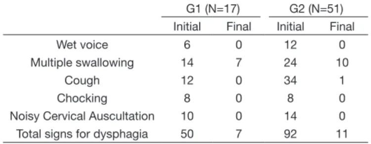
Table 2 presents dysphagia signs found in the groups.
One participant may have presented more than one risk sign
Chart 1. Description of the performed procedures
Procedure Training
Session 1
The participant is guided to say “CA” in high vocal intensity. 3 sets of 10 repetitions. 30-second interval between the sets.
The participant is instructed to perform vertical and horizontal movements with the tongue.
3 sets of 10 repetitions for each movement. 30-second interval between the sets.
It is instructed to the patient to perform maximum tongue protrusion
sustaining for three seconds. 3 sets of 5 repetitions. 30-second interval between the sets.
The participant is guided to say “RA” with sputum movement. 3 sets of 10 repetitions. 30-second interval between the sets.
Session 2
The participant is guided to say “CA” in high vocal intensity, while
performing shove movement with the hands in hooking. 3 sets of 10 repetitions. 30-second interval between the sets.
The participant is instructed to perform vertical and horizontal movements with the tongue, sustaining for three seconds at each movement.
3 sets of 10 repetitions for each movement. 30-second interval between the sets.
It is instructed to the patient to perform maximum tongue protrusion
sustaining for five seconds. 3 sets of 10 repetitions. 30-second interval between the sets.
The participant is guided to say “RA” with sputum movement. 3 sets of 10 repetitions. 30-second interval between the sets.
Session 3
The participant is guided to say “ZA”, extending “Z” the maximum
possible. 3 sets of 10 repetitions. 30-second interval between the sets.
The participant is guided to position the tongue in cheek, with occluded lips and perform counter resistance with the forefinger.
3 sets of 5 repetitions for each side. 30-second interval between the sets.
It is instructed to the patient to perform vocal glissando, with “I”,
starting with low pitched tone and evolving to high pitched. 3 sets of 5 repetitions. 30-second interval between the sets.
The participant is guided to hold the tongue among the teeth and
swallow saliva (tongue-hold). 3 sets of 5 repetitions. 30-second interval between the sets.
Session 4
The participant is guided to say ”Z”, the longest the possible. 3 sets of 10 repetitions. 30-second interval between the sets.
The participant is guided to position the tongue in cheek, with
occluded lips and perform counter resistance with the forefinger. 3 sets of 10 repetitions. 30-second interval between the sets.
It is instructed to the patient to perform vocal glissando, with “I”,
starting with low pitched tone and evolving to high pitched. 3 sets of 10 repetitions for each side. 30-second interval between the sets.
The participant is guided to hold the tongue among the teeth and
swallow saliva (tongue-hold). 3 sets of 10 repetitions. 30-second interval between the sets.
Table 1. Distribution of the individuals according to the level of ASHA NOMS scale
Groups ASHA NOMS
Initial level ≤ 5 Final level ≥ 6
G1 (N=17) 17 14
G2 (N=51) 51 42
Table 2. Comparison of the clinical signs of dysphagia, initial and final
G1 (N=17) G2 (N=51)
Initial Final Initial Final
Wet voice 6 0 12 0
Multiple swallowing 14 7 24 10
Cough 12 0 34 1
Chocking 8 0 8 0
Noisy Cervical Auscultation 10 0 14 0
Total signs for dysphagia 50 7 92 11
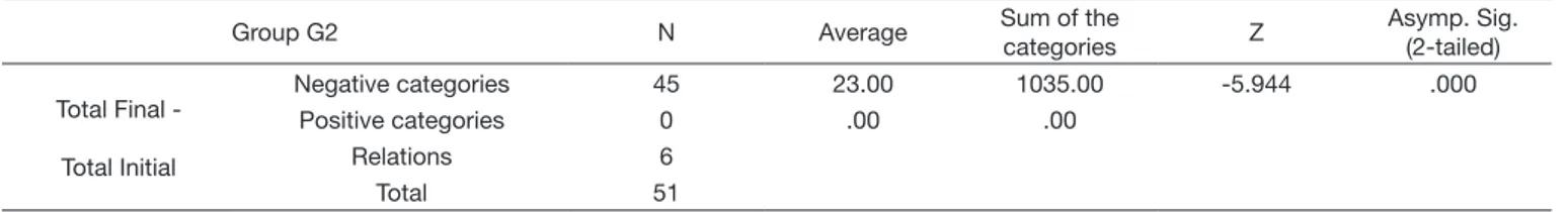
For the analyses of the initial and inal standard, it was
performed Wilcoxon statistic test, according to Tables 3 and 4 that
considered the presence of clinical signs of penetration/aspiration
as negative categories for the analyses; they were: wet voice,
multiple swallowing; cough; choking and noisy cervical
auscultation. When these signs were absent, these categories
were considered positive.
For G2 group there was statistically signiicant improvement.
For G1, the relation was insigniicant, despite the intense
change in ASHA NOMS scale, however, in this group there
was a reduced number of individuals due to the proile severity.
DISCUSSION
Dysphagia is a generalized condition and potentially fatal
that can arise from a variety of disorders that affect the neural,
motor and/or sensorial system parts that base the swallowing
function
(1).
Literature describes higher incidence of dysphagia at the
elderly population, resulting from muscle atrophy, cognitive
decline and increase of risk aspiration. In the studied group, the
average age was 57.1, very close to the populations internationally
described, which is 62 years old
(24).
Dysphagia patients are known for presenting lower frequency
of saliva swallowing than other non-dysphagia patients
(25).
The disuse of swallowing mechanism can reduce its cortical
representation and corresponds to a threat to functional recovery
at long term
(26).
Studies
(1,8)already describe that exercises in speciic muscle
groups, which view at swallowing, can have a signiicant
contribution for the function rehabilitation. The signiicant
reduction of clinic signs presence after performing the exercises
relects the functional improvement at feeding. This approach
can potentially result in life quality improvement and cost
reduction at the management of some dysphagia patients at the
coniguration of acute and chronic care.
Through the clinic evaluation performed, it was possible
to observe the cough as the most prevalent sign among the
participants. When analyzed by groups as the most prevalent, we
have veriied in G1 – multiple swallowing, followed by cough
sign; G2 – cough, followed by multiple swallowing. The found
results support the literature, which indicates as main predictor
for dysphagia: multiple swallowing, noisy cervical auscultation,
wet voice quality, cough and asphyxia
(27).
There are potential limitations in our study. It was not
performed objective swallowing evaluation to document silent or
subclinical aspirations, once, at the service where this research
was developed, there is not easy access to this parameter.
The application of this very protocol of exercises in different
dysphagia etiologies and different age ranges is necessary to
deepen the studies at the improvement of swallowing functionality.
Nowadays, there are more questions than answers on
how to approach more effective and eficiently the dysphagia
rehabilitation
(1), but through the use of a group of exercises it was
possible to observe consistent improvement at the swallowing
performance.
CONCLUSION
The objective of this research was to verify if, by means of
oropharyngeal exercises, it was possible to observe functional
improvement at swallowing in individuals identiied with risk
of oropharyngeal dysphagia. The performance of the protocol
of exercises showed to be eficient, allowing the reduction of
clinical signs presented for dysphagia, and improvement at
ASHA NOMS functional scale.
REFERENCES
1. Burkhead LM, Sapienza CM, Rosenbek JC. Strength-training exercise in dysphagia rehabilitation: principles, procedures, and directions for future research. Dysphagia. 2007;22(3):251-65. PMid:17457549. http://dx.doi. org/10.1007/s00455-006-9074-z.
2. Takizawa C, Gemmell E, Kenworthy J, Speyer R. A systematic review of the prevalence of oropharyngeal dysphagia in stroke, Parkinson’s Disease, Alzheimer’s Disease, Head Injury, and Pneumonia. Dysphagia. 2016;31(3):434-41. PMid:26970760. http://dx.doi.org/10.1007/s00455-016-9695-9.
3. Logemann J. Evaluation and treatment of swallowing disorders. Austin, TX: Pro-Ed.; 1983.
Table 3. Analyses of the initial and final signs of Group G1
Group G1 N Average Sum of the
categories Z
Asymp. Sig. (2-tailed)
Total Final -
Total Initial
Negative categories 16 8.50 136.00 -3.575 .000
Positive categories 16 .00 .00
Relations 1
Total 16
Table 4. Analyses of the initial and final signs of Group G2
Group G2 N Average Sum of the
categories Z
Asymp. Sig. (2-tailed)
Total Final -
Total Initial
Negative categories 45 23.00 1035.00 -5.944 .000
Positive categories 0 .00 .00
Relations 6
4. Jang HJ, Leigh JH, Seo HG, Han TR, Oh BM. Effortful swallow enhances vertical hyolaryngeal movement and prolongs duration after maximal excursion. J Oral Rehabil. 2015;42(10):765-73. PMid:26013277. http:// dx.doi.org/10.1111/joor.12312.
5. Park T, Kim Y. Effects of tongue pressing effortful swallow in older healthy individuals. Arch Gerontol Geriatr. 2016;66:127-33. PMid:27318884. http:// dx.doi.org/10.1016/j.archger.2016.05.009.
6. Yeates EM, Steele CM, Pelletier CA. Tongue pressure and submental surface electromyography measures during noneffortful and effortful saliva swallows in healthy women. Am J Speech Lang Pathol. 2010;19(3):274-81. PMid:20543016. http://dx.doi.org/10.1044/1058-0360(2010/09-0040). 7. Byeon H, Koh HW. Comparison of treatment effect of neuromuscular
electrical stimulation and thermal-tactile stimulation on patients with sub-acute dysphagia caused by stroke. J Phys Ther Sci. 2016;28(6):1809-12. PMid:27390421. http://dx.doi.org/10.1589/jpts.28.1809.
8. Hammer MJ, Jones CA, Mielens JD, Kim CH, McCulloch TM. Evaluating the tongue hold manoeuvre using high-resolution manometry and electromyography. Dysphagia. 2014;29(5):564-70. PMid:24969727. http:// dx.doi.org/10.1007/s00455-014-9545-6.
9. Fujiu-Kurachi M, Fujiwara S, Tamine K, Kondo J, Minagi Y, Maeda Y, et al. Tongue pressure generation during tongue-hold swallows in young healthy adults measured with different tongue positions. Dysphagia. 2014;29(1):17-24. PMid:23728858. http://dx.doi.org/10.1007/s00455-013-9471-z. 10. Shaker R, Easterling C, Kern M, Nitschke T, Massey B, Daniels S, et al.
Rehabilitation of swallowing by exercise in tube-fed patients with pharyngeal dysphagia secondary to abnormal UES opening. Gastroenterology. 2002;122(5):1314-21. PMid:11984518. http://dx.doi.org/10.1053/ gast.2002.32999.
11. Yoon WL, Khoo JK, Rickard Liow SJ. Chin tuck against resistance (CTAR): new method for enhancing suprahyoid muscle activity using a Shaker-type exercise. Dysphagia. 2014;29(2):243-8. PMid:24337867. http://dx.doi. org/10.1007/s00455-013-9502-9.
12. Oh JC. Effects of tongue strength training and detraining on tongue pressures in healthy adults. Dysphagia. 2015;30(3):315-20. PMid:25840786. http:// dx.doi.org/10.1007/s00455-015-9601-x.
13. Adams V, Mathisen B, Baines S, Lazarus C, Callister R. A systematic review and meta-analysis of measurements of tongue and hand strength and endurance using the Iowa Oral Performance Instrument (IOPI). Dysphagia. 2013;28(3):350-69. PMid:23468283. http://dx.doi.org/10.1007/s00455-013-9451-3.
14. Aida S, Takeishi R, Magara J, Watanabe M, Ito K, Nakamura Y, et al. Peripheral and central control of swallowing initiation in healthy humans. Physiol Behav. 2015;151:404-11. PMid:26253217. http://dx.doi.org/10.1016/j. physbeh.2015.08.003.
15. Aydogdu I, Tanriverdi Z, Ertekin C. Dysfunction of bulbar central pattern generator in ALS patients with dysphagia during sequential deglutition. Clin Neurophysiol. 2011;122(6):1219-28. PMid:21111672. http://dx.doi. org/10.1016/j.clinph.2010.11.002.
16. Van Roie E, Delecluse C, Coudyzer W, Bautmans I. Strength training at high versus low external resistance in older adults: effects on muscle volume, muscle strength, and force-velocity characteristics. Exp Gerontol.
2013;48(11):1351-61. PMid:23999311. http://dx.doi.org/10.1016/j. exger.2013.08.010.
17. Kraaijenga SAC, Molen L, Stuiver MM, Teertstra HJ, Hilgers FJM, Brekel MWM. Effects of strengthening exercises on swallowing musculature and function in senior healthy subjects: a prospective effectiveness and feasibility study. Dysphagia. 2015;30(4):392-403. PMid:25840788. http:// dx.doi.org/10.1007/s00455-015-9611-8.
18. ASHA: American Speech-Language-Hearing Association. National Outcomes Measurement System (NOMS): Adult Speech-Language Pathology User’s Guide. Rockville: ASHA; 2003.
19. Padovani AR, Moraes DP, Mangilli LD, Andrade CRF. Protocolo fonoaudiológico de avaliação do risco para disfagia (PARD). Rev Soc Bras Fonoaudiol. 2007;12(3):199-205. http://dx.doi.org/10.1590/S1516-80342007000300007.
20. Hammond CAS, Goldstein LB, Horner RD, Ying J, Gray L, Gonzalez-Rothi L, et al. Predicting Aspiration in Patients With Ischemic Stroke. Chest. 2009;135(3):769-77. PMid:19017886. http://dx.doi.org/10.1378/ chest.08-1122.
21. Hammond SC, Goldstein LB. Cough and aspiration of food and liquids due to oral-pharyngeal dysphagia: ACCP evidence-based clinical practice guidelines. Chest. 2006;129(1, Suppl):154S-68S. PMid:16428705. http:// dx.doi.org/10.1378/chest.129.1_suppl.154S.
22. Lim K, Lee H, Yoo J, Kwon Y. Effect of Low-Frequency rTMS and NMES on Subacute Unilateral Hemispheric Stroke With Dysphagia. Ann Rehabil Med. 2014;38(5):592-602. PMid:25379488. http://dx.doi.org/10.5535/ arm.2014.38.5.592.
23. Park DH, Chun MH, Lee SJ, Song YB. Comparison of swallowing functions between brain tumor and stroke patients. Ann Rehabil Med. 2013;37(5):633-41. PMid:24231855. http://dx.doi.org/10.5535/arm.2013.37.5.633. 24. Hoy M, Domer A, Plowman EK, Loch R, Belafsky P. Causes of dysphagia in a
tertiary care swallowing center. Ann Otol Rhinol Laryngol. 2012;122(5):335-8. PMid:23815051. http://dx.doi.org/10.1177/000348941312200502012;122(5):335-8. 25. Rajaei A, Ashtari F, Azargoon SA, Chitsaz A, Nilforoush MH, Taheri
M, et al. The association between saliva control, silent saliva penetration, aspiration, and videofluoroscopic findings in Parkinson’s disease patients. Adv Biomed Res. 2015;4:108. PMid:26261810.
26. Robbins J, Butler SG, Daniels SK, Gross RD, Langmore S, Lazarus CL, et al. Swallowing and dysphagia rehabilitation: translating principles of neural plasticity into clinically oriented evidence. J Speech Lang Hear Res. 2008;51(1):276-200. PMid:18230851. http://dx.doi.org/10.1044/1092-4388(2008/021).
27. Medeiros GC, Sassi FC, Mangilli LD, Zilberstein B, Andrade CRF. Clinical dysphagia risk predictors after prolonged orotracheal intubation. Clinics. 2014;69(1):8-14. PMid:24473554. http://dx.doi.org/10.6061/ clinics/2014(01)02.