Bacterial adhesion on conventional and self-ligating metallic brackets
after surface treatment with plasma-polymerized hexamethyldisiloxane
Rogerio Amaral Tupinambá1, Cristiane Aparecida de Assis Claro2, Cristiane Aparecida Pereira3, Celestino José Prudente Nobrega4, Ana Paula Rosifini Alves Claro1
Introduction: Plasma-polymerized film deposition was created to modify metallic orthodontic brackets surface properties in order to inhibit bacterial adhesion. Methods: Hexamethyldisiloxane (HMDSO) polymer films were deposited on conventional (n = 10) and self-ligating (n = 10) stainless steel orthodontic brackets using the Plasma-Enhanced Chemical Vapor Deposition (PECVD) radio frequency technique. The samples were divided into two groups according to the kind of bracket and two subgroups after surface treatment. Scanning Electron Microscopy (SEM) analysis was performed to assess the presence of bacterial adhesion over samples surfaces (slot and wings region) and film layer integrity. Surface roughness was assessed by Confocal Interferometry (CI) and surface wettability, by goniometry. For bacterial adhesion analysis, samples were exposed for 72 hours to a Streptococcus mutans solution for biofilm formation. The values obtained for surface roughness were analyzed using the Mann-Whitney test while biofilm adhesion were assessed by Kruskal-Wallis and SNK test. Results: Significant statistical differences (p < 0.05) for surface roughness and bacte-rial adhesion reduction were observed on conventional brackets after surface treatment and between conventional and self-ligating brackets; no significant statistical differences were observed between self-ligating groups (p > 0.05). Conclusion: Plasma-polymerized film deposition was only effective on reducing surface roughness and bacterial adhesion in conventional brackets. It was also noted that conventional brackets showed lower biofilm adhesion than self-ligating brackets despite the absence of film.
Keywords:Orthodontic brackets. Bacterial adhesion. Hexamethyldisiloxane.
1 Universidade Estadual Paulista, Faculdade de Engenharia, Departamento de
Materiais (Guaratinguetá/SP, Brasil).
2 Universidade de Taubaté, Faculdade de Odontologia, Departamento de
Ortodontia (Taubaté/SP, Brasil).
3 Universidade Estadual Paulista, Faculdade de Odontologia, Departamento de
Microbiologia e Imunologia, Instituto de Ciência e Tecnologia (São José dos Campos/SP, Brasil).
4 New York University, College of Dentistry, Linhart Continuing Education
Program (New York/NY, EUA).
Submitted: June 08, 2016 - Revised and accepted: March 14, 2017
DOI: https://doi.org/10.1590/2177-6709.22.4.077-085.oar
How to cite: Tupinambá RA, Claro CAA, Pereira CA, Nobrega CJP, Claro APRA. Bacterial adhesion on conventional and self-ligating metallic brackets after surface treatment with plasma-polymerized hexamethyldisiloxane. Dental Press J Orthod. 2017 July-Aug;22(4):77-85.
DOI: https://doi.org/10.1590/2177-6709.22.4.077-085.oar
» The authors report no commercial, proprietary or financial interest in the products or companies described in this article.
Contact address: Rogerio Tupinambá
Rua Quero-quero, 161, Socorro – Pindamonhangaba/SP – Brasil CEP: 12.424-880 – E-mail: [email protected]
Introdução: a deposição de filme de polímero a plasma foi criada para modificar as propriedades de superfície dos braquetes ortodônticos metálicos, com o intuito de inibir a adesão bacteriana. Métodos: filmes finos de polímero de hexametildisiloxano (HMDSO) foram depositados em braquetes ortodônticos de aço inoxidável convencionais (n = 10) e autoligáveis (n = 10), utilizan-do a técnica de radiofrequência PECVD (Plasma-Enhanced Chemical Vapor Deposition). As amostras foram divididas em dois grupos, de acordo com o tipo de braquete, e dois subgrupos após o tratamento de superfície. A microscopia eletrônica de varredura (MEV) foi realizada para avaliar a presença de adesão bacteriana sobre as superfícies das amostras (região de ranhura horizontal e aletas) e a integridade da camada de filme. A Interferometria Confocal (CI) avaliou a rugosidade, e a molhabilidade superficial foi avaliada por goniometria. Para análise de adesão bacteriana, as amostras foram expostas durante 72 horas a uma solução de Streptococcus mu-tans, para formação de biofilme. Os valores obtidos para a rugosidade da superfície foram analisados pelo teste de Mann-Whitney, enquanto a adesão do biofilme foi avaliada pelos testes de Kruskal-Wallis e SNK. Resultados: observaram-se diferenças estatis-ticamente significativas (p <0,05) para a rugosidade superficial e redução da adesão bacteriana em braquetes convencionais após o tratamento da superfície, e entre braquetes convencionais e autoligáveis. Não foram observadas diferenças estatísticas significativas entre os grupos autoligáveis (p > 0,05). Conclusão: a deposição de polímero a plasma só foi efetiva na redução da rugosidade su-perficial e adesão bacteriana em braquetes convencionais. Observou-se, também, que os braquetes convencionais apresentaram menor adesão ao biofilme do que os braquetes autoligáveis, apesar da ausência de filme.
INTRODUCTION
The amount of sites available to bacterial growth in oral cavity increases in the presence of orthodontic appliances. Therefore, surfaces traditionally unlikely to develop caries become areas with high incidence
of these lesions.1 In the absence of prophylactic
mea-sures, initial carious lesions (white spots) develop
within four weeks.2 Caries have a multifactorial trait,
being dependent on the presence of the host (teeth), diet (sugars intake), cariogenic bacteria (biofilm) and the biofilm’s development stage, to be sustained. Thus, the absence of one of these factors may inhibit
disease installation and its development.3
Streptococcus mutans is one of the main responsible microorganisms for tooth decay. Its installation is de-pendent on vertical and/or horizontal contamination
and it has acidogenic and acidophilic characteristics4.
Their carbohydrate degradation metabolism produc-es acids that demineralize dental surfacproduc-es, leading to
the development of cavities.5
Bacterial adhesion has special characteristics and depends on direct biofilm interaction with the sub-strate surface to which it relates. The presence of
ac-quired enamel pellicle,6 surface energy,7 roughness,8
and wettability9 play critical roles in this interaction,
not only interfering in adhesion properties, but also in the characteristics of biofilm formation.
The recent use of self-ligating brackets in ortho-dontics has contributed for a reduction in plaque ac-cumulation, when compared to conventional
brack-ets ligated by elastics,10 but the performance of these
brackets may be impaired by salivary calculus accu-mulation over the sliding clip mechanism and into
the horizontal archwire slot.11
Plasma polymerization and plasma surface treat-ment techniques have been developed as antibacterial coatings, such as silver-platinum coating for
orth-odontic appliances12 and TiO
2 nanotubes surfaces
coated with magnetron-sputtered Ag, for dental
ap-plications.13 Recent literature reports strategies in
which plasma polymers have also been used as reser-voirs loaded with antibacterial agents which are
sub-sequently released,14-17 served as a diffusion barrier to
control the release rate of these agents — as sealing
agents for carbon nanotubes filled with medication,18
and as functional coatings for connecting antibiotic
or bacteriostatic molecules.19
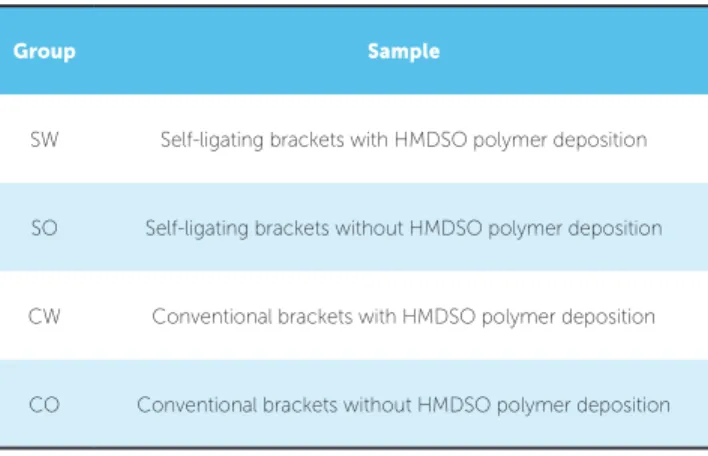
Table 1 - Groups division and names according to surface treatment and bracket type.
This practice, quite common in implantology20
serves as inspiration for orthodontics, where polymer films deposition on orthodontic brackets surface can also be applied to reduce biofilm adhesion and the
risk of enamel lesions during treatment.21
Due to its characteristics of producing nontoxic
ilms,22 having high vapor pressure at room temperature
and being of ease commercial availability,23 the
plas-ma-polymerized hexamethyldisiloxane (HMDSO)
deposition has been largely employed in industry24 and
as biomaterial coating25. HMDSO ilm presents
sev-eral organic components and large hydrophobicity.26
These characteristics have particular importance in
in-hibiting the adherence of Streptococcus mutans.27
The objective of the present study was to compare the performance of the HMDSO film as a surface roughness reduction method and as an inhibiting bar-rier for biofilm formation in two kinds of orthodontic brackets, comparing its efficiency with non-treated brackets. The null-hypothesis was that the presence of HMDSO film would not interfere on biofilm for-mation on the two kind of brackets.
MATERIAL AND METHODS
The study was composed by two groups (n = 34), equally divided by type of bracket, and in two sub-groups, according to ilm deposition (Table 1).
Group Sample
SW Self-ligating brackets with HMDSO polymer deposition
SO Self-ligating brackets without HMDSO polymer deposition
CW Conventional brackets with HMDSO polymer deposition
For the HMDSO deposition and subsequent microbiological tests, the following upper right central incisors metallic brackets (Roth pre-scription, Morelli™, Sorocaba/SP, Brazil) were used: SLI (self-ligating) and Roth Max (con-ventional), both types manufactured by powder injection molding (PIM). The chemical com-position of brackets used was C = 0.20% (max), Cr = 16.5 - 17.5%, Mo = 3.0 - 3.5%, Si = 1.0% (max)
and Ni = 0.90% (max). Self-ligating
brack-ets also presented a sliding clip composed by Ni = 54.5 - 57.0%W and Ti = 45.5 - 43.0%W, which plays an interactive hole in this bracket system.
Each subgroup was composed by 17 samples, in which 2 were chosen to undergo scanning elec-tron microscopy (SEM, Zeiss, model EVO LS15), 5 were used for confocal interferometry (CI) (Leica, DCM3D) and 10 for biofim formation analysis.
Scanning Electron Microscopy (SEM) was used to visualize the surface after polymer deposition. For assessing bacterial adhesion presence over samples surface (slot and wings areas) and back-scattered electrons (BSE) mode assessment of the film layer integrity over the samples, self-ligating brackets had their clips opened. CI was carried out to evaluated surface roughness (Ra, arithmetic av-erage, and Rq, root mean squared) on the wings region. Prior to SEM and CI analysis, 7 brackets of each group were fixed for one hour in 2.5% glutar-aldehyde, and dehydrated at various concentrations of ethanol (10%, 25%, 50%, 75% and 90% for 20 min, and 100% for 1 hour). To complete samples drying, they were incubated in a bacteriological
in-cubator for 48 h at 37oC.
HMDSO films deposition system
Film deposition was performed by Plasma-En-hanced Chemical Vapor Deposition radio frequency method (RF PECVD), using the hexamethyldisilox-ane (HMDSO) monomer as gas source. HMDSO gas plasma was RF-excited, operating in a 13.56 Hz
frequency, and pressure level of 60 x 10-2 Torr, with
20 W power, for 15 minutes. These parameters were chosen as the most adequate by a series of previous tests performed by the authors with different pow-er and time ppow-eriods. The film was deposited on the outer and inner surfaces of the brackets, while their
bases remained facing the surface of the deposition electrode plate on the bottom of the reactor. All self-ligating brackets had their clips closed during the de-position process.
An automated goniometer (Ramé-Hard Instru-ment Co., Advanced Goniometer model 300-F1) was used for evaluating the wettability and surface energy on a stainless steel sheet presenting the same chemical composition of the brackets.
The thickness of HMDSO film was measured in an optical microscope and interferometer (Leica,
DCM3D) on a glass slide substrate,28 which was set
inside the plasma reactor amongst the samples during the deposition process and prepared to present a step on the surface between the film and the substrate.
Biofilm formation
Streptococcus mutans, ATCC #35688 strains were used for biofilm formation, as proposed by Pereira
et al.29 Initially, the strains were seeded in Mitis
Sali-varius to verify its purity, and incubated at 37oC for
24 hours. Standardized suspensions were then
pre-pared with relative optical density at 106 cells/mL.
For this, the strains were grown on brain heart infu-sion agar (BHI, Difco, Detroit, USA) and incubated
at 37oC for 24 hours. After incubation, the growth
was suspended in sterile saline (0.9% NaCl) and the number of cells in each suspension calculated in a spectrophotometer (B582, Micronal, São Paulo, Brazil). Each bracket group was placed in a 12-well plate (Costar Corning, New York, USA) with 1.5 ml BHI plus 5% sucrose, and inoculated with 0.1 mL of bacterial suspension. The samples were incubated at
37oC for 72 hours for the formation of biofilms.
Af-ter this period, the brackets with biofilms were rinsed with phosphate-buffered saline (PBS) and subjected to an orbital shaker for 5 minutes (Solab, Piracicaba, Brazil) for removing non-adhered cells. After proper dilutions, 100-µL aliquots were plated on BHI agar
in Petri dishes. The plates were incubated at 37oC for
72 hours. After that period of incubation, the colony forming units per milliliter (CFU/mL) were quanti-fied on the plates showing from 30 to 300 colonies, and the obtained numbers were converted to their
corresponding logarithm (log10 CFU/mL). Statistical
The Kolmogorov-Smirnov test was carried out to analyze data normal distribution of four groups con-sidering the following assumptions:
» Null hypothesis (H0) = Analyzed data
distribu-tion is similar to a standard normal distribudistribu-tion.
» Alternative hypothesis (Ha) = Analyzed data
distri-bution is not similar to a standard normal distridistri-bution. The Mann-Whitney (surface roughness between subgroups) and Kruskal-Wallis (bacterial adhesion) tests assessed possible differences among groups, con-sidering the following hypotheses:
» Null hypothesis (H0) = Analyzed data are
simi-lar among the groups.
» Alternative hypothesis (Ha) = There is at least
one group different from other groups.
To identify all the possible differences among groups, SNK multiple comparison test was applied.
RESULTS
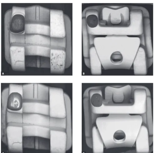
Micrographs of two groups of brackets in BSE mode can be observed in Figure 1, where diferences
in the atomic number of the surface layer molecules create contrast variations, highlighting possible depo-sition defects. Surface visual analysis of the four brack-ets subgroups — self-ligating and conventional, treated and not treated — exhibited a uniform layer deposition pattern on treated samples ater polymer deposition.
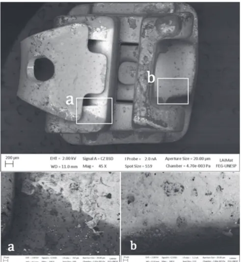
A fairly uniform layer was observed at samples C and D compared to the conditions on untreated sam-ples A and B (Fig. 1). This evidences the proper de-position of the polymer over the outer surface of the brackets. Despite that, SEM analysis of a SW group bracket with its clip open had shown the presence of surface areas without proper coating (Fig 2).
Surface roughness(Ra and Rq) median values of the wings region of the samples are presented in Table 2.
Data analysis by Mann-Whitney test has shown no signiicant statistical diferences on surface roughness
re-duction between subgroups SW and SO for Ra (p = 0.222)
and Rq (p = 0.151). Signiicant statistical surface
rough-ness reduction for Ra (p = 0.008) and Rq (p = 0.008) was
observed between subgroups CW and CO.
A
C
B
D
Figure 1 - Top view of the brackets surfaces:
The presence of the polymer in an uniform layer, with regular thickness, was assessed by the interferometry of the coated layer ilm thickness on the glass slide, ater the deposition of plasma-polymerized HMDSO (Table 4).
Micrographs observed in Figure 3 show the worst
areas of S. mutans bioilm formation on all subgroups,
on the slots (Figs 3A and 3B, lateral view) and wings re-gions (Figs 3C and 3D, frontal view). In SW (Fig 3A) and SO (Fig 3C) groups a greater bacterial adherence was observed, both in the wings and slot regions, when visu-ally compared to CW (Fig 3B) and CO (Fig 3D) groups, even in the presence of the HMDSO ilm (Fig 3).
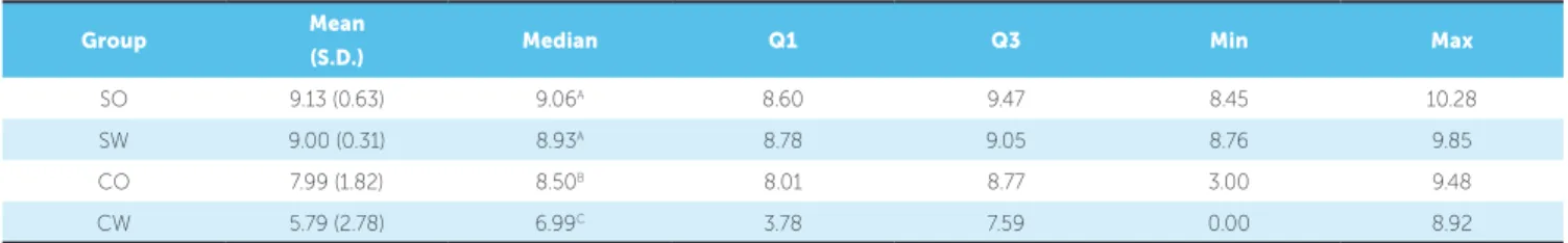
Descriptive statistics of the colony forming units (CFU) data can be observed in Table 5.
The results of Kolmogorov-Smirnov tests identi-ied that SO and CW groups showed normal
distribu-tion (p > 0.05) and that SW and CO groups were not
normally distributed (p < 0.05), whereas the level of
5% signiicance was adopted, so non-parametric sta-tistical analyzes were chosen to analyze the data.
Figure 2 - SEM micrograph shows the pro-jected shadow impressed by the presence of the NiTi clip, creating an interface between the surfaces with deposition and non-deposition (45x magnification). Interface on the wing re-gion (a) and on bracket base region (b) (500x magnification).
Group Ra (median) Rq (median)
CO 3.760 4.963
CW 1.623 2.192
SO 1.749 2.296
SW 1.649 2.177
(p < 0.05)
Table 2 - Confocal Interferometry (CI) evaluation of surface roughness (Ra and Rq).
The result of the Kruskal-Wallis test indicated the presence of at least one group different from the other
groups (H = 18.56, p < 0.001). SNK multiple
com-parison test indicated greater accumulation of bacte-ria in groups SO and SW, and smaller accumulation
in CW group (p < 0.05) (Table 5).
DISCUSSION
HMDSO polymer ilms have been largely used as
surface treatment for biomaterials25 and in dentistry as
carriers for drug delivering16 and for surface wettability
modiication.22 This paper used the HMDSO polymer
to observe its anti-adherence features on bioilm
forma-tion, beneiting from its hydrophobicity characteristics26
and layer thickness, without any associated anti-bacte-rial substance, once in orthodontics, brackets remain in oral cavity for a period of time that varies from 12 to 30 months and most drug delivering properties occur in a brief period of time, varying from 24 to 48 hours. Sur-face properties play an essential hole in bacterial adhe-sion: hidrophobic and high surface energy types of sur-face, as the ones observed in the treated samples, tend to diicult such interaction.
Table 3 - Angles and surface energy measurement observed by the goniometer on the different samples.
Table 4 - Film thickness obtained from plasma deposition parameters.
Sample Contact angle
Water Diiodomethane Polar component Dispersive component Total surface energy Control 79.22 ± 4.43 48.45 ± 0.45 9.49 ± 1.90 36.16 ± 0.22 45.65 ± 1.91 Deposition 1 105.78 ± 1.08 81.67 ± 1.51 2.87 ± 0.44 20.37 ± 0.67 23.24 ± 0.60 Deposition 2 99.87 ± 0.22 72.67 ± 0.47 3.84 ± 0.10 24.40 ± 0.22 28.24 ± 0.18 Deposition 3 103.42 ± 1.15 83.36 ± 0.80 3.92 ± 0.42 19.65 ± 0.34 23.57 ± 0.46
Sample Film thickness (nm)
1 10.36
2 11.3
3 11.25
Mean 10.97
Figure 3 - SEM micrographs of the brackets slots (A, B) and wings region (C, D) demonstrate the bacterial adhesion in both regions of all groups (4,500x magnification).
A
C
B
Table 5 - Descriptive statistics: mean, standard deviation (SD), median, first quartile (Q1), third quartile (Q3), minimum and maximum values, for the values of colony counts (log).
Group Mean
(S.D.) Median Q1 Q3 Min Max
SO 9.13 (0.63) 9.06A 8.60 9.47 8.45 10.28
SW 9.00 (0.31) 8.93A 8.78 9.05 8.76 9.85
CO 7.99 (1.82) 8.50B 8.01 8.77 3.00 9.48
CW 5.79 (2.78) 6.99C 3.78 7.59 0.00 8.92
RF-PECVD technique was chosen for its capabil-ity of controlling film thickness and wettabilcapabil-ity char-acteristic according to determined deposition param-eters, producing uniform and very thin films. Film thickness measurement was facilitated by its
deposi-tion on a glass slide surface,28 once bracket surface
irregularities made interferometry assessment im-practicable. SEM analysis on BSE mode have shown a uniform polymer surface deposition. The deposition process of HMDSO polymer allows samples coating in all dimensions of space, and assures high rates of
deposition.26 Despite that, the base of the brackets did
not receive any coating, once it was facing down the base of the reactor.
The base of the bracket itself had little influence on the amount of adhered biofilm on total bracket surface, once most of the brackets surface and its harsh design are determined by its outer surface and not by the bracket base.
Brackets brand and type choice was based on their composition and manufacturing process, and the same brand was chosen for both brackets types; only brackets geometry and presence of the NiTi clip var-ied in the self-ligation groups.
Literature review10,30 demonstrated that
conven-tional brackets have shown less plaque buildup than self-ligating brackets. Besides that, self-ligating brack-ets as well as ceramic brackbrack-ets provide the formation of a much more pathogenic bioilm, due to their small proportion of anaerobic over aerobic bacteria in colony
forming units (CFU).30 Therefore methods for
reduc-ing plaque adherence, especially on self-ligatreduc-ing brack-ets are very welcome.
Roughness tests were performed only between subgroups once the main concern was the influence of the presence of polimer coating. The low influence
of polymer deposition on biofilm formation in SW and SO groups was directly related to the small re-duction on surface roughness and highly detailed and complex geometry of this type of bracket, which had
a large influence on bacterial adherence.10 Surface
roughness influence overrules surface free energy and
promotes plaque formation and maturation.7
The presence of a NiTi clip as the ligation element in the bracket implies in the presence of an internal longitudinal tunnel, just above the base of the brack-et, for accommodating the NiTi clip. This feature creates a broad contact surface in this type of bracket and results in a perfect site for bacterial adherence and proliferation. In the conventional CO and CW groups, which have a much simpler geometry, this tunnel is not present.
This characteristic, in addition to the untreat-ed surface creatuntreat-ed by the clip shadow, resultuntreat-ed in more bacterial adherence on SW group than on CO. Even in the absence of the polymer coating, CO group has shown lower rates of bacterial adhesion than SO and SW groups, thus demonstrating that the impact of the external geometry and polymer deposi-tion flaws on biofilm formadeposi-tion was bigger than the presence of the polymer.
HMDSO ilm deposition on group SW had serious issues, as shown in Figure 2. This experiment was held with the NiTi clip closed, following the same methods conducted by other authors on their experiments on
bacterial adhesion.10,30-32 This option was made because
present, but this would modify ilm characteristics due
to the risk of sputtering of the initial layer.25 Future
experiments should perform ilm deposition on self-li-gating brackets with the clip open, or without the clip, to observe possible improvement in polymer deposi-tion on inner surfaces of the samples.
Findings have shown that CW group had the best results on reducing bacterial adherence over all groups, demonstrating that the polymer ilm played a fun-damental role in reducing surface roughness and the rate of bacterial adhesion. This very positive outcome unravels a new perspective to surface treatment in or-der to reduce bacterial adhesion in orthodontics, and set HMDSO polymer as a feasible choice for metallic orthodontic brackets coating.
This beneit, not yet commercially available, was also
veriied by Demling et al,21 who conducted an in vivo
study comparing bacterial adhesion in two convention-al brackets, one of them coated with plasma-polymer-ized polytetraluoroethylene. Despite being presented as a case report, the authors observed a much smaller amount of bacteria adhered to the surface of brackets with ilm deposition (4.0 ± 3.6%), compared to brackets without ilm (22.2 ± 5.4%). This primer study can serve as a reference for the indication of plasma surface treat-ment of brackets in orthodontics.
For the special characteristics observed, the au-thors acknowledge the necessity of improvement in self-ligating brackets polymer deposition method, as well as the use of different bracket brands, with differ-ent external geometries and ligation features. The de-velopment of a bacterial adherence inhibiting method is essential for this kind of bracket, once biofilm pres-ence, besides enamel lesions, can also interfere in the opening mechanism of the clip and in the proper in-teraction between the bracket and the archwire, lead-ing to mechanical and operational problems durlead-ing the orthodontic treatment.
Besides this, further studies shall be performed regard-ing possible friction reduction between treated brackets and diferent archwires, due to the surface roughness re-duction observed in polymer deposition groups.
CONCLUSIONS
The results observed in this paper allow the fol-lowing conclusions concerning the HMDSO poly-mer deposition on orthodontic brackets:
» It was more efective in reducing surface roughness
and S. mutans bioilm formation in conventional brack-ets, for their less rugged and more suitable external ge-ometry, which enabled a better polymer ilm deposition.
» Conventional brackets showed lower bioilm
adhe-sion than self-ligating brackets despite the absence of ilm.
» An improved deposition method has to be
em-ployed in self-ligating brackets so that film deposi-tion and hence, the reducdeposi-tion in bacterial adhesion and surface roughness, may be more effective.
Author contributions
1. Tufekci E, Dixon JS, Gunsolley JC, Lindauer SJ. Prevalence of white spot lesions during orthodontic treatment with ixed appliances. Angle Orthod. 2011 Mar;81(2):206-10.
2. Ogaard B, Rølla G, Arends J. Orthodontic appliances and enamel
demineralization. Part 1. Lesion development. Am J Orthod Dentofacial Orthop. 1988 July;94(1):68-73.
3. Selwitz RH, Ismail AI, Pitts NB. Dental caries. Lancet. 2007 Jan 6;369(9555):51-9. 4. Fournier A, Payant L, Bouclin R. Adherence of Streptococcus mutans to
orthodontic brackets. Am J Orthod Dentofacial Orthop. 1998 Oct;114(4):414-7. 5. García-Godoy F, Hicks MJ. Maintaining the integrity of the enamel
surface: The role of dental bioilm, saliva and preventive agents in enamel demineralization and remineralization. J Am Dent Assoc. 2008 May;139 Suppl:25S-34S.
6. Rosan B, Lamont RJ. Dental plaque formation. Microbes Infect. 2000 Nov;2(13):1599-607.
7. Quirynen M, Bollen CM. The inluence of surface roughness and surface-free energy on supra and subgingival plaque formation in man. J Clin Periodontol. 1995 Jan;22(1):1-14.
8. Teughels W, Van Assche N, Sliepen I, Quirynen M. Efect of material characteristics and/or surface topography on bioilm development. Clin Oral Implants Res. 2006 Oct;17 Suppl 2:68-81.
9. Jansen B, Kohnen W. Prevention of bioilm formation by polymer modiication. J Ind Microbiol. 1995 Oct;15(4):391-6.
10. Garcez AS, Suzuki SS, Ribeiro MS, Mada EY, Freitas AZ, Suzuki H. Bioilm retention by 3 methods of ligation on orthodontic brackets: A microbiologic and optical coherence tomography analysis. Am J Orthod Dentofacial Orthop. 2011 Oct;140(4):e193-8.
11. Almeida GA, Ursi W. Ortodontia com braquetes autoligáveis. In: Almeida MR. Ortodontia clínica e biomecânica. Maringá: Dental Press; 2010. p. 561-73. 12. Ryu HS, Bae IH, Lee KG, Hwang HS, Lee KH, Koh JT, et al. Antibacterial efect
of silver-platinum coating for orthodontic appliances. Angle Orthod. 2012 Jan;82(1):151-7.
13. Uhm SH, Lee SB, Song DH, Kwon JS, Han JG, Kim KN. Fabrication of bioactive, antibacterial TiO2 nanotube surfaces, coated with magnetron sputtered
Ag nanostructures for dental applications. J Nanosci Nanotechnol. 2014 Oct;14(10):7847-54.
14. Grössner-Schreiber B, Griepentrog M, Haustein I, Müller WD, Lange KP, Briedigkeit H, et al. Plaque formation on surface modiied dental implants. Clin Oral Implants Res. 2001 Dec;12(6):543-51.
15. Monteiro DR, Gorup LF, Takamiya AS, Ruvollo-Filho AC, Camargo ER, Barbosa DB. The growing importance of materials that prevent microbial adhesion: antimicrobial efect of medical devices containing silver. Int J Antimicrob Agents. 2009 Aug;34(2):103-10.
16. Cortizo MC, Oberti TG, Cortizo MS, Cortizo AM, Fernández Lorenzo de Mele MA. Chlorhexidine delivery system from titanium/polybenzyl acrylate coating: evaluation of cytotoxicity and early bacterial adhesion. J Dent. 2012 Apr;40(4):329-37.
REFERENCES
17. Denes AR, Somers BE, Wong ACL, Denes F. 12-Crown-4–Ether and Tri(ethylene glycol) Dimethyl–Ether plasma-coated stainless steel surfaces and their ability to reduce bacterial bioilm deposition. J App Pol Sci. 2001;81(14):3425-38. 18. Simovic S, Losic D, Vasilev K. Controlled drug release from porous materials by
plasma polymer deposition. Chem Commun (Camb). 2010 Feb 28;46(8):1317-9 19. Su W, Wang S, Wang X, Fu X, Weng J. Plasma pre-treatment and TiO2 coating
of PMMA for the improvement of antibacterial properties. Surfac Coat Technol. 2010;205:465-9.
20. Yoshinari M, Oda Y, Kato T, Okuda K. Inluence of surface modiications to titanium on antibacterial activity in vitro. Biomaterials. 2001 July;22(14):2043-8. 21. Demling A, Elter C, Heidenblut T, Bach FW, Hahn A, Schwestka-Polly R,
et al. Reduction of bioilm on orthodontic brackets with the use of a polytetraluoroethylene coating. Eur J Orthod. 2010 Aug;32(4):414-8. 22. Krasteva NA, Toromanov G, Hristova KT, Radeva EI, Pecheva EV, Dimitrova RP,
et al. Initial biocompatibility of plasma polymerized hexamethyldisiloxane ilms with diferent wettability. J Physics. 2010;253:1-7.
23. Pfeifer J. Hexamethyldisiloxane. In: Encyclopedia of reagents for organic synthesis. New York: L. Paquette; 2004. p. 234-47.
24. Chaiwong C, Rachtanapun P, Sarapirom S, Boonyawan D. Plasma polymerization of hexamethyldisiloxane: investigation of the efect of carrier gas related to the ilm properties. Surf Coat Tech. 2013;229:12-7.
25. Nikiforov A, Xiaolong D, Qing X, Cvelbar U, DeGeyter N, Morent R, et al. Non-thermal plasma technology for the development of antimicrobial surfaces: a review. J Physics D: Appl Physics. 2016;49:204002-9.
26. Förch R, Chifen AN, Bousquet A, Khor HL, Jungblut M, Chu LQ, et al. Recent and expected roles of plasma-polymerized ilms for biomedical applications. Chem Vap Dep. 2007;13(6-7):280-94.
27. Lassen B, Holmberg K, Brink C, Carl A, Olsson A. Binding of salivary proteins and oral bacteria to hydrophobic and hydrophilic surfaces in vivo and in vitro. Col Pol Sci. 1994;272(9):1143-50.
28. Kim SW, Kim GH. Thickness-proile measurement of transparent thin-ilm layers by white-light scanning interferometry. Appl Opt. 1999 Oct 1;38(28):5968-73. 29. Pereira CA, Eskelson E, Cavalli V, Liporoni PC, Jorge AO, do Rego MA.
Streptococcus mutans bioilm adhesion on composite resin surfaces after diferent inishing and polishing techniques. Oper Dent. 2011 May-June;36(3):311-7.
30. van Gastel J, Quirynen M, Teughels W, Pauwels M, Coucke W, Carels C. Microbial adhesion on diferent bracket types in vitro. Angle Orthod. 2009 Sept;79(5):915-21.
31. Baka ZM, Basciftci FA, Arslan U. Efects of 2 bracket and ligation types on plaque retention: A quantitative microbiologic analysis with real-time polymerase chain reaction. Am J Orthod Dentofacial Orthop. 2013 Aug;144(2):260-7.