Assessment of mandibular advancement surgery
with 3D CBCT models superimposition
Alexandre Trindade Simões da Motta*, Felipe de Assis Ribeiro Carvalho**, Lúcia Helena Soares Cevidanes***, Marco Antonio de Oliveira Almeida****
Objectives: To assess surgery and short-term post-surgery changes in the position of the
con-dyles, rami and chin after mandibular advancement. Methodology: Pre-surgery (T1), 1 week post-surgery (T2), and 6 week post-surgery (T3) CBCT scans were acquired for 20 retrog-nathic patients with short or normal face height. 3D models were built and superimposed through a fully automated voxel-wise method using the cranial base of the pre-surgery scan as reference. Anatomic regions of interest were selected and analyzed separately. Within-subject surface distances between T1-T2, T2-T3, and T1-T3 were computed. Color-coded maps and semi-transparent display of overlaid structures allowed the evaluation of displacement direc-tions. Results: After an antero-inferior chin displacement with surgery in all cases (>4 mm in 87.5%), 25% of the patients showed some kind of posterior movement (< 3 mm), and 69% showed an superior movement after splint removal. Comparing T1-T3, an antero-inferior (87.5% of the cases) or only antero-inferior (12.5%) displacement was observed (>4 mm in 80%). Considering all directions of displacement, the surface distance differences for the condyles and rami were small: 77.5% of the condyles moved <2 mm with surgery (T1-T2), and 90% moved <2 mm in the short-term (T2-T3) and in the total evaluation (T1-T3), while the rami showed a <3 mm change with surgery in 72.5% of the cases, and a <2 mm change in 87.5% (T2-T3) and in 82% (T1-T3). Conclusions: Expected displacements with surgery were observed and post-surgery changes suggested a short-term adaptive response toward recovery of condyle and ramus displacements. The changes on the chin following splint removal sug-gested an acceptable adaptation, but with considerable individual variability.
Abstracts
Keywords: Cone-beam computed tomography. Tridimensional image. Computer-assisted surgery. Computer simulation. Orthodontics. Oral surgery.
* DDS, MS, PhD. Professor, Department of Orthodontics, Fluminense Federal University (UFF/Niterói, RJ, Brazil). Visiting Scholar, University of North Caro-lina at Chapel Hill (UNC); CAPES scholarship 382705-4.
** DDS, MS. PhD Student, Department of Orthodontics, State University of Rio de Janeiro (UERJ). Visiting Scholar (UNC). *** DDS, MS, PhD. Professor, Department of Orthodontics (UNC/Chapel Hill).
INTRODUCTION
The application of three-dimensional (3D) imaging of the craniofacial complex in prospec-tive controlled trials can be considered one of the major advances in the search for a complete di-agnosis, treatment planning and outcome evalua-tion.6,19 Investigations using cone-beam computed
tomography (CBCT)15 in Orthodontics and
Oral-Maxillofacial Surgery have shown that this new tool can improve the identification of different patterns of rami and condyles positioning after or-thognathic surgery.4
The hierarchy of stability of surgical proce-dures shows that mandibular advancement is a highly stable procedure in patients with short or normal face height and less than 10 mm advance-ment. It means that there is more than a 90% chance of less than 2 mm change at landmarks and almost no chance of more than 4 mm change during the first post-surgical year. Surgical repo-sitioning of the chin via lower border osteotomy, the most prevalent adjunctive procedure, also is highly stable and predictable.1,17,18
Otherwise, long-term condylar resorption af-ter mandibular advancement and relapse into anterior open bite have been reported as poten-tial clinical problems in some patients.9 It would
occur in 5-10% of patients who have surgery to advance the mandible, but a long-term increase in mandibular length due to residual growth at the condyles is as likely as a decrease because of resorption.13,20 It seems that changes occur in
condylar position after bilateral sagittal split ra-mus osteotomy (BSSO). This however does not seem to be associated with factors such as amount of advancement, degree of proximal rotation, and shape of the mandible.12 Both rotation and
transverse displacement of the condyles related to ramus surgery have been described,2 and the
extent to which mandibular structures rotations/ displacements and bone remodeling/resorption contribute to post-treatment instability are poorly understood at present.
Problems in the identification of anatomic points in conventional cephalometrics have been considered a significant source of error when ob-taining important craniofacial measurements.9
Previous studies based in standardized norms and bidimensional (2D) representation of 3D changes could not answer many questions related to fac-tors influencing treatment response and skeletal remodeling.11 The complex movements during
surgery for dentofacial deformities clearly need to be assessed in three dimensions to improve stabil-ity and reduce symptoms of temporomandibular joint disorder after surgery.5
Orthodontic application of novel 3D imaging techniques include virtual model superimposition to verify growth, treatment changes and stability, soft tissue analysis, and computer simulation of surgical procedures. Previous studies4,5,7 used 3D
models superimpostion in Class III patients, but it is the first experiment to assess surgical changes in Class II mandibular advancement cases through this method.
The purpose of this study was to evaluate the displacements at the chin, condyles and rami be-tween pre-surgery, post-surgery, and splint remov-al through 3D models superimposition.
METHODOLOGY
were also excluded. All patients agreed in having CBCTs in different phases of treatment as it was described in the experimental protocol approved by UNC ethical committee.
The scans were taken one week before surgery (T1), one week after surgery (T2), to assess chang-es with the surgical procedure, and six weeks after surgery (T3), immediately after splint removal to assess short-term adaptive changes. The imaging protocol involved for 16 patients a 36-second head CBCT scanning with a field of view of 230 x 230 mm. All CT scans were acquired with the patient in centric occlusion with the NewTom 3G (Aperio Services LLC, Sarasota, FL, 34236). Four of those patients had at least 1 scan done with the NewTom 9000 (Aperio Services LLC, Sarasota, FL) which has a smaller field of view (F.O.V.), therefore, the chin was not included. A primary reconstruction of tomographic slices was done im-mediately after the exam, with a 0.3 x 0.3 x 0.3 mm voxel resolution. Differently from 2D radio-graphic projection acquisitions that require head positioning standardization, tomographic slices could be obtained without such standards as the whole 3D craniofacial complex was captured.
The following method for showing quantita-tive changes at multiple locations in orthosurgery cases has been validated and used since 20054.
Be-sides, a thorough description of this method is in press in this journal.14
Image archives of 60 scans were exported in DICOM (Digital Imaging and Communication in Medicine) format, converted to GIPL format, and reformatted to a voxel dimension of 0.5 x 0.5 x 0.5 mm, thus reducing size, requiring less com-puting capacity and time during the experiment.
Image segmentation of the anatomic struc-tures of interest and the 3D graphic rendering were done by using the InsightSNAP software (http://www.itksnap.org/), allowing navigation through the slices in axial, sagittal and coronal planes. From a set of more than 300 axial, lat-eral and anteroposterior cross-sectional slices for
each image acquisition, a 3D model of the cranial base, maxilla and mandible was built, allowing navigation between voxels in the volumetric im-age and the 3D graphics with zooming, rotating and panning.
The pre-surgery and post-surgery models were registered based on the cranial base, since this structure is not altered by surgery. A fully automated voxel-wise rigid registration meth-od was used through the Imagine free software (http://www.ia.unc.edu/dev/download/). The soft-ware compares both images using the intensity of gray scale for each voxel of the region, so that the pre-surgical cranial base (T1) was used as ref-erence for the superimposition of post-surgery models (T2 and T3).
To control the cropping for a quantitative analysis of regions of interest such as the condyles, the posterior border of the rami and chin, the 3D models at different time points of each patient were combined also with the Imagine tool. Ana-tomic references were used to determine selection regions: (1) the chin was defined by the long axis of the lower canines post-surgery; (2) the poste-rior border was defined by a plane tangent to the anterior contour of the condyles and parallel to the posterior border of the rami; and (3) the in-ferior limit of the condylar region was defined by the interface of the posterior border cut.
The combined cutted structures were then separated as independent 3D objects, keeping their spacial positioning inside the original tomog-raphy. Each region of interest of each phase was prepared for pair comparisons, and then analyzed separately with MeshValmet (http://www.ia.unc. edu/dev/download/), where measurements of the surface distances between two different time points within the same subject allowed quantifi-cation of rami, condyles and chin displacements that accompanied mandibular surgery.
condyle (n = 20). Surface distances values were obtained and a visual interpretation of the dis-placements was made in all directions: superior, inferior, lateral, medial, anterior, and posterior. A graphical display of superimposed structures was done through color-coded maps, based in the direction and magnitude of each region dis-placement. Basically, they indicate inward (blue) or outward (red) displacement between overlaid structures, with negative or positive values, re-spectively. The absence of surgical displacement is indicated by the green color code.
Figure 1 shows the displacement of a right con-dyle between T1 and T2. The superior, posterior and medial displacements are shown in red, op-posed to blue anterior and lateral surfaces show-ing inward movements. The visualization through semi-transparencies allowed an additional display of the surgical changes, clearly identifying the
lo-calization, magnitude and direction of mandibular displacements.
Descriptive statistics is illustrated in graph-ics of direction and amount of displacement for each anatomic region. Graphics of direction of displacement show, in number of subjects, move-ments in the anterior, posterior, inferior, superior, lateral and medial directions between T1-T2, T2-T3 and T1-T3, and situations without move-ment. Graphics of amount of displacement were based on categorizations of structural movements (intervals in millimeters), between the same time points, and also shown in number of subjects. Val-ues were differently categorized for the chin, rami and condyles, regarding each region movement magnitude. The results between T1-T2 show displacements with surgery; T2-T3 the response after splint removal; and T1-T3 the overall short-term effects of mandibular advancement surgery.
FIGURE 1 - Visualization of a right condyle displaced posterior-superior-medially between T1 and T2; Left: Color coded maps indicate outward displace-ments in red and inward displacedisplace-ments in blue. Right: Semi-transparencies with T1 in solid white and T2 in transparent red (A = anterior; P = posterior).
A P
A P
medial view posterior view
medial view posterior view
-+
+ + + + + +
RESULTS
All the cases showed an anterior-inferior dis-placement of the chin with surgery (Graph 1), over 4 mm in 87.5% of the cases (14 patients be-tween T1-T2). A vertical improvement was one of the treatment objectives in reduced facial height retrognathic patients. Comparing T2-T3, 25% (4 patients) showed some posterior movement (< 3 mm), and 69% (11 patients) an anterior-superior movement. Vertical changes with counterclock-wise rotation and additional anterior movement in most of the cases might be related to splint removal (acrylic thickness). The comparison be-tween T1-T3 showed a chin advancement in all cases, over 4 mm in more than 80% (13 patients), but 12.5% (2 patients) did not present an inferior displacement (Graph 2).
Condyles tended to move latero-posteriorly with surgery (T1-T2), but equally distributed in the vertical direction, compared to a medio-ante-rior movement with a slight supemedio-ante-rior tendency in most of the cases between T2-T3 (Graphs 3 and 4). Surprisingly, post-surgical (T2-T3) amount of displacement was comparable to changes with sur-gery, suggesting an important adaptive response. Considering all directions of displacement, the
surface distance differences were small: 77.5% of the condyles moved less than 2 mm with surgery (T1-T2), while 90% moved less than 2 mm in the short-term between T2-T3 (Graphs 5 and 6). An observation between T1-T3 showed a medio-pos-terior-superior displacement resultant, with 90% of the condyles presenting less than 2 mm, and only 5% less than 3 mm of displacement.
The rami exhibited a tendency to outward movements with surgery (inferior-latero-posteri-or), especially lateral, and inward (superior-me-dio-anterior) displacements after splint removal (T2-T3), also suggesting an important adaptive response, but smaller than 2 mm (Graps 7 and 8). Results suggested that the rami were pushed back with surgery, followed by a muscular adaptation toward the original position. Less than 3 mm of change with surgery was observed in 72.5% of the cases, and less than 2 mm of change in 87.5% of the cases after 6 weeks (Graphs 9 and 10). The resultant surgical and adaptive changes showed a superior-latero-anterior tendency (T1-T3), with less than 2 mm in 82% of the rami. Clinically important displacements were observed with surgery in specific cases, where four patients pre-sented displacements over 4 mm.
GRAPH 1 - Direction of displacement of the chin between T1-T2, T2-T3, and T1-T3, expressed in number of subjects (n = 16).
Number of Subjects 0 2 4 6 8 10 12 14 16
Ant. None Post. None Sup.
T1 - T2 T2 - T3 T1 - T3 Direction of Displacement
CHIN
Inf.
GRAPH 2 - Categorization of the amount of displacement of the chin be-tween T1-T2, T2-T3 e T1-T3, expressed in number of subjects (n = 16).
Number of Subjects 0 2 4 6 8 10 12 14 16
<2 2-4 4-6
T1 - T2 T2 - T3 T1 - T3 Amount of Displacement (mm)
CHIN
DISCUSSION
This paper presents skeletal changes in man-dibular advancement cases immediately after surgery and in a short-term observation at splint removal through the method of tridimensional CBCT models superimposition. Previous avail-able data for comparison have been all based in 2D analysis1,2,9,10,12,13,16,17,18,20 using lateral
cephalo-metric projections and observer-dependent refer-ence points. Class III studies have been carried out with this method, comparing surgical results and stability between combined maxillary advance-ment and mandibular set-back versus maxillary surgery only.5,7,8
The visibility of superimposed 3D models and surface distances measurements clearly showed the localization, magnitude and direction of man-dibular rotations with surgery, allowing A-P, trans-verse and vertical quantification of rami and con-dyles movements. The method used in this study is based in an automatic voxel-wise rigid registra-tion that compares the cranial base grayscale be-tween two CTs, thus avoiding landmark location on complex curving structures.4,5 This
methodol-ogy uses relatively low dose radiation, advanced imaging methods, and public software specifically developed for research purposes.
Our results were comparable to mandibular GRAPH 3 - Direction of displacement of the left condyle between T1-T2,
T2-T3, and T1-T3, expressed in number of subjects (n = 20).
GRAPH 4 - Direction of displacement of the right condyle between T1-T2, T2-T3, and T1-T3, expressed in number of subjects (n = 20).
GRAPH 5 - Categorization of the amount of displacement of the left con-dyle between T1-T2, T2-T3 and T1-T3, expressed in number of subjects (n = 20).
GRAPH 6 - Categorization of the amount of displacement of the right condyle between T1-T2, T2-T3 and T1-T3, expressed in number of sub-jects (n = 20).
Number of Subjects 02 4 6 8 10 12 14 16 18 20
T1 - T2 T2 - T3 T1 - T3 Direction of Displacement
LEFT CONDYLE
Ant. None Post. Inf. None Sup. Lat. None Med.
Number of Subjects 02 4 6 8 10 12 14 16 18 20
T1 - T2 T2 - T3 T1 - T3
Amount of Displacement (mm) Amount of Displacement (mm) LEFT CONDYLE
<1 1-2 >2 <1 1-2 >2
Number of Subjects 02 4 6 8 10 12 14 16 18 20
T1 - T2 T2 - T3 T1 - T3 Direction of Displacement
RIGHT CONDYLE
Ant. None Post. Inf. None Sup. Lat. None Med.
Number of Subjects 02 4 6 8 10 12 14 16 18 20
advancement literature findings1,17 and had the
potential to highlight the associations between alterations in the three planes of space. It also brings into discussion questions regarding bone remodeling and relapse tendency, which will be better observed in long-term follow-ups. This kind of surgical procedure is considered highly stable1,17,18 and in this short-term observation 25%
of the patients showed some posterior movement at the chin after splint removal. These few cases showed less than 3 mm displacements between T2-T3 (Graph 2), and when compared to super-impositions T1-T2 and T1-T3 a high similarity in the color maps (area and intensity of red
sur-faces corresponding to mandibular advancement) could be visually observed, as well as in the dif-ference between solid white and transparent red in the semi-transparencies (Fig 2).
Chin anterior displacement was associated to clockwise rotation and lower face height increase in some patients of the sample (Fig 3), what can be a goal of treatment in patients presenting skel-etal Class II with normal or horizontal pattern. It could be even observed more vertical than hori-zontal changes in cases with small overjet, and the influence of pre-surgery deep overbite should be also considered as a factor of vertical sliding dur-ing BSSO technique. A clockwise rotation is likely GRAPH 10 - Categorization of the amount of displacement of the right ramus between T1-T2, T2-T3 and T1-T3, expressed in number of subjects (n = 20).
GRAPH 7 - Direction of displacement of the left ramus between T1-T2, T2-T3, and T1-T3, expressed in number of subjects (n = 20).
GRAPH 8 - Direction of displacement of the right ramus between T1-T2, T2-T3, and T1-T3, expressed in number of subjects (n = 20).
GRAPH 9 - Categorization of the amount of displacement of the left ra-mus between T1-T2, T2-T3 and T1-T3, expressed in number of subjects (n = 20).
Number of Subjects 02 4 6 8 10 12 14 16 18 20
T1 - T2 T2 - T3 T1 - T3 Direction of Displacement
LEFT RAMUS
Ant. None Post. Inf. None Sup. Lat. None Med.
Number of Subjects 02 4 6 8 10 12 14 16 18 20
T1 - T2 T2 - T3 T1 - T3
Amount of Displacement (mm) Amount of Displacement (mm) LEFT RAMUS
<2 2-3 >3 <2 2-3 >3
Number of Subjects 02 4 6 8 10 12 14 16 18 20
T1 - T2 T2 - T3 T1 - T3 Direction of Displacement
RIGHT RAMUS
Ant. None Post. Inf. None Sup. Lat. None Med.
Number of Subjects 02 4 6 8 10 12 14 16 18 20
to be more stable since muscles inserted at the posterior part of the mandibular body and ramus tend to be shortened, whereas counter-clockwise rotation is associated to muscular stretching. The acrylic splint seems to play an important role in the maintenance of occlusal height immediately after surgery, and visual comparisons of model superimpositions between 1 week and 6 weeks post-surgery showed a counter-clockwise rotation tendency with bite closure and an additional ante-rior movement in some cases (Fig 4).
This comparisons also suggested smaller post-surgery short-term changes at the chin when the mandibular advancement was truly horizontal, but the sample was not big enough to be divided into subgroups to statistically assess vertical dif-ferences. Excellent stability16 was found for
man-dibular deficiency patients treated by means of mandibular advancement only or associated to maxillary impaction, with less than 2 mm changes during the first postsurgical year, and larger (>2 mm) changes due to skeletal remodeling from one to five years post-surgery. Also, interesting findings showed that in patients with anterior open bite pre-treatment a long-term relapse is rarely ob-served, even if skeletal changes due to the verti-cal growth pattern occur, because compensatory
changes in the incisors position tend to maintain the overbite or even increase it. A future com-parison with a group of vertical Class II patients submitted to maxillary impaction and mandibular advancement can tridimensionally highlight out-come and stability differences, as well as condylar remodeling and resorption following clockwise or counterclockwise mandibular rotations. It is im-portant to mention that genioplasty, performed in seven patients of this sample, is a highly stable adjunctive procedure,1,17,18 therefore it can
ad-equately improve the quality of treatment if some A-P or vertical additional change is needed.
Rami and condyles displacements were consid-ered clinically relevant, even so were concentrated under 3 mm and 2 mm, respectively, and chin po-sition was kept stable. Actually, the results sug-gest that an important recovery tendency toward preoperative positions occurred after splint was removed and mandibular function resumed. Oth-erwise, some cases showed a larger displacement in the proximal segment that could be maintained even after a long-term follow-up. Since condylar displacements concentrated under 2 mm, clinical effects are questionable and long-term follow-ups are needed to show if changes were small enough to allow adaptive remodeling without side effects.
FIGURE 2 - Superimpositions between pre-surgery to immediately post-surgery with splint in place (A), immediately post-surgery to splint removal (B), and pre-surgery to splint removal (C). Some posterior (inward) movement of the chin is noted in B, shown by the blue color code. It can also be noted by the comparison between different area and density of red surfaces representing the anterior (outward) displacement in A and C. Still, the resultant superim-position in C shows an acceptable maxillomandibular relation at splint removal, considered a short-term stability. The right ramus shows a slight lateral movement in A (outward), a recovery tendency in B (inward), and green surfaces in C confirming the adaptive response.
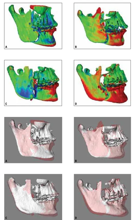
FIGURE 3 - Above: Color maps of surface distances between T1 and T2 models for 4 different patients showing an antero-inferior displacement of the chin post-surgery with the splint in place. Patients A and C showed prevalence of vertical displacement as shown in red at the anterior alveolar process and inferior surface of the symphysis. Patients B and D showed a vertical change with a remarkable chin advancement, shown by red mandibular anterior surfaces. Patient D also showed backward ramus displacement. Below: Semi-transparencies of these cases with superimposed T1 (solid white) and T2 (transparent red).
A B
C D
A B
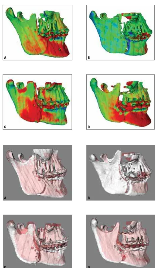
FIGURE 4 - Short-term follow-up (T2-T3) of cases shown in the previous figure. After splint removal, an antero-superior tendency of movement was noted in most of the cases in the sample. Additional anterior mandibular displacement can be seen in color maps red surfaces and in semi-transparencies differ-ences between white (T2) and red (T3) models. A rami comparison between Figures 3 and 4 (T1-T2 x T2-T3) suggests a recovery of surgery displacements after six weeks, with a medio-lateral movement in cases B and C, and antero-posterior movement in case D.
A
C
B
D A
C
B
The direction of displacement assessment showed individual variability, especially at the condyles, but more remarkable direction tenden-cies at the rami. Since the sample was small, it was hard to establish straight relations between the surgical procedure and skeletal response, so some directional tendencies were suggested. Condyles latero-posterior and rami latero-posterior-inferior changes between T1-T2 could result from pres-sure during the BSSO, fragment sliding and rigid fixation, followed by anterior and medio-anterior-superior displacements between T2-T3 as a recovery toward initial positions due to mus-cular stretch.
An increase in inter-gonial and inter-ramus width was verified through lateral and frontal cephalometric radiographs.2 The present study
did not use measurements between bilateral land-marks, but superimpositions between different time-points that allowed the visualization and quantification of an outward movement tendency following surgery, thus agreeing to previous data. Studies using submentovertex x-rays showed that structural rotations around 5 to 10 degrees nor-mally occur and do not necessarily lead to func-tional problems. Also, the amount of rotation ap-parently decrease with time due to remodeling process. Otherwise, pain and in some cases restric-tion of movement could be associated to an ante-rior or medial condyle displacement.1,16
The analysis of the displacement at the chin showed an obvious anterior direction with sur-gery and showed slight variation after splint
removal. Since the sample included only sym-metrical patients, lateral movements were not expected. A correlation between changes at the condyles and chin suggest that an anterior dis-placement of the former after splint removal could partially cause, together with the counter-clockwise rotation and bite closure, some ante-rior movement of the chin at T3, and the short-term stability of the correction.
Future studies with larger samples, long-term follow-ups and improved methodologies will probably be able to show additional find-ings regarding bone remodeling and resorption. Therefore, the sample used in this study is being monitored with progress CBCTs which will allow stability assessment.
CONCLUSION
Superimposition of 3D virtual surface models allowed clear visualization and quantification of outcomes of mandibular advancement surgery. Important displacements with surgery were ob-served in the rami and condyles, but changes at splint removal suggested an adaptive response to-ward recovery of pre-surgery positions, especially the medio-lateral movement of the rami. The changes on the chin after six weeks suggested an overall acceptable adaptation, but with consider-able individual variability.
Submitted in: July 2009
Contact address
Alexandre Trindade Simões da Motta
Av. das Américas, 3500 - Bloco 7/sala 220 - Ed. Hong Kong 3000 CEP: 22.640-102 - Barra da Tijuca - Rio de Janeiro/RJ
E-mail: [email protected]
1. Bailey LJ, Cevidanes LH, Proffit WR. Stability and predictability of orthognathic surgery. Am J Orthod Dentofacial Orthop. 2004 Sep;126(3):27-37.
2. Becktor JP, Rebellato J, Becktor KB, Isaksson S, Vickers PD, Keller EE. Transverse displacement of the proximal segment after bilateral sagittal osteotomy. J Oral Maxillofac Surg. 2002 Apr;60(4):395-403.
3. Bettega G, Cinquin P, Lebeau J, Raphaël B. Computer-assisted orthognathic surgery: clinical evaluation of a mandibular condyle repositioning system. J Oral Maxillofac Surg. 2002 Jan;60(1):27-34.
4. Cevidanes LH, Bailey LJ, Tucker GR Jr, Styner MA, Mol A, Phil-lips CL, Proffit WR, Turvey T. Superimposition of 3D cone-beam CT models of orthognathic surgery patients. Dentomaxillofac Radiol. 2005 Nov;34(6):369-75.
5. Cevidanes LH, Bailey LJ, Tucker SF, Styner MA, Mol A, Phillips CL, Proffit WR, Turvey T. Three-dimensional cone-beam com-puted tomography for assessment of mandibular changes after orthognathic surgery. Am J Orthod Dentofacial Orthop. 2007 Jan;131(1):44-50.
6. Cevidanes L, Motta AT, Styner M, Phillips C. 3D imaging for early diagnosis and assessment of treatment response. In: McNamara Jr JA, Kapila SD. Early orthodontic treatment: is the benefit worth the burden? 33rd Annual Moyers Symposium, Ann Arbor; 2007. 44:305-21.
7. Cevidanes L, Oliveira A, Phillips C, Motta AT, Styner M, Tyndall D. Three dimensional short-term mandibular displacements following Class III surgery. J Dent Res. (Spec Iss A): 1827, 2007. 8. Cevidanes L, Oliveira A, Phillips C, Motta AT, Styner M. 3D
assessment of surgical changes at splint removal for Class III patients. Book of Abstracts of the AAO 107th Annual Session; 2007. 15:32.
9. De Clercq CA, Neyt LF, Mommaerts MY, Abeloos JV, De Mot BM. Condylar resorption in orthognathic surgery: a retrospective study. Int J Adult Orthod Orthognath Surg. 1994;9(3):233-40.
10. Epker BN, Wylie GA. Control of the condylar-proximal mandibular segments after sagittal split osteotomies to
REFERENCES
advance the mandible. Oral Surg Oral Med Oral Pathol. 1986 Dec;62(6):613-7.
11. Harrell WE Jr, Hatcher DC, Bolt RL. In search of anatomic truth: 3-dimensional digital modeling and the future of orthodontics. Am J Orthod Dentofacial Orthop. 2002 Sep;122(3):325-30. 12. Harris MD, Van Sickels JE, Alder M. Factors influencing
condy-lar position after the bilateral sagittal split osteotomy fixed with bicortical screws. J Oral Maxillofac Surg. 1999 Jun;57(6):650-4. 13. Mihalik CA, Proffit WR, Phillips C. Long-term follow-up of Class
II adults treated with orthodontic camouflage: a comparison with orthognathic surgery outcomes. Am J Orthod Dentofacial Orthop. 2003 Mar;123(3):266-78.
14. Motta AT, Carvalho FR, Oliveira A, Cevidanes LHS, Almeida MA. Superposição automatizada de modelos tomográficos tridimensionais em cirurgia ortognática. Dental Press J Orthod. in press.
15. Mozzo P, Procacci C, Tacconi A, Martini PT, Andreis IA. A new volumetric CT machine for dental imaging based on the cone-beam technique: preliminary results. Eur Radiol. 1998;8(9):1558-64.
16. Proffit WR, Bailey LJ, Phillips C, Turvey TA. Long-term stability of surgical open-bite correction by Le Fort I osteotomy. Angle Orthod. 2000 Apr;70(2):112-7.
17. Proffit WR, Turvey TA, Phillips C. Orthognathic surgery: a hierarchy of stability. Int J Adult Orthod Orthognath Surg. 1996;11(3):191-204.
18. Proffit WR, Turvey TA, Phillips C. The hierarchy of stability and predictability in orthognathic surgery with rigid fixation: an update and extension. Head Face Med. 2007 Apr; 30(3):21. 19. Sarment DP. Dental applications for cone-beam computed
tomography. In: McNamara Jr JA, Kapila SD. Digital radiog-raphy and three-dimensional imaging. 32nd Annual Moyers Symposium; 2006. 43: 43-58.