r e v b r a s o r t o p . 2016;51(5):597–600
SOCIEDADE BRASILEIRA DE ORTOPEDIA E TRAUMATOLOGIA
w w w . r b o . o r g . b r
Case
Report
Tibial
shaft
fracture
and
ankle
injury
–
Case
report
夽
Caio
Zamboni
a,∗,
Felipe
Augusto
Garcez
de
Campos
a,
Noel
Oizerovici
Foni
a,
Rafael
Carboni
Souza
a,
Ralph
Walter
Christian
a,
Marcelo
Tomanik
Mercadante
a,baIrmandadedaSantaCasadeMisericóridiadeSãoPaulo,SãoPaulo,SP,Brazil
bFaculdadedeCiênciasMédicasdaSantaCasadeSãoPaulo,SãoPaulo,SP,Brazil
a
r
t
i
c
l
e
i
n
f
o
Articlehistory:
Received14August2015 Accepted31August2015 Availableonline10August2016
Keywords:
Internalfracturefixation Jointinstability
Ankle
a
b
s
t
r
a
c
t
Theauthorsreportonacaseoftibialshaftfractureassociatedwithankleinjury.Theclinical, radiologicalandsurgicalcharacteristicsarediscussed.Assessmentofassociatedinjuriesis oftenoverlookedandtheseinjuriesarehardtodiagnose.Whentorqueoccursinthelower limb,theanklebecomessusceptibletosimultaneousinjury.Itisessentialtomakecareful assessmentbasedonclinical,radiographic,intraoperativeandpostoperativecharacteristics inordertoattainfunctionalrecovery.
©2016SociedadeBrasileiradeOrtopediaeTraumatologia.PublishedbyElsevierEditora Ltda.ThisisanopenaccessarticleundertheCCBY-NC-NDlicense(http:// creativecommons.org/licenses/by-nc-nd/4.0/).
Fratura
diafisária
da
tíbia
e
lesão
do
tornozelo
–
Relato
de
caso
Palavras-chave:
Fixac¸ãointernadefraturas Instabilidadearticular Tornozelo
r
e
s
u
m
o
Osautoresrelatamumcasodefraturadiafisáriadetíbiaassociadoàlesãodotornozelo. Ascaracterísticasclínicas,radiológicasecirúrgicassãodiscutidas.Aavaliac¸ãodelesões associadassãomuitasvezesnegligenciadasededifícildiagnóstico.Quandoumtorqueno membroinferiorocorre,otornozeloficasuscetívelaumalesãosimultânea.Éessencialuma avaliac¸ãocuidadosabaseadanoaspectoclínico,radiográfico,intraepós-operatóriopara recuperac¸ãofuncional.
©2016SociedadeBrasileiradeOrtopediaeTraumatologia.PublicadoporElsevierEditora Ltda.Este ´eumartigoOpenAccesssobumalicenc¸aCCBY-NC-ND(http:// creativecommons.org/licenses/by-nc-nd/4.0/).
Introduction
Thefirst description ofthe associationof diaphyseal tibial fractureswithadditionalankle injurywasmadebyWeber1
夽
StudyconductedattheSantaCasadeSãoPaulo,DepartamentodeOrtopediaeTraumatologia,GrupodeCirurgiadoTrauma,São Paulo,SP,Brazil.
∗ Correspondingauthor.
E-mail:caiozamboni@hotmail.com(C.Zamboni).
in1972.Asthetibialinjuryisvisibleandobvious,apotential associatedankleinjurymaybeneglected.Distaltibiofibular syndesmosisinstabilitymayleadtosubluxationofthetalus. Onceundiagnosed,anklearthrosismaytakeplaceevenifthe
http://dx.doi.org/10.1016/j.rboe.2016.08.003
598
rev bras ortop.2016;51(5):597–600Fig.1–Imageoftheopendiaphysealfractureofthelegwithnoevidenceofinjuryintheanklejoint.
treatmentforthediaphysealtibialfracturehasprovided excel-lentreduction,stabilization,andconsolidation.2
Clinical
report
Male patient, 28 years old, involved in a motorcycle acci-dentwithopenfractureoftherightleg(Fig.1)classifiedas GustiloIIIA.3Heunderwentcleaning,woundlavage, debride-mentoftissuelesions,andtransarticularexternalfixationof thelegbonesattheanklejointaimingtoprovidelocaldamage control.
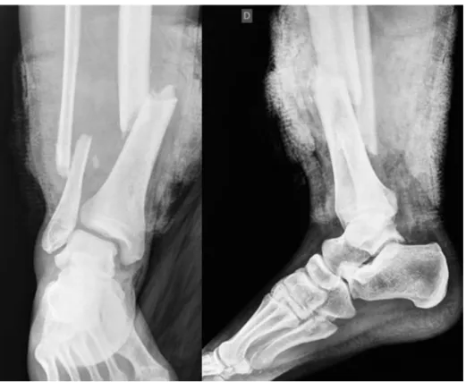
Onthesixthdayafterthetrauma,withtheimprovement of the soft-tissueenvelopeof the left leg, internalfixation wasperformedwithalockedintramedullarynailforthetibial fracture. Duringthesurgicalprocedure,anteriordislocation oftheanklejointwasobserved(Fig.2AandB).Theauthors opted foropenreductionandinternalfixationofthe ankle fracture-dislocationwithplateandscrewsinthefibula; insta-bilityofthe anklesyndesmosiswasprovenwithapositive Cotton4test.Weassociatedthestabilizationofthetibiofibular mortise using apositioning screw through the fibular cor-texandthelateralcortexofthetibia,proximaltothedistal tibiofibularjoint,withoutadirectapproachtotheformer.Final
rev bras ortop.2016;51(5):597–600
599
Fig.3–(A)Radiographyafteropenreductionandinternalfixationofthefibulawithasuprasyndesmotictricorticalscrewon anteroposteriorview.(B)AxialplaneCTscanshowingtheincongruityofthedistaltibiofibularjointanditssubluxation.
radiographyrevisionshowedajointincongruityofthefibula withamultifragmentaryfracturelineonthefirst postopera-tiveday.Axialcomputedtomographyoftheankleconfirmed theexistenceofaprevioustibiofibular subluxation(Fig. 3A andB).
Duringhospitalization,sixdaysaftersurgery,thepatient underwentthethirdsurgicalprocedureaimingopen reduc-tion,ligamentreconstruction,andstabilizationofthedistal tibiofibularjointafterrevisionofpreviousosteosynthesis.
During surgery, avulsion ofthe articular capsule ofthe anklejointatthesuperiorandlateralregionswasobserved, aswellasoftheanteriortibiofibularligament(Fig.4).The sur-gicaltechniqueadoptedwastheremovalofthefibularplate inordertoreviewthereductionoftheanklefracture.
Underdirectview,theauthorsproceededtothereduction ofthedistalfibulatothefibularnotchofthetibia,with tempo-raryfixationofthejointusingsmoothKirschnerwire(Fig.5). Oncethejointreductionwasattested,theosteosynthesisof thefibularfracturewasaddressedusingalongreconstruction plate,asabonedefectwasobservedintheareaofthefracture fragmentation.Autologouscancellousbonegraftwasusedfor defectreconstruction.Sutureofthearticularcapsule,anterior tibiofibularligamentandanteriorsyndesmosiswasexecuted
Fig.4–Intraoperativeclinicalpictureshowingtorn articularcapsuleandanteriortibiofibularligament.
attheankle.Forprotectingtheligamentreconstruction,two positioningscrewswereapplied.Thelockedintramedullary nailusedforthetreatmentofthediaphysealtibialfractures didnotpresentcomplicationsandwasmaintained.
Activeanklemotionwasstimulatedimmediatelyafterthe procedure.Eightweeksafterthelastprocedure,the position-ingscrewsofthedistaltibiofibularjointwereremoved.
Currently, the patient presents no pain complaints and walkswithfullweightbearingandwithoutassistance.The rangeofmotionattheendoftreatmentwas20◦ofdorsiflexion
and40◦ofplantarflexion,symmetricaltothecontralateral.
The final functional evaluation was excellent, totaling 99 pointsintheAmericanOrthopaedicFootandAnkleSociety (AOFAS)Ankle-HindfootScalequestionnaire.
600
rev bras ortop.2016;51(5):597–600Discussion
Diaphysealtibialfracturesassociatedwithligamentinjuriesin theanklepresentahighpotentialforinstabilityandareoften neglected,posingariskofcomplicationssuchasthe develop-mentofsecondaryosteoarthritisandunfavorablefunctional performancewhenundiagnosedanduntreated.2,5
For the reported patient, intraoperative evaluation with Cottontestprovedsufficientandefficienttoassessthe syn-desmosis,waivingtheneedforotherteststoproveligament incompetence.6
Control radiographies after the second surgery showed subluxationoftheankle,althoughthiswasnotobservedin thefinalmomentsofsurgery.ACTscanoftheankleconfirmed thepoorreductionandmadeitpossibletoidentifythe inade-quateroutetakenbythepositioningscrew.Itwasshowntobe effectiveauxiliarytool,notonlyforelucidatingpossible diag-nosticuncertaintiesintheassessmentoftheaxialsections,7 butalsoforhelpingtoplanthedefinitivetreatment.
Inliterature,intraoperative temporary stabilization with Kirschnerwiresisanalternativetoclamps,withareported decrease in the rates ofpoor reductionsofneglected syn-desmosis,asthetechniqueemployedinthethirdoperative procedure in this case.7 Adequate reduction of the distal tibiofibular joint hasbeen shown to be animportant pro-gnosticfactorforfunctional outcomeinankleinjurieswith syndesmoticinjury.7–9
Thepossiblevariationsintheuse ofpositioningscrews, whichprotecttheligamentrepairsoftheanklejointduring freemovement,arethesubjectofdebate.Thenumber, diam-eter,andfixationinthreeorfourcorticesarealsostilldebated inthe literature.10 Inthepresentpatient,theoptiontouse two3.5-mmscrewswasduetothepoorqualityofthefixation ofthefirstscrewinstalled,themostdistalbeingtricortical, whichreachedtothetibiainthemetaphysealareawherethe lateralcortexwasthin.
Theliterature suggests that there are no differences in outcomebetweenpatientswhohaveorhavenotundergone removalofthesupra-syndesmoticpositioningscrewsbefore weightbearinggait.11 Withthe present patient,the screws wereremovedaftereightweeks.
Intheshort-termfollow-up,jointfunctionisadequateand symmetric,andthepatientpresentsnocomplaints.Wethink ofnoreasonforadiverseevolutionofanklefractureswhenthe physiologicalandbiomechanicalrelationshipsaremaintained afterconsolidation.
Conclusion
Theliteraturedescribesthat ankleinjuriesassociatedwith diaphysealfracturesofthetibiaarefrequentlyneglecteddue toitsdifficultdiagnosis,suchasinthepresentcase.Careful pre-andintraoperativeassessmentsbaseduponboth clini-calpracticeandradiography arerequired;thepossibilityof associatedinjuryshouldbekeptinmind.
Conflicts
of
interest
Theauthorsdeclarenoconflictsofinterest.
r
e
f
e
r
e
n
c
e
s
1.WeberBG.Injuryoftheanklejoint.Huber:BernStuttgart Wien;1972.
2.GeorgiadisGM,EbraheimNA,HoefllingerMJ.Displacementof theposteriormalleollusduringintramedullarytibianailing.J Trauma.1996;41(6):1056–8.
3.GustiloRB,AndersonJT.Preventionofinfectioninthe treatmentofonethousandandtwenty-fiveopenfracturesof longbones:retrospectiveandprospectiveanalyses.JBone JointSurgAm.1976;58(4):453–8.
4.CottonFJ.Fracturesandjointdislocations.Philadelphia:WB Saunders;1910.
5.StuermerEK,StuermerKM.Tibialshaftfractureandankle jointinjury.JOrthopTrauma.2008;22(2):107–12.
6.StoffelK,WysockiD,BaddourE,NichollsR,YatesP. Comparisonoftwointraoperativeassessmentmethodsfor injuriestotheanklesyndesmosis.Acadavericstudy.JBone JointSurgAm.2009;91(11):2646–52.
7.SchwarzN,KöferE.Postoperativecomputed
tomography-basedcontrolofsyndesmoticscrews.EurJ Trauma.2005;31(3):66–70.
8.NimickCJ,CollmanDR,LagaayP.Fixationorientationinankle fractureswithsyndesmosisinjury.JFootAnkleSurg. 2013;52(3):315–8.
9.SagiHC,ShahAR,SandersRW.Thefunctionalconsequence ofsyndesmoticjointmalreductionataminimum2-year follow-up.JOrthopTrauma.2012;26(7):439–43.
10.MooreJAJr,ShankJR,MorganSJ,SmithWR.Syndesmosis fixation:acomparisonofthreeandfourcorticesofscrew fixationwithouthardwareremoval.FootAnkleInt. 2006;27(8):567–72.
11.SchepersT.Toretainorremovethesyndesmoticscrew:a reviewofliterature.ArchOrthopTraumaSurg.