UNIVERSIDADE FEDERAL DA PARAÍBA
CENTRO DE CIÊNCIAS EXATAS E DA NATUREZA
PROGRAMA DE PÒS-GRADUAÇÃO EM BIOLOGIA CELULAR E MOLECULAR
ALANNE RAYSSA DA SILVA MELO
INFLUÊNCIA DA EXPOSIÇÃO SOLAR NO PERFIL DE
METILAÇÃO DE DNA DOS GENES
MMP9
E
MIR137
EM AMOSTRAS DE PELE HUMANA
ALANNE RAYSSA DA SILVA MELO
INFLUÊNCIA DA EXPOSIÇÃO SOLAR NO PERFIL DE METILAÇÃO
DE DNA DOS GENES
MMP9
E
MIR137
EM AMOSTRAS DE PELE
HUMANA
Dissertação apresentada ao Programa de Pós-Graduação em Biologia Celular e Molecular, da Universidade Federal da Paraíba, como parte dos requisitos para obtenção do título de Mestre em Biologia Celular e Molecular.
Orientador: Profa. Dra. Naila Francis Paulo de Oliveira
M528i Melo, Alanne Rayssa da Silva.
Influência da exposição solar no perfil de metilação de DNA dos genes MMP9 e MIR137 em amostras de pele humana / Alanne Rayssa da Silva Melo.- João Pessoa, 2015.
49f.
Orientadora: Naila Francis Paulo de Oliveira Dissertação (Mestrado) – UFPB/CCEN
1. Biologia celular e molecular. 2. Epigenética. 3. Metilação de DNA. 4. Radiação solar. 5. MMP. 6. MicroRNA.
ALANNE RAYSSA DA SILVA MELO
DissertaçãodeMestradoavaliadaem27 / 02 / 2015
BANCA EXAMINADORA
_____________________________________________ Profa. Dra. Naila Francis Paulo de Oliveira
Programa de Pós-Graduação em Biologia Celular e Molecular – Universidade Federal da Paraíba
Orientador - UFPB
______________________________________________ Profa. Dra. Darlene Camati Persuhn
Departamento de Biologia Molecular – Universidade Federal da Paraíba Examinador interno - UFPB
______________________________________________ Profa. Dra. Simone Silva dos Santos Lopes
Departamento de Biologia – Universidade Estadual da Paraíba Examinador externo- UEPB
______________________________________________ Profa. Dra. Tatiane Santi Gadelha
Departamento de Biologia Molecular – Universidade Federal da Paraíba Examinador suplente interno- UFPB
______________________________________________ Profa. Dra. Hilzeth de Luna Freire Pessôa
AGRADECIMENTOS
A Deus. Não existe descrição nem agradecimento suficiente para sua atuação em minha vida; Aos Meus pais Arnaud Melo e Crisonete Melo, por compartilharem os meus ideais e os investirem, incentivando-me a prosseguir nesta jornada, pelos grandiosos ensinamentos que
fizeram de mim a pessoa de hoje e pelo laço de amor que nos une; Aos meus irmãos Amanda Raylla, Arnaud Neto (netinho) e Raquel Alexandre, pela verdadeira amizade que
cultivamos, pelo amparo em todos os momentos e por estarmos sempre juntos; Ao meu namorado André Luiz pelo imenso carinho e companheirismo, por respeitar profundamente
a minha maneira de ser, apoiando-me em meus ideais mesmo que para isso fiquemos separados e pelos ótimos momentos que temos passado juntos;
Aos professores do mestrado Naila Francis, Darlene Camati, Mirella Scardua, Krystina Lira, Plínio Delatorre, Joab Lima, Juscélio Cardoso, Eleonidas Moura, Rafael Rosa, Tiago Pereira
e Lucas Lopes, por terem me mostrado, com muita ética o caminho da ciência, pela orientação, pela contribuição imensurável durante a minha pós graduação na forma intelectual e pessoal e, principalmente, pela amizade e o respeito que temos. Vocês são
pessoas incríveis; A todos do LAGEM por terem aberto as portas do laboratório para o desenvolvimento desse trabalho e, principalmente, pela amizade. As “meninas da genética”:
Haline Barroso, Isabelle Borba, Ludimila Araújo, Mikaelly Batista, Laura Souto, Sabrina Rocha; e aos “Y” do grupo: Daniel Uchôa, Bruno Mariz e Dasaiev Dutra. Por todos os bons momentos dentro e fora do laboratório, por todo o apoio na pesquisa e pela grande amizade,
que a cultivemos sempre. Vocês são muito especiais; Aos funcionários Bosco Carlos e Regina Miranda, pela competência e carisma de sempre; Aos familiares dos doadores de pele, meu profundo respeito e gratidão pela gentileza ao doar as amostras para o estudo;
A todos vocês, amigos que amo imensamente, a minha gratidão e o meu
reconhecimento, pois nos méritos desta conquista há muito de vossas presenças: Roberta, Natalina, Thalles, Rayner, Dalliane, Giuseppes, Sâmia, Pamella, Allane, Rubistênia, Patrick,
Janyely, Allisson, Adson e aos maravilhosos amigos do EJC;
A minha orientadora Prof.ª Dr.ª Naila Francis Paulo de Oliveira, excelente pessoa e profissional, na qual eu sou imensamente grata por acreditar em mim, confiando-me o projeto. A secretária Ludmilla Maul e a coordenadora do programa Tatiane Santi-Gadelha, a
pesquisa só foi possível graças à competência e ao apoio incondicional de vocês. A banca examinadora Simone Lopes e Darlene Camati pela amizade, colaboração científica na
pesquisa, gentileza e disponibilidade.
A Universidade Federal da Paraíba e a Coordenação de Aperfeiçoamento de Pessoal de Nível Superior – CAPES, pela presença nesses dois anos de mestrado e pelo apoio
RESUMO
A metilação do DNA constitui um dos principais processos para a regulação da expressão gênica através da inativação reversível dos genes. Fatores ambientais tais como a radiação solar, podem alterar o perfil de metilação de DNA. A proteína MMP-9 faz parte de uma família de colagenases cuja função é a remodelação da matriz extracelular, apresentando-se muito ativa em fases iniciais do câncer e do fotoenvelhecimento. Os microRNAs são moléculas de RNA não codificantes que agem como reguladores pós-transcricionais da expressão gênica através da degradação ou repressão do RNA mensageiro alvo. Estima-se que cerca de 10% da expressão de microRNAs é controlada via metilação de DNA. O microRNA-137 possui função de supressor tumoral em vários tipos de câncer, incluindo o carcinoma espinocelular e melanoma. O objetivo do trabalho foi analisar a influência da exposição solar sobre o perfil de metilação de DNA dos genes da metaloprotease de matriz-9 (MMP9)
e microRNA-137 (MIR137) em células da pele. Para isso, foram analisadas amostras de pele obtidas
de uma área exposta e não exposta ao sol de 28 cadáveres de ambos os sexos, com idade entre 30-89 anos sem histórico de doenças de pele obtidas no Serviço de Verificação de Óbitos da Paraíba (SVO). O DNA genômico foi extraído dos tecidos utilizando-se Trizol e após homogeneização com auxílio de um homogeneizador de tecidos. A análise de metilação de DNA foi realizada pela técnica de PCR Específica para Metilação (MSP), através da prévia modificação do DNA com bissulfito de sódio. As amostras amplificadas foram submetidas à eletroforese em gel de poliacrilamida seguidas de coloração por nitrato de prata. A análise estatística foi realizada pelo teste pareado McNemar ao nível de significância de 5%. A análise revelou que não há diferença significativa entre as regiões exposta e não exposta ao sol, sendo a condição parcialmente metilada a mais frequente para ambos os genes MMP9 (96,4% das amostras) e MIR137 (60,4% das amostras) (p>0,05). Deste modo, concluímos que não há influência da exposição solar no perfil de metilação de DNA nos sítios CpG estudados.
ABSTRACT
DNA methylation is a key process for the regulation of gene expression by reversible inactivation of genes. Environmental factors such as solar radiation, can alter the DNA methylation profile. MMP-9 protein is part of a collagenases family whose function is to remodel the extracellular matrix, which presents itself very active in the early stages of cancer and photoaging. MicroRNAs are non-coding RNA molecules that act as post-transcriptional regulators of gene expression by degrading or repressing the target messenger RNA. It is estimated that about 10% of microRNA expression is controlled via DNA methylation. The microRNA-137 has tumor suppressor function in various types of cancer, including squamous cell carcinoma and melanoma. The aim of our study was to analyze the influence of sun exposure on the DNA methylation profile of matrix metalloprotease-9 (MMP9) and
microRNA-137 (MIR137) genes of skin cells. To this purpose, skin samples were analyzed, which
were obtained from sun-exposed and sun-protected areas from 28 corpses of both sexes, aged 30-89 years with no history of skin diseases obtained from the Brazilian Service of Death Investigation. Genomic DNA was extracted using Trizol and with the aid of a tissue homogenizer. The DNA methylation analysis was performed using Methylation Specific PCR (MSP) by previous modification of the DNA with sodium bisulfite. The amplified samples were subjected to electrophoresis on polyacrylamide gel followed by staining with silver nitrate. Statistical analysis was performed using the McNemar paired test at a significance level of 5%. No differences were found among the areas (p>0.05), with the partially methylated condition found to be a common event in skin for both MMP9
(96.4% of samples) and MIR137 (60.4% of samples). We conclude that sun exposure does not induce
changes in DNA methylation status in the studied CpG sites.
LISTA DE ABREVIATURAS E SIGLAS
5-MeC – 5-Metilcitosina CDH1 – Cadherin 1
CH3 – Grupamento metil
CpG – Citosina/ligação fosfodiester/guanina
DDAH2 – Dimethylarginine dimethylaminohydrolase 2
DNA – Ácido Desoxirribonucleico DNMT – DNA metiltransferase HAT – Histona acetiltransferase HDAC – Histona desacetilase
KRT75 – Queratin 75
MBP – Methyl binding protein MIR – Gene de microRNA
MIR137 – miRNA-137
miRNA – microRNA
MMP – Metaloprotease de matriz
MMP9 – Metaloproteinase de matriz 9
mRNA – RNA mensageiro
MSP – PCR específica para metilação PCR – Reação em cadeia da polimerase pré-miRNA – microRNA precursor pri-miRNA – RNA primário
RISC – RNA - Induced silencing complex RNA – Ácido ribonucleico
RNAi – RNA de interferência SE – sun-exposed
SEC31L2 – SEC3 homólogo a B
SP – Sun-protected
SVO – Serviço de verificação de óbitos
TET2 – Tet methylcytosine dioxygenase 2
LISTA DE FIGURAS
Figura 1 – Esquema do processo de formação da 5-Metilcitosina pela DNA
metiltransferase 01 Figura 2 – Mecanismo pelo qual a metilação do DNA inibe a transcrição gênica 02 Figura 3 – Biogênese e via de silenciamento do miRNA 08 Figure 1 – DNA methylation analysis of the MMP9 gene promoter of skin cells 24
SUMÁRIO
1. INTRODUÇÃO...01
1.1. Metilação de DNA...01
1.2. Radiação Solar...03
1.3. Fotoenvelhecimento e câncer...04
1.4. Metaloproteases de matriz (MMP)...05
1.5. MicroRNAs (miRNA)...06
1.6. Justificativa...11
2. CAPÍTULO 1- Influence of sun exposure on DNA methylation status of the MMP9 and MIR137 genes of human skin sample...13
- Abstract………..………..……….…......…15
- Introduction…..……….……...…16
- Methods………...17
- Results and discussion...19
- Conclusion...23
- Declaration of interest…..………...…..…23
- Figure 1………......24
- Figure 2………....…25
- References...26
3. CONCLUSÃO...30
REFERÊNCIAS BIBLIOGRÁFICAS...31
1. INTRODUÇÃO
Epigenética é o estudo da alteração estável e hereditária da expressão gênica resultante das modificações na fita de DNA e histonas sem alteração da sequência de bases do DNA (BERGER et al., 2009). As alterações epigenéticas correspondem à uma série de modificações no DNA e nas histonas promovidas por enzimas como DNA metiltransferases (DNMTs), histona acetiltransferases (HATs), histona desacetilases (HDACs) e histonas metiltransferases, além disso, proteínas MBPs (Methyl Binding Proteins) como a MECP2 interagem com grupos metil do DNA aumentando ainda mais a compactação do DNA pelas histonas. Em conjunto, essas enzimas regulam a expressão tecido-específica através da metilação do DNA e das modificações nas histonas como: metilação, fosforilação e acetilação (WAGGONER, 2007; DHASARATHY; WADE, 2008). Outro mecanismo de regulação gênica que vem sendo amplamente estudado são as moléculas de MicroRNAs (miRNAs) que agem como reguladores pós-transcricionais da expressão gênica, atuando sobre o RNA mensageiro (mRNA) em genes-alvo (KIM, 2005). Os genes de miRNAs podem ainda ter sua expressão regulada via metilação de DNA (WEBER et al., 2007).
.
1.1 Metilação do DNA
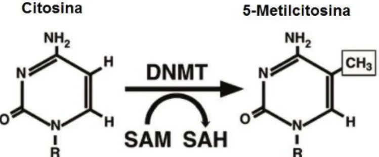
A metilação consiste em uma modificação covalente do DNA na qual um grupamento metil (CH3) é transferido da 5-adenosilmetionina para o carbono 5 de uma citosina que geralmente precede uma guanina (dinucleotídeo CpG) pela ação das DNMTs (BIRD, 2002) (Figura 1).
Figura 1. Esquema do processo de formação da 5-Metilcitosina (5-MeC) pela DNA metiltransferase. Fonte: WEIDMAN et al.,2007.
silenciamento de elementos repetitivos no genoma, sendo ela randômica ou sítio específica (FEINBERG; TYCKO, 2004). Os dinucleotídeos CpG aparecem esparsos pelos genomas eucariotos ou agrupados em regiões definidas como ilhas CpG. Estas ilhas consistem em regiões do DNA maior que 200 pares de base contendo aproximadamente 50% de bases C e G e com uma presença esperada de aproximadamente 60% de dinucleotídeos CpG, sendo frequentes em regiões promotoras de certos genes, incluindo genes de manutenção (LI; DAHIYA, 2002).
A metilação é geralmente associada com um estado repressivo da cromatina e a inibição da expressão gênica. Isto é feito através do bloqueio da ligação de fatores de transcrição ao DNA. Além disso, as proteínas MBPs com afinidade pelo grupo metil, ligam-se às regiões CpGs localizadas nos promotores e impedem o acesso dos fatores de transcrição aos seus sítios (ATTWOOD; YUNG; RICHARDSON, 2002), podendo ainda recrutar repressores transcricionais incluindo HDACs (KLOSE; BIRD, 2006) (Figura 2).
Figura 2. Mecanismo pelo qual a metilação do DNA inibe a transcrição gênica. A. Região promotora desmetilada permitindo a ligação dos fatores de transcrição. B. Metilação impedindo a ligação dos fatores de transcrição. C. Proteínas que se ligam à metilcitosina em ilhas CpG bloqueiam a ligação dos fatores de transcrição. Fonte: ATTWOOD et al., 2002.
às perturbações ambientais (ZHU, 2009).
O genoma está particularmente susceptível à desregulação por fatores ambientais desde a gestação e durante toda a vida do indivíduo. Tal desregulação é provocada por alterações na fita de DNA e histonas, sendo que, alterações epigenéticas são mais frequentes que mutações (USHIJIMA; ASADA, 2010). Padrões de hipermetilação aberrante em promotores de genes representam um dos principais mecanismos associados com a inativação gênica e frequentemente ocorrem em genes supressores tumorais no câncer (WEIDMAN et al., 2007; MOSS; WALLRATH, 2007; FUKUSHIGE; KONDO; HORII, 2009; INBAR-FEIGENBERG et al., 2013).
1.2 Radiação solar
A maior parte da energia irradiada pelo sol está na região do espectro Ultravioleta (UV) (comprimento de 100-400nm) em comparação ao espectro da luz visível (comprimento de 400-700nm) (CRUTZEN, 1992).
A Radiação UV solar é um importante agente carcinogênico ambiente e divide-se em três tipos, dependendo do comprimento de onda: de onda curta UVC (100-280 nm), de onda média UVB (280-320 nm) e de onda longa UVA (320-400 nm). A maior parte da radiação UV é absorvida pela epiderme, com transmissão para derme apenas de UVA (SYED et al., 2013). A radiação UVC é amplamente filtrada pela camada de ozônio, enquanto que, quase todos os raios UVA e até 10% da radiação UVB atingem a superfície da Terra (CRUTZEN, 1992; HAWRYLUK; OZTAN; FISHER, 2014).
Exposição aguda da pele à radiação UV causa queimaduras, alteração da pigmentação, inflamação, imunossupressão e danos no tecido conjuntivo da derme (AMBACH; BLUMTHALER, 1993; BRASH et al., 2001; DAMIAN et al., 2011). A exposição crônica à UV altera a arquitetura normal da pele causando o fotoenvelhecimento e câncer de pele (FISHER et al., 2002; PFEIFER; BESARATINIA, 2012; SYED et al., 2013).
Absorção da radiação solar UV pelas células da pele altera a estrutura química do DNA e causa estresse oxidativo (BAUMANN, 2007; ICHIHASHI et al., 2003; YAAR; GILCHREST, 2007). Essas alterações ativam vias de sinalização celular que regulam múltiplas funções celulares na pele. As células dos mamíferos desenvolveram muitos mecanismos protetores contra os danos da radiação UV, chamados genes de resposta à UV. Estes incluem enzimas de reparo de DNA, transdução de sinal, defesa antioxidante, apoptose, parada do ciclo celular (HOLBROOK; LIU; FORNACE, 1996; SCHARFFETTER-KOCHANEK et al., 2000; SHAULIAN et al., 2000).
A análise de expressão gênica global tempo-dependente de RNAm e proteínas após 24 horas de exposição da pele à radiação UV demonstrou que a maioria dos genes induzidos foram aqueles envolvidos na remodelação da matriz extracelular (metaloproteases de matriz - MMPs) (QUAN et al., 2009; TEWARI et al., 2014). Atualmente, está claro que a radiação UV é capaz de alterar também a expressão de miRNAs (DZIUNYCZ, 2010) e na pele exposta à UV, foi demonstrado que os miRNAs contribuiram para a regulação do ciclo celular e apoptose (SYED et al., 2013).
1.3 Fotoenvelhecimento e câncer
Está bem estabelecido que o fotoenvelhecimento é o fator predominante da aparência prematuramente envelhecida da pele exposta ao sol (YAAR; GILCHREST, 1998).
A pele envelhecida pela luz pode ser caracterizada pela
aparência ressecada, presença
de rugas, flacidez e despigmentação (RABE et al., 2006).
A matriz extracelular é o principal componente da derme e consiste principalmente de proteoglicanos, glicoproteínas e colágeno, os quais têm importante papel na arquitetura da pele e afetam a aparência da pele fotoenvelhecida (CHUNG et al., 2001; WAKATSUKI; ELSON, 2003). A fragmentação do colágeno e a inibição da sua produção prejudica a integridade estrutural da derme, o que é um aspecto proeminente da patofisiologia do envelhecimento prematuro da pele (fotoenvelhecimento) (FISHER et al., 2002; FLIGIEL et al., 2003). Porém, os mecanismos moleculares pelos quais a radiação UV altera a homeostase do colágeno ainda são pouco conhecidos.
a partir dos 40 anos, aumentando sua prevalência de acordo com o avanço da idade, porém, pode ocorrer também em indivíduos jovens. Já o gênero parece ser um fator balanceado. Um estudo brasileiro analisou 364 pacientes com melanoma, registrando a média de idade dos pacientes em 59 anos. Sendo 86,3% pacientes de pele branca, 9,3% dos pacientes de pele negra e 3,8% em pacientes de pele morena (FERRARI JÚNIOR et al., 2008).
Uma classe de genes importantes no desenvolvimento do câncer são os supressores tumorais e em muitos tipos de câncer já foi identificado a metilação aberrante em ilhas CpG como mecanismo epigenético de silenciamento transcricional (JONES; AND; BAYLIN, 2007). Esses mecanismos epigenéticos têm emergido como possuindo um papel central na regulação gênica do melanoma humano, incluindo a identificação de muitos supressores tumorais e oncogenes (HOWELL et al., 2009) e estima-se que o número de genes metilados em cada tipo de câncer seja muito alto ocorrendo em padrões distintos (BAYLIN et al., 1998; COSTELLO et al., 2000; ESTELLER, 2002).
1.4 Metaloproteases de matriz (MMP)
As metaloproteinases de matriz (MMP) são endopeptidases neutras, da família das proteinases zinco-dependentes somando um total de 23 enzimas no homem que se encontram divididas em quatro classes principais: colagenases, gelatinases, estromelisinas e metaloproteases tipo-membrana. Elas desempenham um importante papel nos processos fisiológicos, como embriogênese, remodelação de tecido normal, involução pós-parto e cicatrização de feridas, mantendo um balanço com os inibidores teciduais de metaloproteinases. Estão também envolvidas em condições patológicas nas quais existe a ruptura deste equilíbrio, provocando doenças como artrites, retinopatia diabética, psoríase e contribuir para a invasão de tumores malignos e metástase no câncer (WEEKS; HALME; WOESSNER, 1976; DECLERCK et al., 1992; CURRAN; MURRAY, 2000; RUNDHAUG, 2005; CLARK et al., 2008; FU; PARKS; HEINECKE, 2008).
mecanismo (YE, 2000).
Em particular, a MMP-9 (92-kDa, gelatinase B) é uma importante colagenase que contribui para a digestão do colágeno tipo IV e dos componentes primários da membrana basal (SCHMALFELDT et al., 2001). É produzida por queratinócitos, monócitos, macrófagos, neutrófilos e muitas células tumorais (BIRKEDAL-HANSEN et al., 1993) e tem sua expressão regulada em fibroblastos e queratinócitos durante a remodelação fisiológica da matriz extracelular ou durante destruição tecidual em uma patologia (YE, 2000).
A MMP-9 apresenta importante papel na angiogênese, progressão tumoral e disseminação metastática do câncer (BERGERS et al., 2000; HESLIN et al., 2001; SCHMALFELDT et al., 2001). A hiperexpressão de MMP-2 e MMP-9 tem sido identificada
como parte de um evento inicial na tumorigênese, associado à degradação da membrana basal e à transformação maligna (OHASHI et al., 2000; SAMANTARAY et al., 2004), tendo sido observado em vários tumores (CLARK et al., 2008; TURPEENNIEMI-HUJANEN, 2005). O gene MMP9 possui 13 éxons e está localizado no cromossomo 20 (20q11.2-q13.1)
(GENE ID: 4318). A região 5‟ flanqueadora do gene MMP9 contém muitas regiões consenso
motivos de ligação para os fatores de transcrição AP-1, NF- B, Sp-1 e Ets ( S AT O ; S E I KI , 1 9 9 3 ) . Essa região ainda, é rica em dinucleotídeos CpG, sugerindo que a metilação de DNA pode ser um fator regulador da expressão gênica. Em células de linfoma foi observada uma correlação inversa entre a metilação do promotor do gene MMP9 e o nível da expressão dessa proteína (CHICOINE et al., 2002).
1.5 MicroRNAs (miRNA)
Para tentar compreender os mecanismos de regulação e padrões de alterações da expressão gênica, uma nova classe de moléculas regulatórias conhecidos como microRNAs (miRNAs), está recebendo muita atenção como mediadores de mudanças na expressão gênica e consequentemente nas respostas fisiológicas induzidas pela radiação UV (JAYANTHY; SETALURI, 2015).
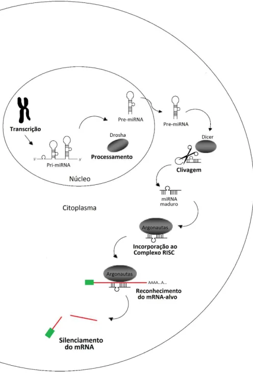
Essas moléculas completamente endógenas fazem parte de uma classe de reguladores de RNA não codificantes denominados RNA de interferência (RNAi) e têm emergido como reguladores-chave durante a oncogênese. Eles foram observados regulando a proliferação celular, diferenciação e apoptose (BARTEL, 2004; FARH et al., 2005; KATO; SLACK, 2008). A biogênese dos miRNAs (Figura 3) envolve inicialmente a transcrição pela RNA polimerase II em um longo RNA primário (pri-miRNA) que é clivado por um complexo proteico chamado Drosha. A clivagem resulta no microRNA precursor (pré-miRNA), com cerca de 70 pares de bases, contendo um trecho de fita dupla e uma alça de fita simples, formando uma estrutura denominada hairpin. O pré-miRNA é exportado para o citoplasma,
onde é clivado pela RNase Dicer, gerando um miRNA maduro com cerca de 22 nucleotídeos (RICARTE FILHO; KIMURA, 2006). Em seguida, o miRNA é incorporado à um complexo proteico denominado RISC (RNA-Induced Silencing Complex) do qual faz parte juntamente
proteínas Argonautas (MACFARLANE; MURPHY, 2010). Essas proteínas são caracterizadas pela presença de domínios conservados (PAZ e PIWI). Elas apresentam atividade de endonuclease dirigida contra o mRNA alvo, apresentando fundamental importância durante o silenciamento via miRNA (HÖCK; MEISTER, 2008). O complexo RISC pareia o miRNA com o mRNA-alvo por complementaridade de bases. Quanto maior for a complementaridade, maior a chance de o mRNA ser degradado, caso contrário, pode ocorrer apenas a repressão da tradução (BARTEL, 2004; RIVAS et al., 2005;
MACFARLANE; MURPHY, 2010).
Abordagens computacionais estimam que 1-5% dos genes codificantes possuem informação para miRNAs em suas sequências intrônicas (LEWIS; BURGE; BARTEL, 2005; BENTWICH et al., 2005). Além disso, estima-se que 30%-60% de todos os genes humanos sejam alvos potenciais de um ou mais miRNAs (LEWIS; BURGE; BARTEL, 2005; FRIEDMAN et al., 2009). Análises bioinformáticas estimam ainda que, cerca de 50% dos genes de miRNAs são associados à ilhas CpG, podendo ter sua expressão regulada via metilação de DNA (WEBER et al., 2007).
Figura 3. Biogênese e via de silenciamento do miRNA. O gene de miRNA é transcrito para gerar um miRNA primário (pri-miRNA), este por sua vez, sofre clivagem no núcleo para formar o percussor do miRNA (pré-miRNA). O pré-miRNA é transportado para o citoplasma e clivado para gerar um duplex de miRNA. Em seguida, o miRNA maduro é incorporado ao complexo RISC, onde liga-se à sequência-alvo regulando o gene. A clivagem do mRNA ou repressão da tradução depende do nível de complementaridade entre o miRNA e o RNAm-alvo. Fonte: Baseado em HAN et al., 2014.
gênica via miRNA em resposta à danos no DNA induzida pela radiação UV, opera desde rápidas modificações na proteína que ocorrem em poucos minutos até a transcrição muito mais lenta podendo levar várias horas ou dias para se desenvolver (SYED et al., 2013).
Um estudo observou que a radiação UV altera a expressão de miRNAs, demonstrando inclusive que diferentes comprimentos de onda podem alterar diferentes miRNAs. Neste estudo foi observado o aumento da expressão dos miRNAs 203, 205 e 21 pela radiação UVA, este último MIR possui propriedades oncogênicas. Já a radiação UVB foi responsável pela diminuição da expressão de miR-205 e não teve efeito sobre o miR-21. Conferindo assim, diferentes propriedades carcinogênicas aos diferentes comprimentos de onda da radiação solar (DZIUNYCZ, 2010).
Muitos microRNAs que são ativados em resposta à diferentes comprimentos de onda tem demonstrado um importante papel na regulação da produção da melanina por melanócitos. Este pigmento é uma importante defesa contra radiação solar que podem causar danos potenciais ao DNA levando ao aparecimento do câncer (SYED et al., 2013; JAYANTHY; SETALURI, 2015).
O gene MicroRNA-137 (MIR137) está localizado no cromossomo 1 (1p21.3) (Gene
ID: 406928) e possui extensa ilha CpG. Estudos tem demonstrado que este MIR possui função de supressor tumoral em vários tipos de câncer, incluindo o carcinoma espinocelular (LANGEVIN et al., 2010) e melanoma, através do controle do ciclo celular ao regular negativamente múltiplos genes-alvo (LUO et al., 2013).
A hipermetilação do gene MIR137 já foi observado em inúmeros cânceres como
glioblastoma (VISANI et al., 2014; BIER et al., 2013), câncer oral (KOZAKI et al., 2008), câncer ovariano (GUO et al., 2013), câncer de pulmão (ZHU et al., 2013), câncer gástrico (Chen et al., 2011), câncer colorretal (BALAGUER et al., 2010; LIU et al., 2011; LIANG et al., 2013) e melanoma (LUO et al., 2013). Neste último trabalho a baixa expressão de MIR137
foi correlacionada à baixa sobrevida de pacientes com melanoma em estágio IV.
O MIR137 inibiu a migração e proliferação do melanoma in vitro através da indução
do tumor à apoptose em vários tipos de câncer (LUO et al., 2013; ZHU et al., 2013) e em outro estudo, a transfecção ectópica do microRNA-137 reduziu o crescimento celular in vitro
de carcinoma de células escamosas oral, deixando claro que este gene é caracterizado como um supressor tumoral silenciado epigeneticamente durante a carcinogênse oral. (KOZAKI et al., 2008). Além disso, foi encontrada diferença significativa no perfil de metilação do MIR137 entre amostras de mucosa de pacientes com câncer oral e indivíduos
Trabalhos confirmam que o MIR137 também está envolvido na regulação da
1.6 Justificativa
Os estudos no padrão de metilação de DNA têm emergido como um importante campo de pesquisa e revelam como o meio ambiente pode afetar nossos genes e alterar drasticamente a expressão gênica resultando em doenças inflamatórias e tumorais, ou mesmo no envelhecimento (ATTWOOD; YUNG; RICHARDSON, 2002).
Já está bem estabelecido que a radiação UV solar pode causar mutações no DNA e aumentar o risco para o desenvolvimento de câncer de pele (PFEIFER; BESARATINIA, 2009), entretanto, as alterações epigenéticas induzidas pela UV nessa patologia constituem uma delicada interação de regulação gênica que ainda não foi completamente compreendida (NANDAKUMAR et al., 2011).
Esses mecanismos são ainda menos esclarecidos quando se trata da regulação por miRNAs (SYED et al., 2013). Além disso, a maioria das evidências de que a radiação UV causa alterações epigenéticas em amostras não tumorais são oriundas de trabalhos experimentais com ratos ou linhagem celulares (MITTAL et al., 2003; NANDAKUMAR et al., 2011; Chen et al., 2012).
Raros são os estudos utilizando amostras de pele obtidas de humanos. Um desses estudos mostrou hipermetilação no gene CDH1 (cadherin 1) em regiões expostas
ao sol (SATHYANARAYANA et al., 2007). Outro estudo revelou tendência à hipometilação em amostras de pele exposta ao sol no promotor do gene KRT75 (queratin 75), e em adição, mostrou que essas alterações são mais acentuadas em indivíduos
idosos (GRÖNNIGER et al., 2010).
Metaloproteases de Matriz e microRNAs já foram observados desregulados em amostras não tumorais de pele exposta a radiação solar (QUAN et al., 2009; JAYANTHY; SETALURI, 2015; SYED et al., 2013) e em amostras tumorais (KERKELÄ; SAARIALHO-KERE, 2003; VILEN et al., 2008; HADLER-OLSEN et al., 2011; SYED et al., 2013). Contudo, nada se sabe sobre o mecanismo molecular pelo qual a expressão desses genes sofreram desregulação.
Os genes MMP9 e MIR137 são ricos em dinucleotídeos CpG e dessa forma são
2. CAPÍTULO 1
Influence of sun exposure on DNA methylation status of the MMP9 and MIR137 genes
of human skin sample
ALANNE RAYSSA DA SILVA MELO1, HALINE BARROSO1, DANIEL UCHÔA DE ARAÚJO2, FRANCISCO RUIDOMAR PEREIRA3, NAILA FRANCIS PAULO DE OLIVEIRA1,2
1 Programa de Pós Graduação em Biologia Celular e Molecular, Centro de Ciências Exatas e da Natureza, Universidade Federal da Paraíba, João Pessoa, PB-Brazil
2 Departamento de Biologia Molecular, Centro de Ciências Exatas e da Natureza, Universidade Federal da Paraíba, João Pessoa, PB-Brazil
3 Departamento de Morfologia, Centro de Ciências da Saúde, Universidade Federal da Paraíba, João Pessoa, PB-Brazil
Corresponding Author:
Dra. Naila Francis Paulo de Oliveira Universidade Federal da Paraíba
Centro de Ciências Exatas e da Natureza Departamento de Biologia Molecular Cidade Universitária – Campus I João Pessoa-PB
Brazil
CEP 58051-900
ABSTRACT
Studies have shown that a variety of environmental factors and habits are associated with epigenetic changes. In addition, various genes are also found to respond to UV radiation. We hypothesised that sun exposure could alter the DNA methylation profile on the matrix metalloprotease-9 (MMP9) and microRNA-137 (MIR137) genes of skin cells of subjects with
no history of skin diseases. Skin biopsies (5mm) were obtained using a punch technique of sun-exposed (outer forearm) and sun-protected areas (inner arm) from 28 corpses from the Brazilian Service of Death Investigation. Genomic DNA was extracted and a DNA methylation analysis was performed using Methylation Specific PCR (MSP) of sun-exposed and sun-protected skin areas. No differences were found among the areas (p>0.05; McNemar), with the partially methylated condition found to be a common event in skin for both MMP9 and MIR137. We conclude that sun exposure does not induce changes in DNA
methylation status in MMP9 and MIR137 genes.
Introduction
Studies show that the environment influences the epigenetic profile, and epigenetic changes may be more common than are mutations (Ushijima et al. 2010). Epigenetic information is defined as information other than the DNA sequence that is faithfully replicated upon somatic cell replication. It is carried by DNA methylation, post-translational histone modifications, and non-coding RNAs, all of which are related to gene expression and chromatin structure. In particular, DNA methylation is characterised by the addition of a methyl group in cytosines that precedes guanine (CpG). CpG dinucleotides are typically clustered in what is termed CpG islands, which are areas rich in CpG sites and are often found in the promoter regions of the genes (Portela & Esteller 2010). Aberrant hypermethylation usually is an early event in carcinogenesis that is associated with gene inactivation, and often occurs in tumour suppressor genes (Sharma et al. 2010). Studies have shown that a variety of factors and habits, such as fungicides, metal ions, drugs, diet, alcohol and smoking habits, are associated with epigenetic changes (Christensen & Marsit 2011, Terry et al. 2011).
The human skin is an organ with many advantages for the study of changes induced by aging and environmental factors because is directly exposed. It is well-established that chronic exposure of human skin to UV solar radiation results in photoaging and can also lead to skin cancer (Pfeifer & Besaratinia 2012). The incidence and mortality rates from skin cancer, especially melanoma, continue to increase worldwide and its
incidence is equivalent to the incidence of malignancies in all other organs combined
(Housman et al. 2003).
respond to UV radiation are comprised of many proteases, like matrix metalloprotease (MMP) (Quan et al. 2009), which degrade components of the extracellular matrix (Ho et al. 2005), as well as reduce the production of type I procollagen (Fisher et al. 2000). In particular, MMP9 is located on chromosome 20 and its protein products have a MW of 92
kDa. It is associated with the invasiveness of tumour cells (Sato & Seiki 1993). The promoter of this gene has a CpG island and aberrant methylation that has already been found in cancer and inflammation (Sato et al. 2003, Roach et al. 2005).
Recently, studies show that the microRNAs (MIR) also respond to UV radiation (Kraemer et al. 2013, Syed et al. 2013). MicroRNAs are a group of endogenous small noncoding RNAs (20–24 nucleotides) that negatively regulate gene expression at the post-transcriptional level, mainly via binding to the 3´-untranslated region (3´-UTR) of the target gene (Bartel 2004). In particular, MIR137 is a short non-coding RNA that is located on
chromosome one and acts like a tumour suppressor. It has a widespread CpG island and hypermethylation of MIR137 have been shown in a variety of types of cancer (Chen et al.
2011, Bier et al. 2013, Zhu et al. 2013, Perez-Carbonell et al. 2014).
Several carcinogenic factors have been investigated for their ability to induce epigenetic changes, but sun radiation has been neglected. Most pieces of evidence in non-tumour samples include results of studies with rodents and cultured cells (Mittal et al. 2003, Nandakumar et al. 2011, Chen et al. 2012). Studies with human skin samples are scarce (Sathyanarayana et al. 2007, Grönniger et al. 2010). Based on these facts, we hypothesised that sun exposure could alter the DNA methylation profile of the MMP9 and MIR137 genes of
skin cells in subjects with no history of skin disease.
Methods
The study was performed in accordance with the current recommendations of the National Health Council – Ministry of Health of Brazil for research in human subjects and with the approval of the Ethics Committee in Research of the Federal University of Paraiba (protocol number 430/2011). Corpses of both genders of individuals that were older than 30 years at the time of their death were included. The corpses were obtained from the Brazilian Service of Death Investigation. Written informed consent for participation was obtained from all families. The demographic data and general health were obtained via record and did not include individuals with a medical history of skin diseases. Skin samples were collected and ranked according to Fitzpatrick‟s (1988) criteria. Samples from 28 subjects were included, and were divided into two conditions according to sun exposure: sun-exposed area (SE) and sun-protected area (SP).
Sample Collection and Genomic DNA Isolation
Biopsies were collected by punch (5mm diameter) from the outer forearm (sun-exposed area) and inner arm (sun-protected area), up to 10 hours after death. Immediately after removal, the biopsies (epidermis and dermis) were stored in a tube containing 800 µL of RNAholder (Bioagency, São Paulo, SP, Brazil) and frozen at -20°C until analysis. Afterwards, the genomic DNA of skin biopsies was purified using the TRIZOL reagent (Invitrogen, Carlsband, CA, USA) following the manufacturer‟s recommendation, using a tissue
homogeniser. DNA quantification was performed using a NanoDrop spectrophotometer (Thermo Scientific). The samples were then frozen at -20°C until the DNA methylation analysis.
DNA Methylation Analysis
Start Green Master Mix (Promega Corporations, Madison, WI, USA) in a final reaction of 20 µL. Fragments of MMP9 were amplified with specific primers for either methylated (F:5‟
-gaagttcgaaattagtttggttaac-3‟, R:5‟ tcccgaataactaatattataaacgta-3‟) or unmethylated targets (F: 5‟-agtttgaaattagtttggttaatgt-3‟, R:5‟-cctcccaaataactaatattataaacata-3‟) with 179 bp. Methylated and unmethylated cycle conditions were as follows: 95°C x 5 minutes; 40 cycles (95°C x 30s, 55°C x 30s, 72°C x 30s) and 72°C x 5 min. (Yang et al. 2009). Fragments of
MIR137 were amplified with specific primers for either methylated (F:5‟
-gcggtagtagtagcggtagc-3‟, R:5‟-acccgtcaccgaaaaaaa-3‟) or unmethylated targets (F:5‟ -ggtggtagtagtagtggtagt-3‟, R:5‟-tacccatcaccaaaaaaaa-3‟) with 86 bp. Methylated and unmethylated cycle conditions were as follows: 95°C x 5 minutes; 40 cycles (95°C x 1min, 58°C (met), 51°C (unm) x 1min, 72°C x 1 min) and 72°C x 5 min. (Dang et al. 2013). Methylated DNA (Methylated CpG Jurkat Genomic DNA, New England Biolabs) and unmethylated (5-Aza-dc Treated Jurkat Genomic DNA, New England Biolabs) were modified, as previously quoted, and amplified by PCR as control reactions with primers for the methylated and unmethylated condition, respectively. Amplified PCR samples (6µl) were loaded in 6% polyacrylamide gels and subjected to electrophoresis. DNA bands were detected after silver staining.
Statistical Analysis
We performed a paired non-parametric McNemar Test at a level of 5% in order to evaluate differences in DNA methylation status among sun-exposed and sun-protected areas of skin cells using BIOESTAT 5.0 software (Ayres et al. 2007).
Results and Discussion
ability to cause skin cancer is directly linked to cause mutations, however, the ability of solar radiation to cause epigenetic changes is poorly known. It is known that epigenetic changes are associated with several types of cancer, non-cancerous conditions and aging (Mau & Yung 2014, Daniel & Tollefsbol 2015). Studies have shown that skin tumours are associated with epigenetic changes and we would like to know if it is possible to observe changes in healthy cells exposed to sun. This knowledge can help to contribute to early diagnosis and therapeutic interventions. In the present study, we investigated the influence of sun exposure on the DNA methylation status of non-tumuoral skin samples, focusing on MIR and MMP
gene family because there is evidence that these genes respond to UV radiation (Quan et al. 2009, Kraemer et al. 2013, Syed et al. 2013).
MMP9 – Samples from 28 corpses that were aged 30–89 years (60.7±17.6;
mean±SD) prior to death were used for this study. Of these, 15 were females and 13 were males. Regarding skin type (Fitzpatrick, 1988), nine were classified as type III (light brown), 12 as type IV (moderate brown) and seven as type V (dark brown).
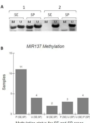
The studied CpG site was previously described (Yang et al. 2009). Out of the total of 28 samples, no sample showed differences between sun-exposed and sun-protected areas. We found that 27 of the skin samples were positive for the partially methylated condition in both SE and SP areas and one was unmethylated for both areas. Partial DNA methylation was considered when samples were positive for both methylated and unmethylated conditions, indicating that DNA methylation in the CpG studied did not occur in all cells, and/or all of the alleles (Figure 1) (p>0.05). MMP9 demethylation was found in the samples
of individuals with osteoarthritis (Roach et al. 2005). An almost complete methylation of CpG sites in the MMP9 gene promoter was observed in the five cancer cell lines (Sato et al.
2003).
MIR137 – Samples from 24 corpses that were aged 30–89 years (61.0±18.1;
males. Regarding skin type (Fitzpatrick 1988), nine were classified as type III (light brown), 12 as type IV (moderate brown) and three as type V (dark brown).
The studied CpG site was previously described (Dang et al. 2013). We found that out of a total of 24 samples, only seven samples showed differences between sun-exposed and sun-protected areas (three partially methylated in SE and unmethylated in SP and four unmethylated in SE and partially methylated in SP) and most had the same condition for both areas (11 partially methylated for SE and SP, four unmethylated for SE and SP and two methylated for SE and SP (Figure 2) (p>0.05)). Partial DNA methylation was considered when samples were positive for both methylated and unmethylated conditions, indicating that DNA methylation in the CpG studied does not occur in all cells, and/or all of the alleles.
Dang et al. (2011) found methylation frequency of 25% up to 58.3% in patients with oral lichen planus and oral squamous cell carcinoma, suggesting MIR137 methylation as a
biomarker for malignant prediction in patients with those diseases. However, MIR137
methylation was only found in the epithelium and not in the connective tissue. In the present study, it was not possible to separate the dermis and epidermis, and so perhaps, we did not find differences between the two areas because the DNA of keratinocytes and fibroblasts were analysed together. MIR137 methylation was also found in gastric cancer (Chen et al.
2011), glioblastoma (Bier et al. 2013), pulmonary cancer (Zhu et al. 2013), colorectal cancer (Perez-Carbonell et al. 2014) and others.
Speculation on a possible connection between UV irradiation and epigenetic changes is growing but is still scarce. Few studies show this association in skin samples obtained from humans. CDH1 methylation was highly significantly related to UV-exposed
skin (Sathyanarayana et al. 2007). Another study revealed hypomethylation in the promoter of the KRT75 gene (queratin 75) in skin samples exposed to the sunlight. Meanwhile, the
promoters of important genes for skin cell homeostasis SEC31L2 (SEC31 homolog a B (S.
cerevisiae), DDAH2 (dimethylarginine dimethylaminohydrolase 2) and TET2 (tet
pronounced than age-related hypermethylation. In addition, the effects appeared stronger in the epidermis than in the dermis (Gronniger et al. 2010). However, the same study showed no differences between the sun-protected and sun-exposed areas in other CpG sites studied. A recent study showed that exposure to solar UV radiation may reduce DNA methylation in circulating lymphocytes (Nair-Shalliker et al. 2014).
Most pieces of evidence in non-tumour samples include results of experimental studies with rodents or cultured cells (Mittal et al. 2003, Nandakumar et al. 2011, Chen et al. 2012, Prasad & Katiyar, 2013, Yang et al. 2014). These studies contrast with another one that reveals that UVB irradiation of keratinocytes has no recognisable global effect on DNA methylation patterns (Lahtz et al. 2013). They suggest that changes in DNA methylation, as observed in skin cancers (Lee et al. 2014, Wu et al. 2014), are not immediate consequences of human exposure to solar UVB irradiation.
It appears that the major mechanism by which UV radiation causes skin tumuorigenesis is via mutations of the TP53 gene. The mutations are predominantly the
UV-signature mutation, i.e., a C→T transitions at dipyrimidine sites, which result from UV radiation-induced cyclobutane pyrimidine dimer formation (Tommasi et al. 1997, Rochette et al. 2009). Methylation of cytosine has been shown to increase the pyrimidine dimer formation that results from cell exposure to solar UV radiation, and many of the mutations occur at the CpG sites (Tommasi et al. 1997).
Regarding the analysis of gene expression, metalloproteinases and microRNAs were deregulated in tumours (Kerkelä et al. 2003, Vilen et al. 2008, Hadler-Olsen et al. 2011, Perera & Ray 2012), and the same was observed in non-tumour samples exposed to UV radiation, but the molecular mechanism is still unknown (Quan et al. 2009, Kraemer et al. 2013). Kraemer et al. (2013) also showed that UVA and UVB irradiation regulate microRNA expression differentially in human primary keratinocytes. In addition, it was already shown that enhanced levels of Dnmts in the UV-exposed mouse skin and UV-induced skin tumours, in addition to its distribution patterns, suggest that the de novo synthesis of Dnmt3a and
UVB-exposure of the skin and within UVB-induced skin tumours. In this study, DNA hypermethylation was consistent with the Dnmts expression findings (Nandakumar et al. 2011).
Age and gender are also associated with different DNA methylation statuses among individuals (El-Maarri et al. 2007, Thompson et al. 2010, Bezerra et al. 2015).Although our demographic data show variations in these aspects, in addition to skin type, this analysis was hampered by the small sample size, although we note that the partially methylated profile for
MIR137 included 10 women out of a total of 11 individuals. The variation in methylation
status, in particular for MIR137 (we have found five statuses, Figure 2B), could be also due
to another intrinsic factor, such as genetic polymorphisms (Arakawa et al. 2012).
Epigenetic changes represent an additional pathway by which environmental factors influence aging and the development of diseases with a tissue-specific manner. In particular, studies involving sun exposure and experiments with UVA and UVB radiation are needed to better understand the relationship between these factors and the epigenome, which can contribute to clinical applications in preventive and personalised medicine settings.
Conclusion
Our data show that the partially methylated condition is a common feature of skin cells for MMP9 and MIR137 genes. So, we conclude that sun exposure does not induce
changes in the DNA methylation status in MMP9 and MIR137 genes.
Declaration of interest
Figure 1 – DNA methylation analysis of the MMP9 gene promoter of skin cells. (A) Bands of two
representative samples obtained after the polymerase chain reaction (179 bp). (B) Methylation status of the MMP9 gene for SE and SP areas (n=28; p>0.05; McNemar). P – Partially methylated, M –
Figure 2 – DNA methylation analysis of the MIR137 gene promoter of skin cells. (A) Bands of two
representative samples obtained after the polymerase chain reaction (86 bp). (B) Methylation status of the MIR137 gene for SE and SP areas (n=24; p>0.05; McNemar). P – Partially methylated, M –
REFERENCES
Arakawa Y, Watanabe M, Inoue N, Sarumaru M, Hidaka Y, Iwatani Y. 2012. Association of polymorphisms in DNMT1, DNMT3A, DNMT3B, MTHFR and MTRR genes with global
DNA methylation levels and prognosis of autoimmune thyroid disease. Clin Exp Immunol 170:194–201.
Ayres M, Jr. Ayres M, Ayres DL, Santos AAS. 2007. Bioestat: aplicações estatísticas nas áreas das Ciências Biomédicas. V. 5.0., 324.
Bartel DP. 2004. MicroRNAs: genomics, biogenesis, mechanism, and function. Cell.
116:281–97.
Bezerra SFO, Costa LA, Freitas PAN, Oliveira NFP. 2014. Age-related changes in DNA methylation status of hTERT gene promoter of oral epithelial cells. Brazilian Arch Biol Technol. 58:82-89.
Bier A, Giladi N, Kronfeld N, Lee HK, Cazacu S, Finniss S, Xiang C, Poisson L, de Carvalho AC, Slavin S, Jacoby E, Yalon M, Toren A, Mikkelsen T, Brodie C. 2013. MicroRNA-137 is downregulated in glioblastoma and inhibits the stemness of glioma stem cells by targeting RTVP-1. Oncotarget. 4:665–76.
Chen IP, Henning S, Faust A, Boukamp P, Volkmer B, Greinert R. 2012. UVA-induced epigenetic regulation of P16(INK4a) in human epidermal keratinocytes and skin tumor
derived cells. Photochem Photobiol Sci. 11:180–90.
Chen Q, Chen X, Zhang M, Fan Q, Luo S, Cao X. 2011. miR-137 is frequently down-regulated in gastric cancer and is a negative regulator of Cdc42. Dig Dis Sci. 56:2009–
16.
Christensen BC, Marsit CJ. 2011. Epigenomics in environmental health. Front Genet. 2:84.
Dang J. Bian YQ, Sun JY, Chen F, Dong GY, Liu Q, Wang XW, Kjems J, Gao S, Wang QT, 2013. MicroRNA-137 promoter methylation in oral lichen planus and oral squamous
cell carcinoma. J Oral Pathol Med. 42:315–21.
Daniel M, Tollefsbol TO. 2015. Epigenetic linkage of aging, cancer and nutrition. J Exp Biol.
218:59–70.
El-Maarri O, Becker T, Junen J, Manzoor SS, Diaz-Lacava A, Schwaab R, Wienker T, Oldenburg J. 2007. Gender specific differences in levels of DNA methylation at selected loci from human total blood: a tendency toward higher methylation levels in males. Hum Genet. 122:505–14.
Fisher GJ, Datta S, Wang Z, Li XY, Quan T, Chung JH, Kang S, Voorhees JJ. 2000. c-Jun-dependent inhibition of cutaneous procollagen transcription following ultraviolet irradiation is reversed by all-trans retinoic acid. J Clin Invest. 106:663–70.
Fitzpatrick TB. 1988. The validity and practicality of sun-reactive skin types I through VI. Arch Dermatol. 124:869–71.
Hadler-Olsen E, Fadnes B, Sylte I, Uhlin-Hansen L, Winberg JO. 2011. Regulation of matrix metalloproteinase activity in health and disease. FEBS J. 278:28–45.
Ho JN, Lee YH, Park JS, Jun WJ, Kim HK, Hong BS, Shin DH, Cho HY. 2005. Protective effects of aucubin isolated from Eucommia ulmoides against UVB-induced oxidative
stress in human skin fibroblasts. Biol Pharm Bull. 28:1244–8.
Holbrook NJ, Liu Y, Fornace A.J. 1996. Signaling events controlling the molecular response to genotoxic stress. EXS. 77:273–88.
Housman TS, Feldman SR, Williford PM, Fleischer AB, Goldman ND, Acostamadiedo JM, Chen GJ. 2003. Skin cancer is among the most costly of all cancers to treat for the Medicare population. J Am Acad Dermatol. 48:425–9.
Kerkelä E, Saarialho-Kere U. 2003. Matrix metalloproteinases in tumor progression: focus on basal and squamous cell skin cancer. Exp Dermatol. 12:109–25.
Kraemer A, Chen IP, Henning S, Faust A, Volkmer B, Atkinson MJ, Moertl S, Greinert R. 2013. UVA and UVB irradiation differentially regulate microRNA expression in human primary keratinocytes. PLoS One. 8(12):e83392.
Lahtz C, Kim SI, Bates SE, Li AX, Wu X, Pfeifer GP. 2013. UVB irradiation does not directly induce detectable changes of DNA methylation in human keratinocytes.
F1000Research. 2:45.
Lee JJ, Murphy GF, Lian CG. 2014. Melanoma epigenetics: novel mechanisms, markers, and medicines. Lab Invest. 94:822–38.
Mau T, Yung R. 2014. Potential of epigenetic therapies in non-cancerous conditions. Front Genet. 5:438.
Mittal A, Piyathilake C, Hara Y, Katiyar SK. 2003. Exceptionally high protection of photocarcinogenesis by topical application of (--)-epigallocatechin-3-gallate in hydrophilic cream in SKH-1 hairless mouse model: relationship to inhibition of UVB-induced global DNA hypomethylation. Neoplasia. 5:555–65.
Nair-Shalliker V, Dhillon V, Clements M, Armstrong BK, Fenech M. 2014. The association between personal sun exposure, serum vitamin D and global methylation in human lymphocytes in a population of healthy adults in South Australia. Mutat Res Fundam Mol Mech Mutagen. 765:6–10.
Nandakumar V, Vaid M, Tollefsbol TO, Katiyar SK. 2011. Aberrant DNA hypermethylation patterns lead to transcriptional silencing of tumor suppressor genes in UVB-exposed skin and UVB-induced skin tumors of mice. Carcinogenesis. 32:597–604.
Perera RJ, Ray A. 2012. Epigenetic regulation of miRNA genes and their role in human melanomas. Epigenomics. 4:81–90.
Perez-Carbonell L, Balaguer F, Toiyama Y, Egoavil C, Rojas E, Guarinos C, Andreu M, Llor X, Castells A, Jover R, Boland CR, Goel A. 2014. IGFBP3 methylation is a novel
diagnostic and predictive biomarker in colorectal cancer. PLoS One. 9:e104285.
Portela, Esteller M. 2010. Epigenetic modifications and human disease. Nat Biotechnol.
28:057–68.
Prasad R, Katiyar SK, 2013. Prostaglandin E2 Promotes UV radiation-induced immune suppression through DNA hypermethylation. Neoplasia. 15:795–804.
Quan T, Qin Z, Xia W, Shao Y, Voorhees JJ, Fisher GJ. 2009. Matrix-degrading metalloproteinases in photoaging. J Investig Dermatol Symp Proc. 14:20–4.
Roach HI, Yamada N, Cheung KSC, Tilley S, Clarke NMP, Oreffo ROC, Kokubun S, Bronner F. 2005. Association between the abnormal expression of matrix-degrading enzymes by human osteoarthritic chondrocytes and demethylation of specific CpG sites in the promoter regions. Arthritis Rheum. 52:3110–24.
Rochette PJ, Lacoste S, Therrien JP, Bastien N, Brash DE, Drouin R. 2009. Influence of cytosine methylation on ultraviolet-induced cyclobutane pyrimidine dimer formation in genomic DNA. Mutat Res. 665:7–13.
Sathyanarayana UG, Moore AY, Li L, Padar A, Majmudar K, Stastny V, Makarla, P, Suzuki M, Minna JD, Feng Z, Gazdar AF. 2007. Sun exposure related methylation in malignant and non-malignant skin lesions. Cancer Lett. 245:112–20.
Sato H, Seiki M. 1993. Regulatory mechanism of 92 kDa type IV collagenase gene expression which is associated with invasiveness of tumor cells. Oncogene. 8:395–
405.
Sato N, Maehara N, Su GH, Goggins M. 2003. Effects of 5-aza-2‟-deoxycytidine on matrix metalloproteinase expression and pancreatic cancer cell invasiveness. J Natl Cancer Inst. 95:327–30.
Scharffetter-Kochanek K, Brenneisen P, Wenk J, Herrmann G, Ma W, Kuhr L, Meewes C, Wlaschek M. 2000. Photoaging of the skin from phenotype to mechanisms. Exp Gerontol. 35:307–16.
Sharma S, Kelly TK, Jones PA. 2010. Epigenetics in cancer. Carcinogenesis. 31:27–36.
Shaulian E, Schreiber M, Piu F, Beeche M, Wagner EF, Karin M. 2000. The mammalian UV response: c-Jun induction is required for exit from p53-imposed growth arrest. Cell.
103:897–907.
Syed DN, Khan MI, Shabbir M, Mukhtar H. 2013. MicroRNAs in skin response to UV radiation. Curr Drug Targets. 14:1128–34.
Terry MB, Delgado-Cruzata L, Vin-Raviv N, Wu HC, Santella RM. 2011. DNA methylation in white blood cells: association with risk factors in epidemiologic studies. Epigenetics.
6:828–37.
Thompson RF, Atzmon G, Gheorghe C, Liang HQ, Lowes C, Greally JM, Barzilai N. 2010. Tissue-specific dysregulation of DNA methylation in aging. Aging Cell. 9:506–18.
Tommasi S, Denissenko MF, Pfeifer GP. 1997. Sunlight induces pyrimidine dimers preferentially at 5-methylcytosine bases. Cancer Res. 57:4727–30.
Vilen ST, Nyberg P, Hukkanen M, Sutinen M, Ylipalosaari M, Bjartell A, Paju A, Haaparanta V, Stenman UH, Sorsa T, Salo T. 2008. Intracellular co-localization of trypsin-2 and matrix metalloprotease-9: possible proteolytic cascade of trypsin-2, MMP-9 and enterokinase in carcinoma. Exp Cell Res. 314:914–26.
Wu J, Zhang JR, Qin J. 2014. Clinical significance of methylation of E-cadherin and p14ARF
gene promoters in skin squamous cell carcinoma tissues. Int J Clin Exp Med. 7:1808–
12.
Yang AY, Lee JH, Shu L, Zhang C, Su ZY, Lu Y, Huang MT, Ramirez C, Pung D, Huang Y, Verzi M, Hart RP, Kong ANT. 2014. Genome-wide analysis of DNA methylation in UVB- and DMBA/TPA-induced mouse skin cancer models. Life Sci. 113:45–54.
Yang S, Wen H, Zhang G, Zhao S, Luo Y, Lu Q. 2009. [Triptolide evaluates DNA methylation level of matrix metalloproteinase 9 gene in human fibrosarcoma HT-1080 cells].
Zhongguo Zhong Yao Za Zhi. 34:611–4.
Zhu X, Li Y, Shen H, Li H, Long L, Hui L, Xu W. 2013. miR-137 inhibits the proliferation of lung cancer cells by targeting Cdc42 and Cdk6. FEBS Lett. 587:73–81.
_______________________
3. CONCLUSÃO
Nossos dados mostram que a condição parcialmente metilada é um aspecto comum dos genes MMP9 e MIR137 em células da pele. Assim, concluímos que a exposição solar
REFERÊNCIAS*
AMBACH, W.; BLUMTHALER, M. Biological effectiveness of solar UV radiation in humans.
Experientia, v. 49, n. 9, p. 747–53, 1993.
ATTWOOD, J. T.; YUNG, R. L.; RICHARDSON, B. C. Cellular and Molecular Life Sciences Review DNA methylation and the regulation of gene transcription. v. 59, p. 241–257, 2002. BALAGUER, F. et al. Epigenetic silencing of miR-137 is an early event in colorectal carcinogenesis. Cancer research, v. 70, n. 16, p. 6609–18, 2010.
BARTEL, D. P. MicroRNAs: genomics, biogenesis, mechanism, and function. Cell, v. 116, n. 2, p. 281–97, 2004.
BARTEL, D. P. MicroRNAs: target recognition and regulatory functions. Cell, v. 136, n. 2, p. 215–33, 23, 2009.
BARTEL, D. P.; CHEN, C.-Z. Micromanagers of gene expression: the potentially widespread influence of metazoan microRNAs. Nature reviews. Genetics, v. 5, n. 5, p. 396–400, 2004. BAUMANN, L. Skin ageing and its treatment. The Journal of pathology, v. 211, n. 2, p. 241–51, 2007.
BAYLIN, S. B. et al. Alterations in DNA methylation: a fundamental aspect of neoplasia.
Advances in cancer research, v. 72, p. 141–96,1998.
BENTWICH, I. et al. Identification of hundreds of conserved and nonconserved human microRNAs. Nature genetics, v. 37, n. 7, p. 766–70, 2005.
BERGER, S. L. et al. An operational definition of epigenetics. Genes & development, v. 23, n. 7, p. 781–3, 2009.
BERGERS, G. et al. Matrix metalloproteinase-9 triggers the angiogenic switch during carcinogenesis. Nature cell biology, v. 2, n. 10, p. 737–44, 2000.
BIER, A. et al. MicroRNA-137 is downregulated in glioblastoma and inhibits the stemness of glioma stem cells by targeting RTVP-1. Oncotarget, v. 4, n. 5, p. 665–76, 2013.
BIRD, A. DNA methylation patterns and epigenetic memory. Genes & development, v. 16, n. 1, p. 6–21, 2002.
BRASH, D. E. et al. The DNA damage signal for Mdm2 regulation, Trp53 induction, and sunburn cell formation in vivo originates from actively transcribed genes. The Journal of investigative dermatology, v. 117, n. 5, p. 1234–40, 2001.
CHEN, I.-P. et al. UVA-induced epigenetic regulation of P16(INK4a) in human epidermal keratinocytes and skin tumor derived cells. Photochemical & photobiological sciences : Official journal of the European Photochemistry Association and the European Society for Photobiology, v. 11, n. 1, p. 180–90, 2012.
CHEN, Q. et al. miR-137 is frequently down-regulated in gastric cancer and is a negative regulator of Cdc42. Digestive diseases and sciences, v. 56, n. 7, p. 2009–16, 2011.
CHICOINE, E. et al. Evidence for the role of promoter methylation in the regulation of MMP-9 gene expression. Biochemical and biophysical research communications, v. 297, n. 4, p. 765–72, 2002.
CHUNG, J. H. et al. Modulation of skin collagen metabolism in aged and photoaged human skin in vivo. The Journal of investigative dermatology, v. 117, n. 5, p. 1218–24, 2001. CLARK, I. M. et al. The regulation of matrix metalloproteinases and their inhibitors. The international journal of biochemistry & cell biology, v. 40, n. 6-7, p. 1362–78, 2008.
COSTELLO, J. F. et al. Aberrant CpG-island methylation has non-random and tumour-type-specific patterns. Nature genetics, v. 24, n. 2, p. 132–8, 2000.
CRUTZEN, P. J. Ultraviolet on the increase. Nature, v. 356, n. 6365, p. 104–105, 1992. CURRAN, S.; MURRAY, G. Matrix metalloproteinases. European Journal of Cancer, v. 36, n. 13, p. 1621–1630, 2000.
DAMIAN, D. L. et al. An action spectrum for ultraviolet radiation-induced immunosuppression in humans. The British journal of dermatology, v. 164, n. 3, p. 657–9, 2011.
DANG, J. et al. MicroRNA-137 promoter methylation in oral lichen planus and oral squamous cell carcinoma. Journal of oral pathology & medicine : official publication of the International Association of Oral Pathologists and the American Academy of Oral Pathology, v. 42, n. 4, p. 315–21, 2013.
DECLERCK, Y. A. et al. Inhibition of invasion and metastasis in cells transfected with an inhibitor of metalloproteinases. Cancer research, v. 52, n. 3, p. 701–8, 1992.
DHASARATHY, A.; WADE, P. A. The MBD protein family-reading an epigenetic mark?
Mutation research, v. 647, n. 1-2, p. 39–43, 2008.
DZIUNYCZ, P. Squamous Cell Carcinoma of the Skin Shows a Distinct MicroRNA Profile Modulated by UV Radiation. The Journal of investigative dermatology, v. 130, n. 11, p. 2686–2689, 2010.
ESTELLER, M. CpG island hypermethylation and tumor suppressor genes: a booming present, a brighter future. Oncogene, v. 21, n. 35, p. 5427–40, 2002.
FARH, K. K.-H. et al. The widespread impact of mammalian MicroRNAs on mRNA repression and evolution. Science (New York, N.Y.), v. 310, n. 5755, p. 1817–21, 2005. FEINBERG, A. P.; TYCKO, B. The history of cancer epigenetics. Nature reviews. Cancer, v. 4, n. 2, p. 143–53, 2004.
FERRARI JÚNIOR, N. M. et al. Cutaneous melanoma: descriptive epidemiological study.
Sao Paulo Medical Journal, v. 126, n. 1, p. 41–47, 2008.
FISHER, G. J. et al. Mechanisms of photoaging and chronological skin aging. Archives of dermatology, v. 138, n. 11, p. 1462–70, 2002.
FLIGIEL, S. E. G. et al. Collagen degradation in aged/photodamaged skin in vivo and after exposure to matrix metalloproteinase-1 in vitro. The Journal of investigative dermatology, v. 120, n. 5, p. 842–8, 2003.
FRIEDMAN, R. C. et al. Most mammalian mRNAs are conserved targets of microRNAs.
Genome research, v. 19, n. 1, p. 92–105, 2009.
FU, X.; PARKS, W. C.; HEINECKE, J. W. Activation and silencing of matrix metalloproteinases. Seminars in cell & developmental biology, v. 19, n. 1, p. 2–13, 2008.
FUKUSHIGE, S.; KONDO, E.; HORII, A. Methyl-CpG targeted recruitment of p300 reactivates tumor suppressor genes in human cancer cells. Biochemical and biophysical research communications, v. 379, n. 4, p. 1021–6, 2009.
GILHAR, A. et al. Cutaneous Biology Ageing of human epidermis : the role of apoptosis , Fas and telomerase. British Journal of Dermatology, v. 150, p. 56–63, 2004.
GRÖNNIGER, E. et al. Aging and Chronic Sun Exposure Cause Distinct Epigenetic Changes in Human Skin. Plos genetics, v. 6, n. 5, 2010.
GROUP CONSORTIUM. Identification of risk loci with shared effects on five major psychiatric disorders: a genome-wide analysis. Lancet, v. 381, n. 9875, p. 1371–9, 2013.
GUO, J. et al. miR-137 suppresses cell growth in ovarian cancer by targeting AEG-1.
Biochemical and biophysical research communications, v. 441, n. 2, p. 357–63, 2013.
HADLER-OLSEN, E. et al. Regulation of matrix metalloproteinase activity in health and disease. The FEBS journal, v. 278, n. 1, p. 28–45, 2011.
HAN, C. et al. Role of microRNA-1 in human cancer and its therapeutic potentials. BioMed research international, v. 2014, p. 428371, 2014.
HAWRYLUK, E. B.; OZTAN, A.; FISHER, D. E. Effects of Ultraviolet Exposure Behaviors on Skin Pigmentation and Melanoma. Pigmentary disorders, 2014.
HESLIN, M. J. et al. Role of matrix metalloproteinases in colorectal carcinogenesis. Annals of surgery, v. 233, n. 6, p. 786–92, 2001.
HÖCK, J.; MEISTER, G. The Argonaute protein family. Genome Biol v. 9, n.2, p. 210, 2008. HOLBROOK, N. J.; LIU, Y.; FORNACE, A. J. Signaling events controlling the molecular response to genotoxic stress. EXS, v. 77, p. 273–88, 1996.
HOWELL, P. M. et al. Epigenetics in human melanoma. Cancer control : journal of the Moffitt Cancer Center, v. 16, n. 3, p. 200–18, 2009.
ICHIHASHI, M. et al. UV-induced skin damage. Toxicology, v. 189, n. 1-2, p. 21–39, 2003. INBAR-FEIGENBERG, M. et al. Basic concepts of epigenetics. Fertility and sterility, v. 99, n. 3, p. 607–15, 2013.
JAYANTHY, A.; SETALURI, V. Light-Regulated MicroRNAs. Photochemistry and photobiology, v. 91, n. 1, p. 163–72, 2015.
JOHN, B. et al. Human MicroRNA Targets. Plos biology, v. 2, n. 11, 2004.
JONES, P. A.; AND; BAYLIN, S. B. The Epigenomics of Cancer. Cell, p. 70, 2007.
KATO, M.; SLACK, F. J. microRNAs: small molecules with big roles - C. elegans to human cancer. Biology of the cell / under the auspices of the European Cell Biology Organization, v. 100, n. 2, p. 71–81, 2008.
KERKELÄ, E.; SAARIALHO-KERE, U. Matrix metalloproteinases in tumor progression: focus on basal and squamous cell skin cancer. Experimental dermatology, v. 12, n. 2, p. 109–25, 2003.
KIM, A. H. et al. Experimental validation of candidate schizophrenia gene ZNF804A as target for hsa-miR-137. Schizophrenia research, v. 141, n. 1, p. 60–4, 2012.