Cardíaco Hospital and PROCEP/Teaching and Research Center of Pró-Cardíaco Hospital, Rio de Janeiro
Mailing address: Roberto Bassan - Hospital Pró-Cardíaco - Rua Dona Mariana, 219 - 22280-020 - Rio de Janeiro, RJ - Brazil
Purpose – To evaluate the efficacy of a systematic
mo-del of care for patients with chest pain and no ST segment elevation in the emergency room.
Methods – From 1003 patients submitted to an
algo-rithm diagnostic investigation by probability of acute ische-mic syndrome. We analyzed 600 ones with no elevation of ST segment, then enrolled to diagnostic routes of median (route 2) and low probability (route 3) to ischemic syndrome.
Results – In route 2 we found 17% acute myocardial
infarction and 43% unstable angina, whereas in route 3 the rates were 2% and 7%, respectively. Patients with nor-mal/non–specific ECG had 6% probability of AMI whe-reas in those with negative first CKMB it was 7%; the asso-ciation of the 2 data only reduced it to 4%. In patients in route 2 the diagnosis of AMI could only be ruled out with serial CKMB measurement up to 9 hours, while in route 3 it could be done in up to 3 hours. Thus, sensitivity and ne-gative predictive value of admission CKMB for AMI were 52% and 93%, respectively. About one-half of patients wi-th unstable angina did not disclose objective ischemic changes on admission.
Conclusion - The use of a systematic model of care in
patients with chest pain offers the opportunity of hindering inappropriate release of patients with ACI and reduces un-necessary admissions. However some patients even with normal ECG should not be released based on a negative first CKMB. Serial measurement of CKMB up to 9 hours is necessary in patients with medium probability of AMI.
Key wors: acute myocardial infarction, chest pain, emer-gency room, unstable angina
Arq Bras Cardiol, volume 74 (nº 5), 412-417, 2000
Roberto Bassan, Roberto Gamarski, Lúcia Pimenta, André Volschan, Marcelo Scofano, Hans Fernando Dohmann, Monica Araujo, Cristina Clare, Marcelo Fabrício, Carlos Henrique Sanmartin,
Kalil Mohallem, Renato Macaciel, Sergio Gaspar
Rio de Janeiro, RJ - Brazil
Efficacy of a Diagnostic Strategy for Patients with Chest
Pain and No ST-Segment Elevation in the
Emergency Room
The diagnostic management of patients arriving at the emergency room with chest pain is one of the great challen-ges of medical practice. This is due not only to the fact that several thoracic and nonthoracic diseases can be the cause of the symptom but also because some of these patholo-gies may have a very high mortality rate, as is the case with aortic dissection, pulmonary embolism and acute myocar-dial infarction. Therefore, emergency physicians usually are extremely cautious when they see these patients and try to identify and hospitalize those with high-risk diseases. Al-though aortic dissection and pulmonary embolism are infre-quently seen in the emergency room (less than 1% of chest pain patients), acute myocardial infarction and unstable angina are more common (approximately 10% and 20%, res-pectively) 1-4 .
Acute coronary insufficiency has the electrocardio-gram as its diagnostic method of choice. However, several studies have demonstrated that this tool has low sensitivity for the diagnosis of this syndrome (about 50%) 5,6. The
pre-sent study tries to establish a rapid and accurate diagnostic strategy for patients seen in the emergency room with chest pain who do not have the typical electrocardiographic featu-re of acute myocardial infarction ( ST segment elevation ).
Methods
Pro-Cardiaco Hospital is a primary- and tertiary-care private institution for clinical and cardiologic patients loca-ted in the center of the city of Rio de Janeiro, Brazil. It has an active 9-bed emergency room and a cardiologist-staffed am-bulance service for house-calls.
by consensus of a group of investigators after thorough review and discussion of the medical literature 2,3,7,8. By taking
into account the type of chest pain and the electrocardiogra-phic changes present on admission, patients were triaged to diagnostic pathways - called routes - in which the need for further investigation and its duration in searching for a possi-ble ischemic cause was decided according to the estimated probability of coronary artery disease.
Chest pain was considered any pain or discomfort lo-cated between the nose level and umbilicus spontaneosly complained by the patient. To enter the systematic inves-tigation, the patient did not need to have chest pain at the time of hospital arrival, but it was necessary that it had oc-curred in the last 12 hours and had been important enough to make the patient seek emergency care.
All chest pain characteristics were carefully and tho-roughly prospectively obtained from the patient by the cardio-logist on-call in the emergency room at hospital arrival. After that - and before obtaining an electrocardiogram - the chest pain was classified by the cardiologist who saw the patient into one of the following four types: Type A (definitely angina): chest pain whose characteristics gave the physician the certainty of the diagnosis of acute coronary insufficiency, independently of test results; Type B (probably angina): chest pain whose characteristics would make acute coronary insufficiency the main diagnostic hypothesis, but requiring tests to confirm the diagnosis; Type C (probably not angina): chest pain whose characteristics would not make acute coronary insufficiency the main diagnostic hypothesis, but requiring tests to rule out the diagnosis; Type D (definitely not angina): chest pain whose characteristics gave the physician the certainty that acute coronary insufficiency was not the cause of the symptom (D1= with undetermined cause on admission; D2= with determined cause on admission).
After chest pain classification, an 18-lead electrocar-diogram (12 conventional plus 4 right precordial and 2 dor-sal leads) was performed and ECG was classified as follows: ST segment elevation: when positive J-ST shift greater than 0.1mV occurred in at least 2 contiguous leads in the frontal plane, or greater than 0.2 mV in the horizontal plane; 2) ST segment depression or T wave inversion: when negative J-ST shift equal to or greater than 0.1 mV occurred in at least 2 contiguous leads, or isolated T wave inversion occurred in at least 2 contiguous leads; 3) Left bundle branch block: when, in the presence of sinus rhythm, duration of QRS complexes was equal to or greater than 120 msec, with QS or rS morphology in lead V1 and QRS intrinsecoid deflection was equal to or greater than 60 msec in leads 1, V5 and V6, associated or not with Q waves in those leads (9) ; 4) Normal
or nonspecific: when no changes occurred, or when changes in either QRS duration and morphology, or J-ST shifts, or both were of a lesser degree than the aforementio-ned ones, even in the presence of old pathologic Q waves. In those patients who presented with left bundle bran-ch block on the ECG, bran-chest pain was further classified into 2 types: 1) Pain of acute myocardial infarction: chest pain suggestive of acute myocardial infarction due to its clinical
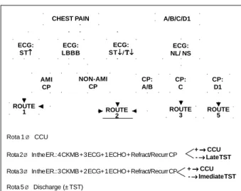
characteristics, especially because of its strong intensity and long duration (equal to or greater than 30 minutes), and as-sociated symptoms (pallor, sweating, nausea, vomiting, dyspnea, etc); 2) Pain of non-acute myocardial infarction: when chest pain did not fulfil the abovementioned properties. Based on chest pain characteristics and ECG type on admission, patients were triaged to diagnostic pathways or routes, in which diagnostic and prognostic procedures and therapeutic measures were pre-established (fig. 1).
Thus, patients were included into route 1 if they had chest pain and ECG strongly suggestive of acute myocar-dial infarction (ST segment elevation or left bundle branch block). Due to the very high probability of acute myocardial infarction, these patients underwent either thrombolytic therapy, primary angioplasty or conservative management while in the emergency room and were sent thereafter to the coronary care unit.
Patients were included into route 2 if they had ST de-pression or T wave inversion, or chest pain suggestive of acute coronary insufficiency (type A or B) but without is-chemic ECG changes, or yet, left bundle branch block and nonacute myocardial infarction-type chest pain. Due to the high probability of unstable angina and intermediate probability of acute myocardial infarction in this subset of patients, they were kept in the emergency room for at least 9 hours to undergo serial ECG and plasma creatine-kinase measurements (every 3 hours) and 2D echocardiogram as soon as possible.
Patients were included into route 3 if they had chest pain not completely exclusive of acute coronary insuffi-ciency (type C) and without ischemic ECG changes. Due to the low probability of unstable angina and acute myocardial infarction, these patients were also kept in the emergency room for at least 6 hours to undergo serial ECG and creatine-kinase measurements ( every 3 hours) and a 2D echocar-diogram.
CHEST PAIN A/B/C/D1
ECG: ST↑↑↑↑↑
ECG: LBBB
ECG: ST↓/Τ↓↓/Τ↓↓/Τ↓↓/Τ↓↓/Τ↓
ECG: NL/ NS
AMI CP
NON-AMI CP
CP: A/B
CP: C
CP: D1
ROUTE
1 ROUTE
2
ROUTE
3 ROUTE5
Fig. 1 - Diagnostic pathways for patients with chest pain (AMI- acute myocardial infarction; CCU- coronary care unit; CKMB- creatine-kinase-MB; CP- chest pain; ECG- electrocardiogram; ECHO- echocardiogram; ER- emergency room; LBBB- left bundle branch block; NL/NS- normal/nonspecific; REFRACT/RECURR-refractory/ recurrent; TST- treadmill stress testing.
Rota 1 Ø CCU
Rota 2 Ø In the ER.: 4 CKMB + 3 ECG+ 1 ECHO + Refract/Recurr CP
Rota 3 Ø In the ER.: 3 CKMB + 2 ECG+ 1 ECHO + Refract/Recurr CP Rota 5 Ø Discharge (± TST)
Patients on route 2 and 3 that later presented any eviden-ce of myocardial necrosis (CK-MB elevation) or persistent/ recurrent myocardial ischemia (see below) were transferred to the coronary care unit for further evaluation and treatment. Other patients in routes 2 and 3 continued under vigilance in the emergency room or intermediate care unit and, if remaining asymptomatic and without evidence of recurrent ischemia, underwent treadmill stress testing (or exercise myocardial scintigraphy) in 24 - 48 hours and 06 - 12 hours, respectively.
Patients were included into route 5 who had nonan-gina chest pain (type D) without ischemic ECG changes. These patients were immediately discharged from the emergency room and instructed to undergo a treadmill stress test as outpatients.
At the time of this study, route 4 was planned to inves-tigate patients with suspected aortic dissection or pulmona-ry embolism, even if not active.
For the purpose of transferring patients initially alloca-ted to routes 2 or 3 to the coronary care unit, myocardial ne-crosis was considered present when plasma CK-MB was elevated above the upper limit of normal in any measure-ment undertaken in the emergency room, even when it did not fulfil the diagnostic criteria of acute myocardial infarc-tion (see below). For the same purpose, persistent or recur-rent myocardial ischemia was considered to exist when new or worsening of old ST depression or T wave inversion de-veloped or when segmental or global systolic dysfunction in the echocardiogram in areas without old Q wave in the ECG occurred. Chest pain refractoriness to appropriate pharmacologic anti-ischemic treatment, as well as recurren-ce, even in the absence of ECG changes, were also determi-nants for transferring route 2 and 3 patients to the coronary care unit.
In relation to the final diagnosis, a patient was designa-ted as having acute myocardial infarction when he or she had, independently of admission, ECG changes, abnormal serial CK-MB levels equal to or greater than 10% of total CPK plus any one of the following criteria: 1) three conse-cutive CK-MB measurements above the upper limit of nor-mal; 2) two consecutive CK-MB measurements at least 50% above the upper limit of normal; 3) one CK-MB measure-ment at least 100% above the upper limit of normal.
A patient was designated as having unstable angina if he or she did not have any of the above mentioned CK-MB elevations and had: 1) Chest pain type A or B with duration greater than 20 minutes, independently of any ischemic changes in the ECG, echocardiogram or stress test; or 2) Chest pain type C or D with duration greater than 20 minu-tes, but associated with ischemic changes in the ECG, echo-cardiogram or stress test.
A patient was designated as not having acute corona-ry insufficiency when no criteria for acute myocardial infarc-tion or unstable angina were fulfilled (ruled out).
Finally, a patient was designated as having an unde-termined diagnosis when acute myocardial infarction or unstable angina could neither be ruled in nor ruled out due to insufficient diagnostic information.
Statistical analysis: Sensitivity, specificity and positi-ve and negatipositi-ve predictipositi-ve values of tests were calculated for acute myocardial infarction. Sensitivity was defined as the rate of positive tests in patients with infarction. Specifi-city was defined as the rate of negative tests in patients wi-thout infarction. The positive predictive value was defined as the rate of patients with positive tests who had infarc-tion. Negative predictive value was defined as the rate of patients with negative tests that did not have infarction. Global accuracy was defined as the rate of correct diagnosis of tests (positive and negative) in the studied population. Significance of difference between proportions was calcula-ted by the chi-square test.
Results
Of 1003 patients with chest pain seen between Novem-ber 1, 1996 and February 28, 1998 in the emergency room, 119 were immediately transferred to the coronary care unit for having ST segment elevation or left bundle branch block plus acute myocardial infarction-type chest pain (route 1). Two-hundred twenty-four patients were discharged from the emergency room for having definitely not angina-type chest pain (type D) plus a normal/nonspecific ECG (route 5). Therefore, 660 patients without ST segment elevation that remained in the emergency room to be investigated in rou-tes 2 and 3 constitute the study sample and followed the diagnostic pathways (fig. 1).
The 433 patients in route 2 presented with the follo-wing ECG patterns on admission: ST segment depression/T wave inversion (n=159), left bundle branch block plus non acute myocardial infarction-type chest pain (n=25) and nor-mal/ nonspecific ECG plus definitely/ probable angina-type chest pain (n=249). Two hundred and twenty-seven patien-ts were allocated in route 3 (normal/ nonspecific ECG plus probably not angina-type chest pain for patients).
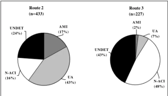
Figure 2 discloses the relationship between allocated routes and patients’ final diagnosis. It can be seen that more than one-half of those patients assigned to route 2 had acute coronary insufficiency (AMI= 73, UA=186) whereas in route 3 this occurred in less than 10% of patients (AMI= 4, UA=16). Similarly, the final diagnosis of absence of acute coronary insufficiency was found in 71 patients (16%) in route 2 and 109 patients (48%) in route 3.
In route 2, acute myocardial infarction was found in 45 of 159 patients (28%) with ST depression/T inversion, in 2 of 25 patients (8%) with left bundle branch block plus non-AMI chest pain, and in 26 of 249 patients (10%) with nor-mal/nonspecific ECG plus type A/B chest pain.
was the first CK-MB negative. In the 249 patients with nor-mal/nonspecific ECG and type A/B chest pain 26 had acute myocardial infarction but the first CK-MB was negative in 14 (54%). Of the 73 patients in route 2 who had acute myo-cardial infarction, the CK-MB became positive only in the sample obtained in the 9th hour postadmission in 3 of the patients. Therefore, the sensitivity of the first CK-MB for the diagnosis of AMI in patients in route 2 was 51% whe-reas negative predictive values of the first and third CK-MB (0 hour and 6th hour) were 89% and 99%, respectively (fig. 3). In route 3, one of the 4 patients who had acute myocardial infarction had a negative CK-MB on admission, and in all CK-MB became positive by the third hour. Therefore, sen-sitivity of the first CK-MB in route 3 was 75% but the nega-tive predicnega-tive value was 99.5% (fig. 3).
Thus, global accuracy of the first CK-MB for the diag-nosis of AMI in patients with chest pain and no ST eleva-tion or left bundle branch block (routes 2 and 3) was 91%, with 52% sensitivity and 93% negative predictive value.
In the analysis of 476 patients with normal/nonspecific ECG in routes 2 and 3, thirty (6%) had acute myocardial in-farction and 124 (26%) had unstable angina. From 395 pati-ents of this group who had negative CK-MB on admission, 15 (4%) had AMI. Fourteen of the 15 had type A or B chest pain and one had type C, making the probability of AMI in these subgroups (type A/B and type C) 7% and 0.5%, respectively
(p<0.005). Therefore, the sensitivity of the first CK-MB for the diagnosis of AMI in patients with normal/ nonspecific ECG was 46% and the negative predictive value was 96%.
Eighty-nine of 202 patients (44%) with unstable angi-na in routes 2 and 3 had some sort of ischemic ECG changes on admission or during the emergency room stay (ST de-pression or T inversion), 10 (5%) had left bundle branch block, 48/156 (31%) had segmental left ventricular systolic dysfunction on the echocardiogram and 23/177 (13%) had troponin-T elevation (several patients had concomitant changes). Therefore, 112 patients (55%) had some evidence of significant myocardial ischemia at rest whereas in 90 (45%) nothing abnormal was found.
Discussion
Patients seen in the emergency room with chest pain and no ST segment elevation in the ECG constitute a signi-ficant diagnostic challenge. The need to rule out acute myo-cardial infarction as the cause of the symptom has driven physicians to adopt precocious and defensive behavior for the sake of patient’s safety, but also for their own. This poli-cy has resulted in large resource expenditures as many of these patients are admitted to high-cost hospital units (co-ronary care and intensive care) where they end up also un-dergoing high-cost diagnostic tests (nuclear medicine, cine-coronary arteriography, etc). The final result of this strate-gy has been disappointing as only 20 to 30% of these pati-ents actually have acute coronary insufficiency (less than one-half having myocardial infarction) 2-4. The cost of these
hospitalizations, that result from 3 to 6 million annual emer-gency room visits for chest pain in the United States, is esti-mated to be 6-8 billion dollars 3.
At the same time, and in spite of physicians’ defensive behavior, it as been observed that about 5% of patients se-en in emergse-ency departmse-ents in the United States with chest pain and acute myocardial infarction are inappropria-tely discharged home 1,10,11. About 25% of these patients
died a few days afterwards. It has also been estimated that 20% of medical malpractice costs in lawsuits paid by phy-sicians or their insurance companies are due to these diag-nostic mistakes 12.
Route 2 (n=433)
Route 3 (n=227)
UNDET (24%)
A M I (17%)
N-ACI
(16%) UA
(43%)
UNDET (43%)
A M I (2%)
UA (7%)
N-ACI (48%)
Fig. 2 - Rates of acute myocardial infarction (AMI), unstable angina (UA), undeter-mined diagnosis (UNDET) and absence of acute coronary insufficiency (N-ACI) in 660 patients with chest pain in routes 2 and 3.
Fig. 3 - Sensitivity (SENS) and negative predictive value (NPV) of serial CK-MB for the diagnosis of acute myocardial infarction according to assigned route in the diagnostic pathway.
50 100
0 %
Route 2 (n = 433)
Route 3 (n = 227)
In Brazil, no measurements or estimates have been ma-de of the amount and quality of emergency medical visits for chest pain. A survey made at our institution between November 1, 1996 and June 30, 1997 disclosed that this was the single most common cause of emergency room visits: 21% of 2490 visits in that period. This rate is higher than the 5-10% rate found in North American hospitals 1,3,4 and it is
certainly due to the peculiar characteristics of our institu-tion that, besides being a community cardiology-oriented hospital, it is also a reference center for emergency cardio-logy care.
In addition, emergency care in many towns in Brazil is precarious, and even in large and cosmopolitan cities the quality is heterogeneous from institution to institution. Thus, it is not illogical to assume that the rate of inappropri-te discharge from the emergency room of patients with acute myocardial infarction in Brazil would be greater than 10% in general 13 .
Concerned with these medical, legal and economic problems, several groups have sought special strategies of care for patients with chest pain.These strategies look for a fast diagnosis (or ruling out some diagnoses), for ideally reducing diagnostic error to zero, and for diminishing the cost of investigation 2,3,6,7,14,15, that is, to act sing the
cost-efficiency binomial. The creation of socalled Chest Pain Units has been proclaimed as the most appropriate way to reach this aim 3,8,16.
The present study demonstrates that patients with chest pain and nondiagnostic ECG (without ST segment ele-vation) must be initially stratified according to the pretest probability of acute myocardial infarction or acute coronary insufficiency. Thus, patients with ST depression or T inver-sion, with left bundle branch block, or with normal/nonspe-cific ECG but with definite or probable angina-like chest pain (route 2 of our diagnostic pathways) had rates of 17% of AMI and 43% of UA. These figures contrast with the rates of 2% and 7%, respectively, found in patients with normal/ nonspecific ECG plus not probable angina-like chest pain (route 3). These results are quite similar to those found by Goldman et al 2 and indicate that, even in route 3 patients
with low probability of AMI this diagnosis should be care-fully scrutinized if one does not wish to erroneously dis-charge the patient.
At the same time one should not fail to mention that about 15% of patients in route 2 had the diagnosis of acute coronary insufficiency ruled out during the emergency ro-om stay whereas this occurred in almost one-half of route 3 patients (fig. 2). Had these patients been hospitalized in the coronary care unit, as is done in many centers, they would have stayed there for 3 or 4 days, undergoing continuous ECG monitoring, serial plasma enzyme measurements and other tests, which would certainly result in a large and unne-cessary expenditure 3.
In addition, this study demonstrates that diagnostic strategies to separate chest pain patients into subgroups of risk does not apply only to determining the probability of AMI. It also establishes how long a patient should be
eva-luated for the correct diagnosis to be made (or to rule out the suspected diagnosis). The study made clear that patients enrolled in route 2 of the diagnostic pathways can only be sent safely to a provocative test of myocardial ischemia after measuring plasma CK-MB 9 hours after hospital ad-mission. Patients designated to route 3 can have it done after the measurement at 3 hours. From patients in route 2 with a first (admission) negative CK-MB 11% had AMI whereas in route 3 AMI was seen in less than 1%.
In patients with chest pain and no ST elevation, the diagnosis of AMI can only be confirmed by a rise in plasma CK-MB. However, several studies have demonstrated a low sensitivity for this cardiac marker when measured on hospital arrival (around 35%) 6,17. This sensitivity increases
as the time interval between chest pain onset and hospital arrival becomes greater 18
.
The diagnostic strategy of our study differs some-what from that of Gibler at al 3. These authors recommend
that all patients with chest pain suggestive of acute corona-ry insufficiency and no ST elevation be evaluated with se-rial CK-MB measurements up to 9 hours after admission. Our diagnostic pathways require that this is only to be done in patients in route 2, thus avoiding greater costs for patients in route 3 (that constitute 23% of the entire chest pain popula-tion or 34% of those without ST elevapopula-tion). In those, acute myocardial linfarction can be ruled out in 3 hours.
Patients with normal/nonspecific ECG constitute the most problematic group, once their probability of having AMI is low but not zero. In our study, 476 patients did not have significant ECG changes on admission; however, 6% of them had acute myocardial infarction and 26% had unstable angina. First CK-MB was negative in one-half (15/ 30) of those with normal/ nonspecific ECG changes and AMI. Conversely, 4% (15/395) of those with normal/ nonspecific ECG plus negative first CK-MB had AMI. The-se data highlight the precaution one has to take in patients without ECG changes in order to avoid the release of pati-ents with unrecognized AMI.
The diagnosis of unstable angina is frequently diffi-cult to establish, due to unclear presenting symptoms or to nondiagnostic ECG changes. Many of these patients could only have the diagnosis ruled out after a provocative test of myocardial ischemia or cinecoronary arteriography. We have recently demonstrated that about one-half of unstable angina patients have a normal or nonspecific ECG 19. In the
present study, most patients underwent treadmill stress testing and, eventually, exercise myocardial scintigraphy; thus, very few patients were sent straight to cinecoronary arteriography for diagnostic purposes.
Our study confirms the results obtained by Goldman et al 2 using an algorithm based on chest pain
probabi-lity) had a rate of 2%, a similar figure to the other study (Gol-dman’s rates obtained from published data).
Beyond the diagnostic accuracy for acute myocardial infarction, the pathway used in this study is also in search of identifying patients with unstable angina and to establish their risk of events. Although patients assigned to route 2 (high probability of UA) had an incidence of 43%, those in route 3 (low probability) had 7%. The lack of concern with the diagnosis of unstable angina is a significant drawback and limitation of Goldman’ s algorithm 20 .
Therefore, the diagnostic pathway used in this study al-lowed the systematic management of chest pain patients, tailoring the diagnostic strategy according to the pretest pro-bability of acute coronary insufficiency, thus permitting re-duction of investigative costs. This is the first published study that proposes a discriminative diagnostic model for chest pain patients based on chest pain type and ECG changes.
At a time when government resources allotted to health care are in their lowest per capita volume in Brazil’s history, and, simultaneously, private health care insurance companies and group health maintenance organizations are trying to progressively reduce their expenses, it is im-perative to make medical practice cost-effective. Syste-matic models of medical care that optimize the binomial quality-cost relationship turn out to be vital mechanisms in this task. The diagnostic pathway used by our group -applied in the context of a Chest Pain Unit - is a tool of great value (as demonstrated in this study) to improve quality and efficiency of medical care and, at the same time, reduce global and per patient costs. This model can be used in any hospital or emergency room, because it does not require special equipment or large investments. What is indispensable to have is a uniform strategy to be practiced by a trained and qualified team.
References
1. Graff LG, Dallara J, Ross MA, et al. Impact on the care of the emergency department chest pain patient from the chest pain evaluation registry (CHEPER) study. Am J Cardiol 1997; 80: 563-8.
2. Goldman L, Cook EF, Brand DA, et al. A computer protocol to predict myocardial infarction in emergency department patients with chest pain. N Engl J Med 1988; 318: 797-803.
3. Gibler WB, Runyon JP, Levy RC, et al. A rapid diagnostic and treatment center for patients with chest pain in the emergency department. Ann Emerg Med 1995; 25: 1-8.
4. Zalenski RJ, Rydman RJ, McCarren M, et al. Feasibility of a rapid diagnostic protocol for an emergency department chest pain unit. Ann Emerg Med 1997; 29: 99-108.
5. Lee TH, Weisberg MC, Brand DA, et al. Candidates for thrombolysis among emergency room patients with acute chest pain. Potential true- and false-positive rates. Ann Intern Med 1989; 110: 957-62.
6. Bassan R, Scofano M, Gamarski R, et al. Dor torácica na sala de emergência: a importância de uma abordagem sistematizada. Arq Bras Cardiol 2000; 74: 13-21. 7. Pozen MW, D’Agostino RB, Mitchel JB, et al. The usefulness of predictive instrument to reduce inappropriate admissions to the coronary care unit. Ann Intern Med 1980; 92: 238-42.
8. Bahr RD. Acute outpatient care and comprehensive management of acute myocardial ischemia in chest pain emergency departments. Md Med J 1995; 44: 691-3. 9. Sgarbossa EB, Pinski SL, Barbagelata A, et al. Electrocardiographic diagnosis
of evolving acute myocardial infarction in the presence of left-bundle branch block. N Engl J Med 1996; 334: 481-7.
10. Lee TH, Ronan GW, Weisberg MC, et al. Clinical characteristics and natural history of patients with acute myocardial infarction sent home from the emergency room. Am J Cardiol 1987; 60: 219-24.
11. Mc Carthy BD, Beshansky JR, Agostino RB, Selker HP. Missed diagnosis of acute myocardial infarction in the emergency department: results from a multicenter study. Ann Emerg Med 1993; 22: 579-82.
12. Karcz A, Holbrook J, Burke MC, et al. Massachusetts emergency medicine closed malpractice claims: 1988-1990. Ann Emerg Med 1993; 22: 553-9.
13. Bassan R, Pimenta L, Scófano M, et al. How many patients with acute myocardial infarction are at risk of being erroneously released from the emergency room? Eur Heart J 1999; 20 (suppl): 283.
14. Gaspoz JM, Lee TH, Cook EF, Weisberg MC, Goldman L. Outcome of patients who were admitted to a new short-stay unit to “rule out” myocardial infarction. Am J Cardiol 1991; 68: 145-9.
15. Tatum JL, Jesse RL, Kontos MC, et al. Comprehensive strategy for the evaluation and triage of the chest pain patient. Ann Emerg Med 1997; 29: 116-25. 16. Graff L, Joseph T, Andelman R, et al. American College of Emergency Physicians
Information Paper: chest pain units in emergency departments - a report from the short - term observation services section. Am J Cardiol 1995; 76: 1036-9. 17. Lee TH, Weisberg MC, Cook EF, et al. Evaluation of creatine kinase and creatine
kinase - MB for diagnosing myocardial infarction: Clinical impact in the emergency room. Arch Intern Med 1987; 147: 115-21.
18. Marin MM, Teichman SL. Use of rapid serial sampling of creatine kinase MB for very early detection of myocardial infarction in patients with acute chest pain. Am Heart J 1992; 123: 354-61.
19. Bassan R, Araujo M, Volschan A, Dohmann HF, Gaspar S, Fabrício M. Does the diagnosis of unstable angina require the presence of ischemic ECG changes? J Am Coll Cardiol 1999; 33(supl): 393-A.