Pró-Cardíaco Hospital, Rio de Janeiro
Mailing address: Roberto Bassan - Hospital Pró-Cardíaco - Rua General Polidoro, 192 - 22280-000 - Rio de Janeiro, RJ – Brazil – E-mail: [email protected]
Roberto Bassan
Rio de Janeiro, RJ - Brazil
Chest Pain Units. A Modern Way of Managing Patients with
Chest Pain in the Emergency Department
It is estimated that 5 to 8 million individuals with
chest pain or other symptoms suggestive of myocardial
ischemia are seen each year in emergency departments
(ED) in the United States
1,2, which corresponds to 5 to
10% of all visits
3,4. Most of these patients are hospitalized
for evaluation of possible acute coronary syndrome (ACS).
This generates an estimated cost of 3 - 6 thousand dollars
per patient
5,6. From this evaluation process, about 1.2
mil-lion patients receive the diagnosis of acute myocardial
in-farction (AMI), and just about the same number have
uns-table angina. Therefore, about one half to two thirds of
the-se patients with chest pain do not have a cardiac cauthe-se for
their symptoms
2,3. Thus, the emergency physician is faced
with the difficult challenge of identifying those with ACS
-a life-thre-atening dise-ase - to tre-at them properly, -and to
discharge the others to suitable outpatient investigation
and management.
To establish the correct diagnosis and the appropriate
treatment for patients with chest pain is one of the most
im-portant problems facing not only physicians and hospitals
but also the payers – government, health-insurance
compa-nies, or health maintenance organizations. Emergency
physicians are traditionally recommended to act on behalf of
patients’ health and safety. Therefore, incapable of
establi-shing with certainty the etiologic diagnosis of chest pain
patients using history, physical examination and
electrocar-diography (ECG) data, physicians are pressured to admit
these patients to the coronary care unit. As most of these
patients do not have cardiovascular disease, this results in
the expenditure of some 5 to 8 billion dollars in cost for
un-necessary hospital admissions in the United States
2,7,8.
However, even with this exaggerated effort to identify
cases of ACS, an average of 2 - 3% of patients with chest
pain who actually have AMI are unintentionally released
from the EDs in the United States, and this rate may go up to
11% at some centers
8-10. This amounts to some 40,000
indi-viduals each year. In countries where emergency physicians
have less expertise in dealing with chest pain patients or are
less aggressive in admitting them to the hospital, this rate
could reach 20%
11.
At the same time, physicians have been pressured by
health insurance companies and hospital managers to avoid
admitting patients who have an unclear diagnosis
12.
Re-trospective denial of payment by insurers for hospitalized
patients who end up not having ACS makes the admission
of low-risk patients problematic. The release of patients with
AMI represents a significant medico-legal risk for
emer-gency physicians, with 20% of malpractice dollar
settle-ments each year in the United States being associated with
the misdiagnosis of AMI
13,14.
For all the previously mentioned reasons, physicians
are faced with the problem of admitting most patients
co-ming to the ED with chest pain, or releasing those that have
a very low likelihood of a life-threatening disease, yet they
may in fact have ACS with a resulting complication. Thus,
most emergency physicians in the United States admit
vir-tually all patients who have any possibility of ACS due to
knowledge of the following information. First, some 15 to
30% of such patients actually do have ACS
15,16. Second,
just about one half of patients with AMI have the classic
change of ST-segment elevation on the admission ECG
17,18.
Third, less than 50% of patients having AMI without
ST-segment elevation have an abnormal admission serum
creatine kinase-MB (CK-MB) level
19-21.
Therefore, the evolution of Chest Pain Units has been
recognized as a reasonable and viable approach to deal with
these patients in the ED in a cost-effective way
12,22, as we
will discuss in this report.
Chest pain units -
Since the early 1960s, coronary care
units have been the ideal setting for managing patients with
a clear-cut diagnosis of AMI. The excellent results
obser-ved in these units, particularly with early recognition and
treatment of cardiac arrhythmias and cardiac arrest, led
phy-sicians to begin admitting patients with suspected ACS
23,24.
The result of this more liberal approach was more than one
half of admitted patients did not have a final diagnosis of
ACS
24. Consequently, high-cost hospital beds were filled
by emergency physicians with low-risk patients, resulting in
saturation or overflow of the coronary care units, with
sub-optimal use of medical resources, and high costs associated
with this evaluation.
Chest Pain Units or Centers can be defined as a new
area of emergency medical care devoted to improving
mana-gement of patients with acute chest pain or any other
symptom suggestive of ACS. The main objectives of these
units are to provide (1) easy and friendly admission for the
patient presenting to the ED, (2) priority and rapid access to
the medical staff in the ED, and (3) an organized and
effici-ent strategy of medical care within the ED, including
diag-nosis and treatment, aimed at dispensing the best possible
medical care at the lowest possible cost.
Chest Pain Units can be located in or adjacent to the
ED, in a true physical area, or just as a working process
wi-thin the emergency center. What is essential is that a group
of trained and qualified personnel act in synchrony when
receiving a patient with chest pain to achieve the previously
mentioned objectives: rapid and efficient evaluation, early
identification of ACS, high-quality care, and
cost-effecti-veness
2,3,25,26.
One of the keys to the success of the Chest Pain Units
is the use of systematic diagnostic algorithms and specific
management protocols
3,26. The use of Chest Pain Units has
resulted in improved care of patients with and without ACS,
as depicted in table I and discussed as follows.
Pre-hospital delay (procrastination of patients with
ACS in coming to the ED) is a worldwide problem and
res-ponsible for about 50% of AMI deaths
27,28. Many studies
have demonstrated that the mean time-interval between
symptom onset and hospital arrival in patients with AMI is
2 to 3 hours
2,29. This delay may lead to prehospital death
and may be the reason for ineligibility for thrombolytic
therapy in many patients with AMI
30,31. Chest Pain Units
can be an instrument for patient education, particularly
for those needing risk factor modification or symptom
re-cognition
2,3.
In-hospital delay, the time interval between hospital
arrival and diagnosis with initiation of specific therapy (also
known as door-to-needle time), is another problem that
af-fects most of the hospitals around the world, even in
deve-loped countries. This time frame is about 1 hour
2,29. One of
the most important reasons for this delay is lack of priority in
the initial assessment of chest pain patients, which are
frequently passed by on behalf of those with trauma,
gas-trointestinal bleeding, etc. Chest Pain Units perform an
important and unique role in reducing this delay through its
action to prioritorize high-risk individuals and to use
proto-cols to evaluate and treat patients
2,3,12,26, as recommended
by the National Heart Attack Alert Program
31.
Inappropriate hospital release of patients with AMI and
unstable angina is a serious problem in emergency medicine
that has been persistent over time
9,10,32,33. As previously
mentioned, diagnostic error in these cases has ranged from
zero to more than 10% in renowned medical institutions
10.
Through the training of its personnel and the use of careful
and systematic diagnostic strategies, Chest Pain Units can
decrease inappropriate AMI release to less than 1%.
Excessive and unnecessary hospitalizations in
high-complexity, high-cost units, such as coronary care units, are
a frequent problem in medical practice, especially when
physicians are in need of a bed for their patients with known
AMI. Chest Pain Units act to buffer the coronary care units
by evaluating patients with an unclear diagnosis, therefore
reducing the rate of low-risk admissions and, consequently,
increasing the availability of beds for those who really need
them
26,34.
The high costs of contemporary medicine have proved
to be an important economic burden for society. Money
used unwisely for the management of low-risk chest pain
pa-tients could be better used in high-risk papa-tients.
Cost-con-tainment measures make Chest Pain Units more attractive
not only to administrators but also to physicians as well,
because low-risk patients can also be thoroughly and
ade-quately evaluated in this setting
10. Chest Pain Units have
been demonstrated to reduce these costs, mainly through
reduction in duration of hospital stay and the number of
diagnostic tests ordered, especially those that have little or
no diagnostic yield
5,7,28,35-38. Diagnostic algorithms or
pro-tocols are important tools to attain such efficiency. As they
also improve the quality of medical care, Chest Pain Units
promote an unquestionable salutary shift in the
cost-bene-fit relationship.
Diagnostic strategies for chest pain patients -
Chest
pain is a symptom associated with multiple pathologic
en-tities, some benign
39. However, emergency physicians are
usually concerned primarily with those that are
life-threa-tening, namely ACS, pulmonary embolism, and aortic
dissection. Although none of these are the most frequent
cause of chest pain in the ED, AMI and unstable angina
are quite common in this setting (10 to 30% of cases).
Pul-monary embolism and aortic dissection have an incidence
of less than 1%.
The history is an extremely valuable tool in the
diffe-rential diagnosis of chest pain
39-43. The association of chest
pain characteristics and admission ECG changes, with or
without the information of patient’s age, risk factors profile,
and past history, has enabled investigators to create
proba-Table I - Aims of chest pain
1. Reduce prehospital delay of chest pain patients.
2. Reduce in-hospital delay for identifying and treating ACS patients. 3. Prevent inappropriate release of ACS patients.
4. Reduce unnecessary hospitalization rate for non-ACS patients 5. Reduce medical costs in the assessment of chest pain patients.
bilistic algorithms or clinical prediction rules to estimate the
chance of ACS or AMI in these patients
16,21,39,44,45. The
diagnostic accuracy of these tools has been confirmed in
several studies
46-49and recommended in 1 guideline
50.
Determination of pretest probability of ACS is
impor-tant in establishing diagnostic strategies that are most
cost-effective. Thus, patients with a high probability should be
thoroughly investigated, whereas patients with a low
proba-bility of ACS may need less extensive and costly
investiga-tion in the emergency setting. Several strategies have been
proposed and used in different centers
8,21,44,51-53, but they all
have in common the need for a diligent and careful
determi-nation of the pretest probability of disease and the proper
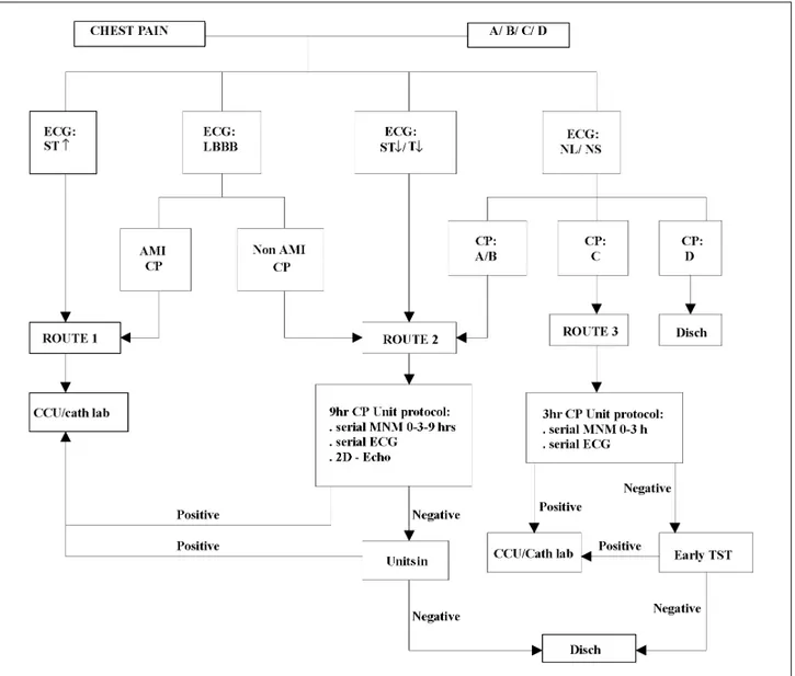
allocation of resources. Figure 1 depicts the diagnostic
stra-tegies used in the Pro-Cardiaco Hospital, establishing
dif-ferent diagnostic pathways according to the pretest
proba-bility of ACS. Characterization and classification of chest
pain type and admission ECG (tabs. II and III) are crucial
steps to correctly stratify the probability of ACS in these
patients. Thus, route 1 is the pathway for high-probability
patients, whereas route 2 is for those with intermediate
pro-bability and route 3 for those with low propro-bability
21,34. A
previously published study from our group validated the
discriminatory property of this model, as depicted by the
observed rates of AMI and unstable angina in those routes
of 74% and 17%, 17% and 43%, and 2% and 7%,
respec-tively
34.
Developing a protocol for the Chest Pain Unit -
Any
diagnostic protocol or algorithm for assessing patients who
come to the ED with chest pain must be based on the
inter-pretation of chest pain characteristics and admission ECG,
which can be made by experienced and trained emergency
physicians and nurses. With these data one can make an
ac-curate estimate of the risk (or probability) of ACS
34,39,40,54.
However, diagnostic confirmation most of the time requires
the use of other laboratory examinations.
Serum markers, such as myoglobin, CK-MB, and
tro-ponins I and T are necessary to detect myocardial necrosis
and to risk-stratify these patients
5,19,55. The duration of
myocardial necrosis markers screening should not be less
than 3 hours and generally between 6 and 9 hours after
ad-mission to the Chest Pain Center. Ideally, 3 serial
measure-ments should be obtained until at least 12 hours after pain
onset
19-21,24,51,55.
Patients with a diagnosis of AMI or high-risk unstable
angina confirmed at this point should be admitted to the
hospital, but their antiischemic therapy should be initiated
in the ED.
Remaining patients with a negative series of
myocar-dial necrosis markers still require an evaluation for acute
cardiac ischemia without infarction. ST-segment trend
mo-nitoring, two-dimensional echocardiography, and rest
myocardial perfusion scintigraphy have been
systematical-ly used
42,51,52.
Sensitivity of rest tetrofosmin or sestamibi SPECT for
detecting AMI has ranged from 90 to 100%, with a negative
predictive value of 99%
44,57-60, whereas the rest
echocardio-gram is between 47 - 93% and 86 - 99 %, respectively
34,51,61-63.
The diagnostic accuracy of ST- segment trend monitoring is
still under investigation
64,65.
Graded exercise testing, with or without myocardial
ra-dionuclide scintigraphy or stress echocardiography, can be
performed to further risk-stratify these patients in whom
AMI or rest myocardial ischemia has been ruled out
34,35,51,66.
Although these tests are important tools to assist in the
diagnosis of residual myocardial ischemia and, therefore,
unstable angina, they also contribute to assess prognosis
in acute chest pain patients. A negative exercise test is
as-sociated with minimal (<2%) chance of death or AMI in the
following year
5,44,57,58,63,67-69.
Thus, provocative testing
be-comes extremely important in completing the Chest Pain
Unit evaluation in these patients.
Therefore, the Chest Pain Units provide a thorough
evaluation for patients with chest pain presenting to the ED.
This investigation is aimed at detecting not only myocardial
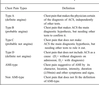
Table II - Classification and definitions of chest pain types used in the Chest Pain Unit of Pró-Cardíaco Hospital
Chest Pain Types Definition
Type A Chest pain that makes the physician certain (definite angina) of the diagnosis of ACS, independently
of other tests.
Type B Chest pain that makes ACS the main (probable angina) diagnostic hypothesis, but needing other
tests to confirm it.
Type C Chest pain that does not make (probable not angina) ACS the main diagnostic hypothesis, but
needing other tests to rule it out. Type D Chest pain that does not include ACS as a (definite not angina) cause (D1= without diagnosis on
admission; D2= with diagnosis)
AMI-type Chest pain suggestive of AMI by its character, location, intensity, duration (≥30min) and other symptoms and signs. Non AMI-type Chest pain that does not fit the definition
of AMI-type.
ACS- acute coronary syndrome; AMI- acute myocardial infarction.
Table III - Classification and definitions of the admission electrocardiogram (ECG) pattern used in the Chest Pain Unit of
Pró-Cardíaco Hospital
ECG Types Definition
ST-segment elevation Positive deviation of J-ST >0.1mV in ≥ 2 leads of the frontal plane or >0.2mV in leads of the horizontal plane.
ST-segment depression Negative deviation of J- ST ≥0.1mV in ≥ 2 leads, or T-wave inversion in ≥2 leads. Left bundle branch block In sinus rhythm, QRS duration ≥ 120 ms
with QS or rS in V1 and intrinsecoid deflection ≥60 ms in L1, V5 or V6 , and absence
of Q-waves in these leads.
necrosis, but also rest and exercise-induced ischemia.
Pa-tients with a negative evaluation that encompasses these 3
objectives have a very low risk for complications after
dis-charge from the Chest Pain Unit.
Chest Pain Units in Brazil -
The first Chest Pain Unit
in our country was developed in 1996 at Pró-Cardíaco
Hos-pital, a private, clinical emergency care institution in the city
of Rio de Janeiro. Chest pain is responsible for some 20% of
all ED visits in that hospital, a rate remarkably higher than
that seen in general EDs.
The Chest Pain Unit was initially seen with restrictions
and disbelief by attending physicians and health insurers
due to the systematic diagnostic protocol, supposedly more
costly and cumbersome than the traditional ED evaluation.
However, a rapid change of opinion occurred as results and
benefits of the new method of patient assessment were
appre-ciated. A data bank, prospectively obtained, allowed
gene-ration of important scientific information that were was
im-mediately released to the medical community
11,18,20,21,34,69,
confirming reports of other institutions and validating its
own efficacy.
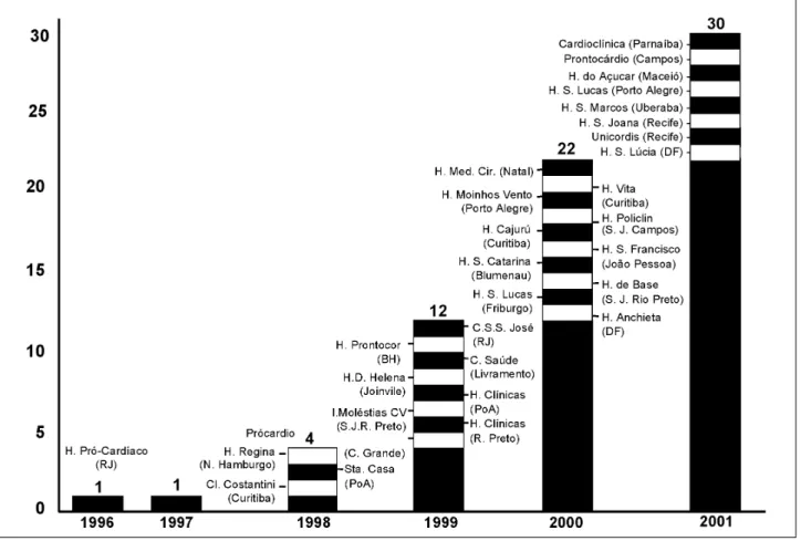
Following this pioneering experience, other
institu-tions established their own Chest Pain Units. In 1998, 4 of
them were functioning in Brazil and in 2001 a total of 30 were
known, located in 13 states, most of them in medium-sized
cities and small hospitals (figs. 2 and 3). It is important to
emphasize that an estimated 1000 Chest Pain Units exist at
the present time in the United States
7, which represents
al-most 20% of all the 4,300 EDs in that country.
Fig. 2 - Development of Chest Pain Units in Brazil (up to July 2001).
References
1. Stussman BJ. National Hospital Ambulatory Medical Care Survey: 1995 emer-gency department summary. Advance data from vital and health statistics. No. 285. Hyattsville, Md: National Center for Health Statistics, 1997 (DHHS publi-cation no. (PHS) 97-1250).
2. Ewy GA, Ornato JP. 31st Bethesda Conference. Emergency Cardiac Care (1999). J
Am Coll Cardiol 2000; 35: 825-80.
3. Graff L, Joseph T, Andelman R, et al. American College of Emergency Physicians Information Paper: chest pain units in emergency departments – a report from the short-term observation section. Am J Cardiol 1995; 76: 1036-9.
4. Zalenski RJ, Rydman RJ, McCarren M, et al. Feasibility of a rapid diagnostic protocol for an emergency department chest pain unit. Ann Emerg Med 1997; 29: 99-108. 5. Farkouh ME, Smars PA, Reeder GS, et al. A clinical trial of a chest pain
observa-tion unit for patients with unstable angina. N Engl J Med 1998; 339: 1882-8. 6. Lee TH, Goldman L. Evaluation of the patient with acute chest pain. N Engl J
Med 2000; 342: 1187-95.
7. Zalenski RJ, Rydman RJ, Ting S, Kampe L, Selker HP. A national survey of emergency department chest pain centers in the United States. Am J Cardiol 1998; 81: 1305-9. 8. Storrow AB, Gibler WB. Chest pain centers: diagnosis of acute coronary
syndro-mes. Ann Emerg Med 2000; 35: 449-61.
9. McCarthy BD, Beshansky JR, D´Agostino RB, Selker HP. Missed diagnosis of a-cute myocardial infarction in the emergency department: results from a multicen-ter study. Ann Emerg Med 1993; 22: 579-82.
10. Pope JH, Aufderheide TP, Ruthazer R, et al. Missed diagnosis of acute cardiac is-chemia in the emergency department. N Engl J Med 2000; 342: 1163-70. 11. Bassan R, Scofano M, Gamarski R, Pimenta L, Volschan A, Clare C. How many
pa-tients with acute myocardial infarction are at risk of being erroneously dischar-ged from the emergency room? Eur Heart J 2000; 21(suppl): 19.
12. Gibler WB. Chest pain units: Do they make sense now? Ann Emerg Med 1997; 29: 168-71.
13. Rusnak RA, Stair TO, Hansen K, Fastow JS. Litigation against the emergency physician: common features in cases of missed myocardial infarction. Ann Emerg Med 1989; 18: 1029-34.
14. Karcz A, Holbrook J, Burke MC, et al. Massachusetts emergency medicine closed malpractice claims: 1988-1990. Ann Emerg Med 1993; 22: 553-9.
15. Lee TH, Juarez G, Cook EF, et al. Ruling out acute myocardial infarction. A pros-pective multicenter validation of a 12-hour strategy for patients at low risk. N Engl J Med 1991; 324: 1239-46.
16. Selker HP, Griffth JL, D’Agostino RB. A tool for judging coronary care admis-sion appropriateness, valid for real-time and retrospective use. A time-insensiti-ve predictitime-insensiti-ve instrument (TIPI) for acute cardiac ischemia: a multicenter study. Med Care 1991; 29: 610-627 (Erratum, Med Care 1992; 30: 188).
17. Rude RE, Poole K, Muller JE, et al. Electrocardiographic and clinical criteria for recognition of acute myocardial infarction based on analysis of 3697 patients. Am J Cardiol 1983; 52: 936-42.
18. Bassan R, Scofano M, Mesquita E, Dohmann HF, Sanmartin C, Clare C. Diagnos-tic accuracy of the ECG for acute myocardial infarction and unstable angina: expe-rience in a chest pain unit. Ann Emerg Med 1999; 34(part 2): S-64.
19. Gibler WB, Young GP, Hedges JR, et al. Acute myocardial infarction in chest pain patients with nondiagnostic ECGs: serial CK-MB sampling in the emer-gency department. Ann Emerg Med 1992; 21: 504-12.
20. Bassan R, Gamarski R, Pimenta L, Scofano M, Fabricio M, Macaciel R. How many CK-MB determinations are necessary to rule out acute myocardial infarction in pa-tients without ST-segment elevation? Ann Emerg Med 1999; 34 (part 2): S-47. 21. Bassan R, Gamarski R, Pimenta L, et al. Eficácia de uma estratégia diagnóstica para
pacientes com dor torácica e sem supradesnível do segmento ST na sala de emer-gência. Arq Bras Cardiol 2000; 74: 405-11.
22. Newby LK, Mark DB. The chest pain unit - ready for prime time? N Engl J Med 1998; 339: 1930-2.
23. Lown B, Vasaux C, Hood Jr WB, Fakhro AM, Kaplinsky E, Roberge G. Unresol-ved problems in coronary care. Am J Cardiol 1967; 20: 494-508.
24. Roberts R, Kleiman NS. Earlier diagnosis and treatment of acute myocardial infarction necessitates the need for a “new diagnostic mind-set”. Circulation 1994; 89: 872-81. 25. Gibler WB. Chest pain evaluation in the ED: beyond triage (editorial). Am J
Emerg Med 1994; 12: 121-2.
26. Gibler WB. Evaluating patients with chest pain in the ED: improving speed, effi-ciency, and cost – effectiveness, or teaching an old dog new tricks (Editorial). Ann Emerg Med 1994; 23: 381-2.
27. Bahr RD. Chest pain center: moving toward proactive acute coronary care. Int J Cardiol 2000; 72: 101-10.
28. McGovern PG, Pankow JS, Shahar E, et al, for the Minnesota Heart Survery In-vestigators. Recent trends in acute coronary heart disease – mortality, mobidity, medical care, and risk factors. N Engl J Med 1996; 334: 884-90.
29. Kereiakes DJ, Weaver WD, Anderson JL, et al. Time delays in the diagnosis and treat-ment of acute myocardial infarction: a tale of eight cities. Report from the prehospital study group and the Cincinnati Heart Project. Am Heart J 1990; 120: 773-80. 30. Anderson HV, Willerson JT. Thrombolysis in acute myocardial infarction. N
Engl J Med 1993; 329: 703-9.
31. National Heart Attack Alert Program Coordinating Committee 60 Minutes to Treatment Working Group. Emergency department: rapid identification and treatment of patients with acute myocardial infarction. Ann Emerg Med 1994; 23: 311-29.
32. Schor S, Behar S, Modan B, Barell V, Drory J, Kariv I. Disposition of presumed coro-nary patients from an emergency room. A follow-up study. JAMA 1976; 236: 941-3. 33. Lee TH, Rouan GW, Weisberg MC, et al. Clinical characteristics and natural his-tory of patients with acute myocardial infarction sent home from the emergency room. Am J Cardiol 1987; 60: 219-24.
34. Bassan R, Scofano M, Gamarski R, et al. Dor torácica na sala de emergência. A im-portância de uma abordagem sistematizada. Arq Bras Cardiol 2000; 74: 13-21. 35. Gomez MA, Anderson JL, Karagounis LA, Muhlestein JB, Mooers FB. An
emer-gency department-based protocol for rapidly ruling out myocardial ischemia re-duces hospital time and expense: results of a randomized study (ROMIO). J Am Coll Cardiol 1996; 28: 25-33.
36. Hoekstra JW, Gibler WB, Levy RC, et al. Emergency – department diagnosis of myocardial infarction and ischemia; a cost analysis of two diagnostic protocols. Acad Emerg Med 1994; 1: 103-10.
37. Roberts RR, Zalenski RJ, Mensah EK, et al. Costs of an emergency department – based accelerated diagnostic protocol vs hospitalization in patients with chest pain: a randomized controlled trial. JAMA 1997; 278: 1670-6.
38. Graff LG, Dallara J, Ross MA, et al. Impact on the care of the emergency department chest pain patient from the chest pain evaluation registry (CHEPER) study. Am J Cardiol 1997; 80: 563-8.
39. Panju AA, Hemmelgarn BR, Guyatt GH, Simel DL. Is this patient having a myo-cardial infarction? JAMA 1998; 280: 1256-63.
40. Diamond GA, Forrester JS. Analysis of probability as an aid in the clinical diag-nosis of coronary artery disease. N Engl J Med 1979; 300: 1350-8.
41. Berger JP. Right arm involvement and pain extension can help to differentiate coro-nary diseases from chest pain of other origin: a prospective emergency ward study of 278 consecutive patients admitted for chest pain. J Intern Med 1990; 227: 165-72. 42. Ryan TJ. Refining the classification of chest pain: a logical next step in the
evalua-tion of patients for acute cardiac ischemia in the emergency department. Ann Emerg Med 1997; 29: 166-8.
43. Hutter Jr AM, Amsterdam EA, Jaffe AS. 31st Bethesda Conference: Emergency
Car-diac Care (1999). Task Force 2: Acute Coronary Syndromes; Section 2B-Chest discomfort evaluation in the hospital. J Am Coll Cardiol 2000; 35: 853-62. 44. Tatum JL, Jesse RL, Kontos MC, et al. Comprehensive strategy for the evaluation
and triage of the chest pain patient. Ann Emerg Med 1997; 29: 116-25. 45. Goldman L, Cook EF, Brand DA, et al. A computer protocol to predict myocardial
infarction in emergency department patients with chest pain. N Engl J Med 1988; 318: 797-803.
46. Pozen MW, D’Agostino RB, Selker HP, Sytkowski PA, Hood Jr WB. A predictive
patients. Conversely, it has allowed individuals with chest
pain from other causes to be investigated in a less complex
and inexpensive environment, releasing them safely from
the hospital. The final consequence of the proper use of
Chest Pain Units has been a significant reduction in
diag-nostic errors, decreased hospital admissions with
instrument to improve coronary care unit admission practices in acute ischemic heart disease. A prospective multicenter clinical trial. N Engl J Med 1984; 310: 1273-8. 47. Goldman L, Cook EF, Johnson PA, Brand DA, Rouan GW, Lee TH. Prediction of
the need for intensive care in patients who come to emergency department with acute chest pain. N Engl J Med 1996; 334: 1498-504.
48. Aufderheide TP, Rowlandson I, Lawrence SW, Kuhn EM, Selker HP. Test of the acute cardiac ischemia time-insensitive predictive instrument (ACI-TIPI) for pre-hospital use. Ann Emerg Med 1996; 27: 193-8.
49. Zalenski RJ, McCarren M, Roberts R, et al. An evaluation of a chest pain diagnos-tic protocol to exclude acute cardiac ischemia in the emergency department. Arch Intern Med 1997; 157: 1085-91.
50. Selker HP, Zalenski RJ, Antman EM, et al. An evaluation of technologies for iden-tifying acute cardiac ischemia in the emergency department: executive report of a National Heart Attack Alert Program Working Group report. Ann Emerg Med 1997; 29: 1-12.
51. Gibler WB, Runyon JP, Levy RC, et al. A rapid diagnostic and treatment center for pa-tients with chest pain in the emergency department. Ann Emerg Med 1995; 25:1-8. 52. Christian TF, Clements IP, Gibbons RJ. Noninvasive identification of myocardium at risk in patients with acute myocardial infarction and nondiagnostic electro-cardiograms with technetium-99m-sestamibi. Circulation 1991; 83: 1615-20. 53. Nichol G, Walls R, Goldman L, et al. A critical pathway for management of
pa-tients with acute chest pain who are at low risk for myocardial ischemia: recom-mendations and potential impact. Ann Intern Med 1997; 127: 996-1005. 54. Lee TH. Chest pain in the emergency department: uncertainty and the test of time.
Mayo Clinic Proc 1999; 66: 963-5.
55. Young GP, Gibler WB, Hedges JR, et al. Serial creatine kinase-MB results are a sensitive indicator of acute myocardial infarction in chest pain patients with nondiagnostic electrocardiograms: the second emergency medicine cardiac re-search group study. Acad Emerg Med 1997; 4: 869-77.
56. The Joint European Society of Cardiology/American College of Cardiology Committee. Myocardial infarction redefined. Eur Heart J 2000; 21: 1502-13. 57. Varetto T, Cantalupi B, Altieri A, Orlandi C. Emergency room technetium-99m
sestamibi imaging to rule out acute myocardial ischemic events in patients with nondiagnostic electrocardiograms. J Am Coll Cardiol 1993; 22: 1804-8. 58. Hilton TC, Thompson RC, Williams HJ, Saylors R, Fulmer H, Stowers SA.
Te-chnetium-99m sestamibi myocardial perfusion imaging in the emergency room evaluation of chest pain. J Am Coll Cardiol 1994; 23: 1016-22.
59. Kontos MC, Jesse RL, Schmidt KL, Ornato JP, Tatum JL. Value of acute rest sesta-mibi perfusion imaging for evaluation of patients admitted to the emergency de-partment with chest pain. J Am Coll Cardiol 1997; 30: 976-82.
60. Heller GV, Stowers SA, Hendel RC, et al. Clinical value of acute rest technetium-99m tetrofosmin tomographic myocardial perfusion imaging in patients with acute chest pain and nondiagnostic electrocardiograms. J Am Coll Cardiol 1998; 31: 1011-7. 61. Sabia P, Afrookteh A, Touchstone DA, et al. Value of regional wall motion
abnorma-lity in the emergency room diagnosis of acute myocardial infarction: a prospective study using two-dimensional echocardiography. Circulation 1991; 84: 185-92. 62. Peels CH, Visser CA, Kupper AJ, Visser FC, Roos JP. Usefulness of two-dimen-sional echocardiography for immediate detection of myocardial ischemia in the emergency room. Am J Cardiol 1990; 65: 687-91.
63. Kontos MC, Arrowood JA, Paulsen WHJ, Nixon JV. Early echocardiography can predict cardiac events in emergency department patients with chest pain. Ann Emerg Med 1998; 31: 550-7.
64. Fesmire FM, Percy RF, Bardoner JB, Wharton DR, Calhoun FB. Usefulness of au-tomated serial 12-lead ECG monitoring during the initial emergency depart-ment evaluation of patients with chest pain. Ann Emerg Med 1998; 31: 3-11. 65. Jernberg T, Lindahl B, Wallentin L. ST-segment monitoring with continuous
12-lead ECG improves early risk stratification in patients with chest pain and ECG nondiagnostic of acute myocardial infarction. J Am Coll Cardiol 1999; 34: 1413-9. 66. Kirk JD, Turnipseed S, Lewis WR, Amsterdam EA. Evaluation of chest pain in low-risk patients presenting to the emergency department: the role of immediate exercise testing. Ann Emerg Med 1998; 32: 1-7.
67. Kamaran M, Teague SM, Finkelhor RS, Dawson N, Bahler RC. Prognostic value of dobutamine stress echocardiography in patients referred because of suspected coronary artery disease. Am J Cardiol 1995; 76: 887-91.
68. Colon III PJ, Mobarek SK, Milani RV, et al. Prognostic value of stress echocardio-graphy in the evaluation of atypical chest pain patients without known corona-ry artecorona-ry disease. Am J Cardiol 1998; 81: 545-51.
69. Macaciel R, Bassan R, Gamarski R, Scofano M, Vivacqua R, Mesquita ET. Prog-nostic value of treadmill stress testing in patients admitted to the emergency room with chest pain. Eur Heart J 2001; 22(suppl): 34.