Ventricular Remodeling in Right Ventricular Apical Pacing
Rodrigo Tavares Silva, Martino Martinelli Filho, Júlio César de Oliveira, Carlos Eduardo Batista de Lima, Daniela Garcia
Moreno Cabral Martins, Cinthya Ibraim Guirao, Silvana Angelina Dório Nishióka, Roberto Costa, Eduardo Argentino
Sosa, José Antonio Franchini Ramires
Hospital das Clínicas Heart Institute – FMUSP - São Paulo, SP, Brazil
Mailing Address: Rodrigo Tavares Silva •
Rua João Moura 870 – 05412-002 – São Paulo – SP - Brazil E-mail: [email protected]
Manuscript received January 31, 2006; revised manuscript received March 7, 2006; accepted May 9, 2006
Summary
Objective: To determine ventricular remodeling (VR) and the role of clinical and functional variables in patients with normal cardiac function who underwent right ventricular apical pacing (RVAP).
Methods: Among the 268 consecutive patients with standard pacemaker due to complete atrioventricular block (CAVB), those with left ventricular ejection fraction (LVEF) < 55% and left ventricular end-diastolic diameter (LVEDD) > 53 mm on Doppler echocardiography were excluded. Ventricular remodeling was defined as echocardiographic changes documented at least six months after implantation, namely, a >10% increase in LVEDD and a > 20% decrease in LVEF. The following variables were analyzed: underlying heart disease, NYHA functional class of heart failure (HF), time of ventricular stimulation, and QRS duration. Statistical analysis included likelihood ratio test, Fisher’s exact test, and Wilcoxon rank-sum test. A p value < 0.05 was considered statistically significant.
Results: The study included 75 patients, mean age 70.9 ± 14, of whom 22.6% were male. Mean time between both evaluations was 80.2 months. Before implantation, mean LVEF was 72% and LVEDD was 46 mm; after implantation this values were 69.7% (p = 0.0025) and 48.5 mm (p = 0.0001), respectively. Mean QRS duration after implantation was 156 ms. Ventricular remodeling was observed only in four patients (5.3%), and no exploratory variable specified this behavior.
Conclusion: In a long-term follow-up, patients without ventricular dysfunction who underwent RV apical pacing (RVAP) showed low VR rate, and no analyzed variable was associated with its occurrence.
Key words: Ventricular remodeling; artificial pacemaker; heart block; ventricular function.
Introduction
Cardiac pacing is known to be a safe and effective therapy in the management of symptomatic bradyarrhythmias1. The development of alternative surgical techniques for implanting cardiac stimulation devices was an outstanding scientific achievement of the second half of the 20th century.
The modern era of cardiac pacing dates back to 1958, when Elmqvist and Senning implanted the first cardiac pacemaker by thoracotomy in a human and, shortly thereafter, Furman and Robinson implanted the first endocardial lead2.
Since then, a number of experiments introduced different configurations as alternative implantation techniques. The right ventricular apex is the preferred site for cardiac stimulation, both because it is easily accessible and provides lead stability, and remains the most widely used in standard indications3,4.
On the other hand, the last decades were marked by technological breakthroughs in the field of implantable devices. The algorithmic functions of electronic devices have grown increasingly sophisticated, and the leads have become
safer and biocompatible.
All these were clearly translated into considerable improvement in the quality of life of patients implanted with these devices.
Despite this evidence, the worsening of clinical conditions experienced by some patients was recently associated to the RV apical pacing itself. In these cases, it was found that disorders in ventricular activation sequence lead to impaired hemodynamic function, similar to that caused by left bundle branch block (LBBB)5-7.
Ventricular dyssynchrony induced by pacing may result in cellular damage, changes in ventricular geometry and systolic function, increase in atrial dimensions, and hemodynamic deterioration8,10. Some authors, however, do not agree with these findings11. In a recent study, Nahlawi et al12 demonstrated that RV pacing has a modest effect on ventricular function depression and suggested that further studies be made to identify its real pathophysiological mechanisms.
In light of these findings, specialists in this field were urged to pay closer attention to individual clinical features and think carefully when choosing surgical alternatives, such as the use of active fixation leads in other sites, like the RV outflow tract or resynchronization pacemaker9.
Therefore, the documentation of ventricular dyssynchrony induced by RV apical pacing may be an important adjunctive tool. Tissue Doppler echocardiography, as well as other imaging techniques, has proved to be of great value in this evaluation19.
However, the critical information necessary to assist decision making on the best surgical approach, that is, the anatomic and functional behavior of the heart related to clinical findings and pacemakers, is yet to be described. This is an analysis of ventricular remodeling (VR) strictly related to the anatomic and functional behavior of the heart in pacemaker-dependent patients3
.
The aim of the present study is two-fold: to evaluate the amount of LV remodeling in patients with normal cardiac function who underwent RV apical pacing and to identify its clinical predictors.
Methods
Case series - Study sample was selected by a single examiner from among 268 consecutive patients seen at the Pacemaker Clinic of the Heart Institute of the University of São Paulo Medical School (HCFMUSP) between August 2004 and February 2005. Those with the following characteristics were eligible for the study: 1) diagnosis of complete AV block (CAVB); 2) transthoracic echocardiogram (TTE) prior to permanent pacemaker implantation showing left ventricular ejection fraction (LVEF) > 55% and left ventricular end-diastolic diameter (LVEDD) < 53 mm; 3) control echocardiography more than six months after implantation; 4) ventricular lead placed in the RV apex, with stimulation thresholds <1.5 V, pulse width of 0.5 ms and R-wave > 5 mV, irrespective of polarity (unipolar or bipolar).
Pacemakers were programmed to meet the individual needs of each patient. Exclusion criteria were: intermittent AV block; the presence of multisite RV pacing, resynchronization pacemaker, or implantable cardioverter-defibrillator; patients who underwent heart transplantation; severe lung disease; muscular dystrophies; non-apical ventricular lead.
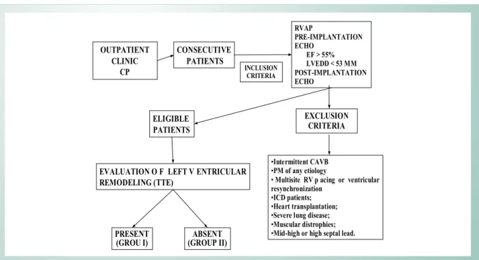
Study design - Figure 1 depicts a schematic representation of the study protocol. The first clinical and functional evaluation was performed before pacemaker implantation and the second, the most recently performed, at least six months after implantation.
Left ventricular remodeling was defined as structural changes found in a comparative analysis of both echocardiographic examinations. The following parameters were considered: increase in LVEDD > 10% and decrease in LVEF > 20%.
All patients were assessed for heart disease, HF functional class (NYHA), time of ventricular stimulation, presence of AV synchrony, and paced-QRS duration on ECG.
Transthoracic echocardiogram - LVEDD was analyzed
in millimeters (mm) by M-mode echocardiography, and LVEF was analyzed in percentages (%) by two-dimensional echocardiography. The Echocardiographic Division of the Heart Institute was entrusted with the examinations. The current Brazilian Echocardiography Society guidelines were followed, as were the instructions provided by the team responsible for outpatient follow-up.
Clinical evaluation - Clinical evaluation was based on the analysis of data from institutional medical records prior to pacemaker implantation and at the time of the last TTE examination. The New York Heart Association (NYHA) criteria for HF functional class were used20.
Time of ventricular stimulation - Defined as time elapsed (in months) between permanent pacemaker implantation and the second TTE examination.
Atrioventricular synchrony - Atrioventricular synchrony was defined as atrial and ventricular sequential depolarization, regardless of artificial stimulation. The presence of synchrony was established at the time of pacemaker implantation, and later changes were not taken into consideration.
QRS complex analysis - The duration of paced-QRS complex (milliseconds) was assessed in a 12-lead surface electrocardiogram (ECG) calibrated at 0.1 mV/mm and paper speed of 25 mm/sec. The time interval from pacemaker spike to the end of the QRS complex was considered.
Statistical analysis - Ventricular remodeling rate was measured by registering the absolute values of occurrences. Functional echocardiographic behavior was assessed by comparing pre- and post-pacemaker implantation means using the Wilcoxon rank-sum test. Fisher’s exact test was performed for associated risk factors for VR, the likelihood ratio test was used for classificatory parameters, and the Wilcoxon rank-sum test, for quantitative parameters. A p value < 0.05 was considered statistically significant.
Results
Of the 268 patients evaluated, 75 were eligible for the study. This population’s characteristics, prior to pacemaker implantation, are described in Table 1. Mean age of the group was 70.9 ± 14, and 22.6% of the subjects were male. The most frequent heart diseases were Chagas heart disease (33%) and hypertensive heart disease (32%). Mean LVEF before PM implantation was 72%, and LVEDD was 46 mm. Sixty-nine patients (92%) were in NYHA functional class I or II; six patients were in class III and no patient was in class IV.
Echocardiographic examinations considered for VR analysis were performed, on average, 1.6 days before and 80.2 months after pacemaker implantation.
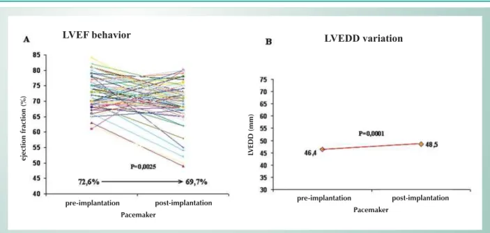
After pacemaker implantation, LVEF decreased from 72.6% to 69.7% (p = 0.0025) and LVEDD increased from 46.4 mm to 48.5 mm (p = 0.0001), and both changes were statistically significant (Fig. 2).
Mean age of the subgroup that evolved with VR was 61.5 ± 16. Three patients had Chagas heart disease, and one patient was hypertensive. All patients were in NYHA functional class II. With reference to echocardiographic parameters, the following was noted: LVEDD increased, on average, 14.6% (48.7 mm pre-implantation to 57 mm post-implantation), and LVEF decreased, on average, 24.2% (69.2% pre-implantation to 52.5% post-implantation). Paced-QRS duration was 170 ms. Atrioventricular synchrony was preserved in three patients. Mean time of ventricular stimulation was 113 ± 4 months.
Discussion
This study’s findings suggest that RV apical pacing virtually does not cause myocardial anatomic and functional changes. In a primary analysis, this information is very practical and useful and, to some extent, surprising, because patients were followed-up on for a long period. Therefore, if RV apical pacing is not considered physiological and, generally speaking, if the ensuing sequence of myocardial electrical activation is deemed deleterious, as it produces hemodynamic, structural, and functional changes in the heart9, why have our patients evolved so favorably?
First and foremost, Pastore et al21 have demonstrated recently, through surface ECG mapping, that pacemaker-induced LBBB does not share exactly the same characteristics as spontaneous LBBB.
In case of cardiac pacing, it is known that the immediate activation of the interventricular septum increases the strain on the LV lateral wall22. Such imbalance of regional forces causes abnormal septal contraction towards the RV, which becomes even more delayed compared to the LV. Finally, there may be an increase in LV end-systolic diameter or decrease in the
interventricular septum regional ejection fraction23,24. However, our observations, drawn from clinical experience at the Pacemaker Clinic of the Heart Institute, suggest that these pathophysiological mechanisms only account for hemodynamic deterioration in the setting of cardiomyopathy.
Fig. 1 -Study design. CP - Cardiac pacing; RVAP - right ventricular apical pacing; ECHO - echocardiogram; CAVB - complete atrioventricular block; PM - pacemaker; RV - right ventricle; ICD - implantable cardiac defibrillator.
CONSECUTIVE PATIENTS OUTPATIENT
CLINIC CP
RVAP
PRE-IMPLANTATION ECHO
EF > 55% LVEDD < 53 MM POST-IMPLANTATION ECHO
INCLUSION CRITERIA
EXCLUSION CRITERIA
•Intermittent CAVB •PM of any etiology
•Multisite RV p acing or ventricular resynchronization
•ICD patients; •Heart transplantation; •Severe lung disease; •Muscular distrophies; •Mid-high or high septal lead.
ELIGIBLE PATIENTS
EVALUATION O F LEFT V ENTRICULAR REMODELING (TTE)
PRESENT (GROU I)
ABSENT (GROUP II)
Total number of patients n=75
Age, years (SD) 70.9 ± 14.3
Sex
Female 58 (77.3%)
Male 17 (22.7%)
Underlying heart disease
Chagasic 25 (33.3%) Hypertensive 24 (32%) Idiopathic 18 (24%) Ischemic 8 (10.6%)
Echocardiographic parameters (SD)
LVEDD (mm) 46 ± 3.18 LVEF (%) 72 ± 4.86
HF functional class (NYHA)
I 26 (34.7%)
II 43 (57.3%)
III 6 (8%)
SD: standard deviation; LVEDD: LV end-diastolic diameter; LVEF: left ventricular ejection fraction; HF: heart failure.
Nielsen et al25 described a decrease in inferior, septal and global myocardial blood flow during RV pacing, when compared to AAI pacing in the same population.
Other studies comparing the AAI(R) vs. DDD(R) and DDD(R)
vs. VVI(R) pacing modes evaluated the role of RV apical pacing in ventricular remodeling 15,26-28. In great part, these studies’ findings suggest impairment of myocardial function.
The DAVID trial13 compared the clinical efficacy of implantable cardioverter-defibrillator (ICD) programmed to the DDDR mode-70 bpm vs. VVI mode – 40 bpm (PM turned off) and showed greater mortality and admission rates for CH in the DDDR group. These authors attempted to correlate clinical behavior and RV apical pacing, but pacemaker dependence was lower than 60% and all patients had LVEF < 35%, reducing considerably the strength of clinical correlation.
Likewise, the MADIT II14 trial, which included only patients with severe heart dysfunction, showed greater HF rate in
the ICD group than in the control group (drug therapy). The worsened behavior (19.9% x 14.9%) was attributed to the ICD ventricular backup pacing feature.
On comparing the characteristics of the population of the studies mentioned above with those of our population,
Fig. 3 -Ventricular remodeling (VR) in the population that underwent RV apical pacing.
Ventricular Remodeling Rate
T
o
ta
l
o
f
p
a
ti
e
n
ts
Ventricular remodeling
( – ) ( + )
Total (%) 71 (94.7%) 4 (5.3%)
Underlying heart disease p = 0.206
Chagasic 22 (88%) 3 (12%) Hypertensive 23 (95,8%) 1 (4,17%) Idiopathic: 18 (100%) 0 Ischemic 8 (100%) 0
Atrioventricular synchrony p = 0.910
Present 55 (94.83%) 3 (5.17%) Absent 16 (94.1%) 1 (5. 88%)
HF functional class (NYHA) p = 0.099
I 39 (100%) 0 II 32 (90.7%) 4 (9.3%)
III 0 0
Paced-QRS p = 0.283
Duration (ms) 156.1 ± 23.5 170 ± 20
TVP (months) 78.3 ± 47.9 113 ± 41.8 p = 0.131
IC: insuficiência cardíaca; ms: milissegundos; TEV: tempo de estimulação ventricular.
Table 2 – Analysis of clinical variables for ventricular remodeling Fig. 2 -Variation of echocardiographic parameters before and after RV apical pacing. Figure A shows LVEF decrease after pacemaker implantation (p = 0.0025); figure B shows the significant increase in LVEDD with pacemaker (p = 0.0001). CP: cardiac pacing; LVEDD: LV end-diastolic diameter; LVEF: left ventricular ejection fraction; RV: right ventricle.
LVEDD variation
LVEF behavior
eje
ct
io
n
fr
ac
ti
o
n
(
%)
pre-implantation post-implantation Pacemaker
pre-implantation post-implantation Pacemaker
LV
EDD
(m
m
)
a clear difference in myocardial function impairment is noted. In our study, myocardial dysfunction was an exclusion criterion, and in the other studies underlying heart disease was markedly severe.
In addition, as far as LBBB is concerned, and drawing an analogy with the clinical behavior of asymptomatic hypertensive patients with normal cardiac function, the importance of conduction disturbance is entirely diverse. Despite showing mechanical changes due to the rapid septal activation relative to the LV free wall, these changes are unable to exhaust the cellular energy supply, and myocardial performance tends to be maintained10, 25.
Nielsen et al.25 findings, which correlated changes in myocardial blood flow with RV apical pacing, should be analyzed in the light of the pathophysiological mechanisms known to be associated with dilated and ischemic cardiomyopathy. In these cases, regional ischemia may account for a deleterious vicious cycle that tends to worsen HF in the presence of prior myocardial dysfunction.
Another documented piece of evidence is the correlation between cardiac pacing and severe mitral regurgitation secondary to papillary muscle lesion by abnormal electromechanical activation19. Of course, for any valve dysfunction of this magnitude to manifest itself, the primary anatomic change must be severe, caused by major cardiac involvement.
This was the only study to evaluate VR in a population characterized by normal cardiac function at study entry and that was totally dependent on apical ventricular pacing.
Conversely, the scientific evidence published thus far on VR related to RV apical pacing is based on comparison of clinical findings with the physiological stimulation mode (AAI) in patients with sinus node disease15,26-28. Well, these patients have a distinctive clinical behavior. In this respect, the subanalyses of all the mentioned studies are subject to criticism, because, regardless of the anatomic and functional changes and the presence of HF, atrial fibrillation was invariably the single predictive event of higher mortality, correlated with high stroke rates and also with ventricular pacing mode. This happened because these trials were not designed to evaluate this primary endpoint.
Our study, on the other hand, defined VR on the grounds of documented echocardiographic changes of clinical and
functional impact. Pathoanatomical changes were not taken into account, since myocardial biopsy is not routinely performed in these patients. The most relevant findings of our study, namely, significant decrease in LVEF from 72.6% to 69.7% (p = 0.0025) and significant increase in LVEDD from 46.4 mm to 48.5 mm (p = 0.0001), do not imply any change in the therapeutic strategy.
However, the criterion used to define VR allowed identifying patients with unfavorable clinical course entirely related to RV apical pacing, because pacemaker dependence was absolute.
In sum, based on the main topics of this discussion, the initial question about the behavior of our patients can be answered: cardiac function is the key element of the analysis, and pacemaker-induced LBBB causes hemodynamic deterioration in the presence of myocardial impairment. Stratification by transthoracic echocardiogram prior to PM implantation (documentation of normal cardiac function) is enough to identify the population at high risk to evolve without VR after RV apical pacing.
The limitation of our study is related to the fact that patients were selected from outpatient visits. Obviously, the patients enrolled in the study were selected previously on the basis of a favorable clinical course. Effects of comorbidities related to PM implantation, that is, complications caused by the cardiac pacing itself, were not considered.
On the other hand, the low VR rate found in our study and the strict criteria to define remodeling, the clinical and functional relevance of which may have underestimated the number of patients that evolved with slight change in cardiac function, precluded identification of VR predictive factors.
Conclusions
Ventricular remodeling prevalence in patients without cardiomyopathy and under RV apical pacing is low (5.3%). Our study did not identify VR associated factor in this subgroup. There is little evidence that HF represents a limiting factor for indication of RV apical pacing to treat bradyarrhythmias in patients with normal cardiac function. Consequently, prospective, randomized, larger studies are needed to confirm these findings.
References
1. Tse HF, Lau CP. Long-term effect of right ventricular pacing on myocardial perfusion and function. J Am Coll Cardiol. 1997; 29: 744-9.
2. Lamas GA, Ellenbogen DA, Hennekens CH, Montanez A. Evidence base for pacemaker mode selection - from physiology to randomized trials. Circulation. 2004; 109: 443-51.
3. Cock CC, Giudici MC, Twisk JW. Comparison of the haemodynamic effects of right ventricular outflow-tract pacing with right ventricular apex pacing. Europace. 2003; 5: 275-8.
4. Melo CS (ed.). Temas de marcapasso. 2a ed. São Paulo: Lemos Editorial; 2004.
5. Tantengco MV, Thomas RL, Karpawich PP. Left ventricular disfunction after long-term right ventricular apical pacing in the young. J Am Coll Cardiol. 2001; 37: 2093-100.
6. Nelson GS, Curry CW, Wyman BT, Kramer A, Declerck J, Talbot M, et al. Predictors of systolic augmentation from left ventricular preexitation in patients with dilated cardiomiopathy and intraventricular condution delay. Circulation. 2000; 101: 2703-9.
7. Prinzen FW, Peschar M. Relation between the pacing induced sequence of activation and left ventricular pump function in animals. Pacing Clin Electrophysiol. 2002; 25: 484-98.
8. Vassalo AJ, Cassidy DM, Miller JM, Buxton AE, Marchlinski EF, Josephson EM. Left ventricular endocardial activation during right ventricular pacing: effect of underlying heart disease. J Am Coll Cardiol. 1986; 7: 1228-33. 9. Vernooy K, Verbeek XA, Peschar M, Prinzen FW. Relation between abnormal
impulse conduction and heart failure. J Interv Cardiol. 2003; 16: 557-62. 10. Lee MA, Dae MW, Lanberg JJ, Grifin JC, Chin MC, Finkbeiner WE, et al. Effects
innervation, function, and histology. J Am Coll Cardiol. 1994; 24: 225-32. 11. Nielsen JC, Andersen HR, Thomsen PEB, Thuesen L, Mortensen PT, Vesterlund
T, et al. Heart failure and echocardiographic changes during long-term follow-up of patients with sick sinus syndrome randomized to single-chamber atrial or ventricular pacing. Circulation.1998; 97: 987-95.
12. Nahlawi M, Waligora M, Spies SM, Bonow RO, Kadish AH, Goldberg JJ. Left ventricular function during and after right ventricular pacing. J Am Coll Cardiol. 2004; 44:1883-8.
13. Wilkoff BL, Cook JR, Epstein AE, Greeme HL, Hallstrom AP, Hsia H, et al. Dual-chamber pacing or ventricular back-up pacing in pacients with as implantable defibrillator: the dual chamber and VVI implantable defibrillator (DAVID) trial. JAMA. 2002; 288: 3115-23.
14. Moss AZ, Zareba W, Hall WJ, Klein H, Wilber DJ, Cannom DS, et al. Prophylatic implatation of a defibrillator in a patients with myocardial infarction and reduced ejection fraction. N Engl J Med. 2002; 346: 877-83. 15. Lamas GA, Lee KL, Sweney MO, Silverman R, Leon A, Yee R, et al. Ventricular pacing or dual-chamber pacing for sinus-node dysfunction. N Engl J Med. 2002; 346: 1854-62.
16. Doshi R, Daoud E, Fellows C, Turk K, Duran A, Hamdan M, et al. Left ventricular-based cardiac stimulation post AV nodal ablation evaluation (The PAVE Study). J Cardiovasc Electrophysiol. 2005; 16: 1160-5.
17. Barold SS, Herweg B, Sweeney MO. Minimizing right ventricular pacing. Am J Cardiol. 2005; 95: 966-9.
18. Barold SS. Adverse effects of ventricular desynchronization induced by long-term right ventricular pacing. J Am Coll Cardiol. 2003; 42: 624-6. 19. Yu CM, Zhang Q, Fung JWH, Chan HCK, Chan YS, Yip GWK, et al. A
novel tool to asses systolic asyncrhrony and identify responders of cardiac resynchronization therapy by tissue synchronization imaging. J Am Coll Cardiol. 2005; 45: 677-84.
20. Criteria Committee, New York Heart Association. Inc. Diseases of the heart and blood vessels: nomenclature and criteria for diagnosis. 6th ed. Boston: Little Brown;1964.
21. Pastore CA, Tobias N, Samesima N, Martinelli Filho M, Pedrosa A, Nishioka S, et al. Body surface potential mapping investigating the ventricular activation patterns in the cardiac resynchronization of patients with left bundle-branch block and heart failure. J Electrocardiol. 2006; 39: 93-102.
22. Rosenqvist M, Isaaz K, Botvinick EH, Dae MW, Cockrell J, Aboot JA, et al. Relative importance of activation sequence compared to atrioventricular syncrony in left ventricular function. Am J Cardiol. 1991; 67: 148-65. 23. Grines LC, Bashore TM, Boudoulas H, Olson S, Shafer P, Wooley CF.
Funcional abnormalities in isolated left bundle branch block: the effect of interventricular assynchrony. Circulation.1989; 79: 845-53.
24. Xiao HB, Gibson DG. Effects of intermittent left bundle branch block on left ventricular diastolic function: a case report. Int J Cardiol. 1994; 46: 85-8. 25. Nielsen J, Bottcher M, Nielsen TT, Pedersen AK, Andersen HR. Regional
myocardial blood flow in patients with sick sinus syndrome randomized to long-term single-chamber atrial or dual chamber pacing - effect of pacing mode and rate. J Am Coll Cardiol. 2000; 35: 1453-61.
26. Nielsen JC, Kristensen L, Andersen HR, Mortensen PT, Pedersen OL, Pedersen AK. A randomized comparison of atrial and dual chamber pacing in 177 consecutive patients with sick sinus syndrome: echocardiographic and clinical outcome. J Am Coll Cardiol. 2003; 42: 614-23.
27. Andersen HR, Nielsen JC, Thomsen PEB, Thuesen L, Mortensen PT, Vesterlund T, et al. Long-term follow-up of patients from a randomized trial of atrial versus ventricular pacing for sick-sinus syndrome. Lancet. 1997; 350: 1210-6.