Case Report
Keywords
Defibrillators, implantable; ventricular septal rupture; electrodes, implantable.
We describe the case of a 62-year-old patient who returned for evaluation nine months after receiving an implantable cardioverter-defibrillator (ICD) with signs of delayed right ventricular (RV) perforation. The clinical signs that allowed the diagnosis of this late presentation to be achieved are discussed herein, as well as the conduct and the frequency of this complication in the literature.
Delayed Right Ventricular Perforation in Patient with Implantable
Cardioverter - Defibrillator
Alexsandro Alves Fagundes, Luiz Pereira de Magalhães, Jussara Pinheiro, Leonardo Flausino, Luciano Rapold Souza
Hospital Português da Bahia, Salvador, BA - Brazil
Mailing address: Alexsandro Alves Fagundes •
Rua Professor Amilcar Falcão, 20/901 - Barra - 40140-480 - Salvador, BA - Brazil
E-mail: [email protected], [email protected]
Manuscript received June 17, 2009; revised manuscript received August 16, 2009; accepted March 4th, 2010.
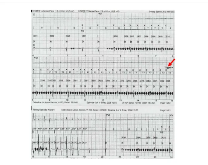
after 90 days. At that time, she complained of “thumps” in the abdomen without the sensation of shock. At the clinical evaluation, the presence of hiccups compatible with diaphragmatic stimulation was observed. The electronic interrogation showed absence of ventricular capture and sensitivity. There were several episodes of inadequate detection of ventricular tachycardia triggered by artifact noise in the sense channel, treated with anti-tachycardia stimulation (ATP) (Figure 1). The differential diagnosis of this finding that of external noise (electric shock, magnetic interference) or internal noise due to problems in the lead conductor. In this case, the findings of loss of ventricular capture and sensitivity ruled out the possibility of external interference. Such data raised the hypothesis of electrode fracture. The patient was referred to emergency hospital admission.
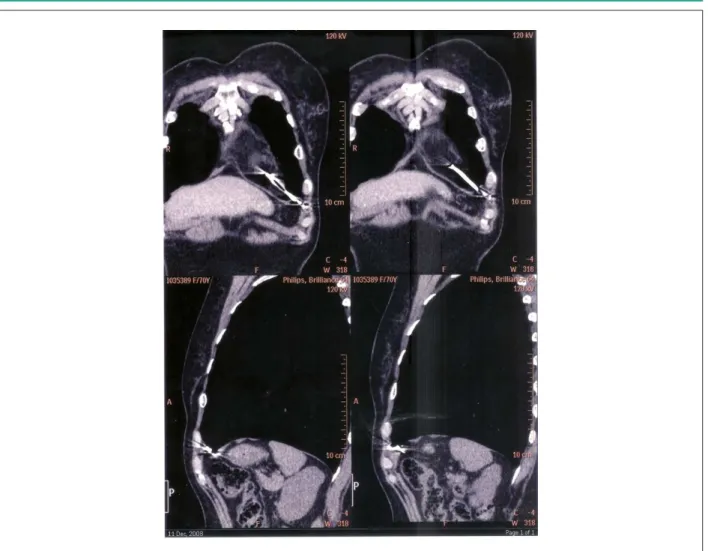
The chest X-ray disclosed that the position of the lead was clearly outside the cardiac area, next to the costal grid. The echocardiogram did not show pericardial effusion and the chest tomography confirmed the presence of the electrode tip in the chest cavity, perforating the right ventricular free wall (Figure 2).
After discussing the case with the cardiac surgical team, it was decided to remove the lead under direct visualization through a thoracotomy, which was carried out uneventfully. During the surgical procedure, the lead was removed without difficulty and no injuries were macroscopically identified in the lead isolation or conductor. A new lead was implanted by endovascular technique under fluoroscopic view in ventricular septum.
The patient presented an uneventful evolution with no arrhythmic events in the last 8 months of follow-up.
Discussion
Acute right ventricular perforation during endovascular electrode implantation is an uncommon complication. Usually, the presentation is catastrophic, with hemodynamic instability due to cardiac tamponade and potentially fatal outcome. Another important information on this complication is that its manifestation is usually recognized in the acute phase of peri-implantation1. Its identification requires a high degree of clinical suspicion in the presence of signs of hemodynamic instability, increased cardiac silhouette, phrenic nerve stimulation and loss of the intracavitary electrogram.
The post-implantation chest X-ray and echocardiogram might be useful in the identification of subclinical presentations. In addition to the potentially fatal hemodynamic complications, the RV perforation can impair the recognition of ventricular
Introduction
Delayed complications can occur in patients that have received an implantable cardioverter-defibrillator (ICD) device. However, late manifestations of myocardial perforation are not always evident. Thus clinical and electronic signs that are characteristic of this complication must be known so that they can be studied.
Case report
A 62-year-old female patient was submitted to a single-chamber ICD implantation, model Atlas VR St Jude, in February 2008 due to an episode of sustained ventricular tachycardia with pre-syncope.
At that time, an echocardiogram was performed, which showed an ejection fraction (EF) of 45% with a small aneurism in the basal region of the LV.
A coronary angiography was performed and showed no signs of atherosclerosis. The serological test for Chagas’ disease was positive.
The 30-day assessment did not show the presence of arrhythmic events. The impedance and the ventricular lead stimulation threshold were normal. The patient remained asymptomatic and was treated with amiodarone, carvedilol and enalapril.
Nine months after the implantation, the patient came for reassessment, although she had been instructed to return
Case Report
Fagundes et al Delayed right ventricular perforation
Figure 1 -ATP therapy hindered by noise. The lower tracing shows high-frequency artifacts. Rate measurement by the ICD is determined by the events identiied by
the letter R. After 12 events (R) felt in the tachycardia zone (T2), an anti-tachycardia pacing (ATP) therapy was started (arrow). Observe that the interference persists even after the therapy.
arrhythmias, preventing the ICD protection potential. There have been reports of delayed RV perforation that was initially identified by the loss of intracavitary potential, even though there was no change in the electrode position at the X-ray2.
Recently, late presentations of cardiac perforation not documented at the acute phase of the implantation have been reported in the literature. In a recent retrospective study, 100 patients that had received a pacemaker or ICD were submitted to a chest tomography. All patients were asymptomatic. Perforations were identified in up to 15% of this series, including atrial perforations. The mean time between the implantation and the chest CT was 43 months (p=0.0002). This fact demonstrates how perforations that go clinically undetected can develop chronically. Factors related to structural cardiopathy, as well as the type of lead used might be related to this complication. All the patients had normally-functioning devices, with no impedance alterations and no cases of cardiac tamponade. The authors point out the benign and asymptomatic characteristics of this complication that often goes undetected and that many times does not require intervention3.
The sense impairment of the intracavitary electrogram hinders the detection of ventricular arrhythmias, preventing the therapy success, as well as being able to determine noise in the ventricular sense circuit and trigger inappropriate shocks. In this patient, fortunately, the noise triggered painless therapy (ATP), which was not perceived by the patient.
The right ventricular perforation is, therefore, a relatively frequent and underdiagnosed complication. The radiological aspect is not always diagnostic. Alterations in phrenic sensitivity and stimulation must be taken into account, which was decisive for the clinical suspicion in our case. Most late presentations present benign evolutions; however, it is necessary to pay special attention to the parameters of sensitivity, impedance and thresholds, which can impair the therapeutic function in ventricular arrhythmias.
Potential Conflict of Interest
No potential conflict of interest relevant to this article was reported.
Case Report
Fagundes et al
Delayed right ventricular perforation
Figure 2 -Chest CT. Note the electrode tip perforating the RV and located in the costal grid.
References
1. Geyfman V, Storm RH, Lico SC, Oren JW 4th. Cardiac tamponade as complication of active-fixation atrial lead perforations: proposed mechanism and management algorithm. Pacing Clin Electrophysiol. 2007; 30 (4): 498-501.
2. Satpathy R, Hee T, Esterbrooks D, Mohiuddin S. Delayed defibrillator lead
perforation: an increasing phenomenon. Pacing Clin Electrophysiol. 2007; 30: 28-32. Pacing Clin Electrophysiol. 2008; 31 (1): 10-2.
3. Hirschl DA, Jain VR, Spindola-Franco H, Gross JN, Haramati LB. Prevalence and characterization of asymptomatic pacemaker and ICD lead perforation on CT. Pacing Clin Electrophysiol. 2007; 30 (1): 28-32.
Arq Bras Cardiol 2010; 95(6) : e148-e150
Sources of Funding
There were no external funding sources for this study.
Study Association
This study is not associated with any post-graduation program.