Spontaneous splenic rupture in a patient receiving
thrombolytic therapy
Ruptura esplênica espontânea em paciente submetida a terapia
trombolítica
Elizabeth Revesz, John A. Grimaldi, Elizabeth T. Clark, Francis J. Podbielski*
Introduction
Spontaneous splenic rupture is an entity that was first described by Atkinson in 1874.1There have been multiple reported cases of spontaneous rupture of the apparently normal spleen.1-10We describe a case of spontaneous sple-nic rupture that was preceded by the administration of tis-sue plasminogen activator (t-PA).
Case report
A 67-year-old female patient previously known to the vascular surgery service presented with a chief com-plaint of the sudden onset of progressive left lower extre-mity pain that began 1 week prior to presentation. This pain was first noticed secondary to exercise intolerance and was exacerbated by physical exertion. The pain was described as sharp, unremitting, and localized to the upper
thigh and calf. There was no associated dyspnea or chest pain. The patient reported non-compliance with her warfa-rin regimen. Past medical history included type II diabetes mellitus, peripheral vascular disease, hypertension, and hypercholesterolemia. Past surgical history was remarka-ble for a left femoral to anterior tibial bypass graft perfor-med at our institution 2 years prior to this presentation. Medications included warfarin, clonidine, fluvastatin, in-sulin, and furosemide.
Physical examination was marked by absent pulses in her bypass graft, posterior tibial, and dorsalis pedis arteri-es. The left foot was notably cooler to the touch when com-pared with the right foot.
An aortography with bilateral lower extremity views revealed occlusion of the bypass graft. The interventional
274
J Vasc Bras. 2009;8(3):274-276.
Copyright © 2009 by Sociedade Brasileira de Angiologia e de Cirurgia Vascular * Department of Surgery, Resurrection, St. Joseph Hospital, Chicago, Illinois, USA.
No conflicts of interest declared concerning the publication of this article. Manuscript received Nov 10 2008, accepted for publication Mar 11 2009.
CASE REPORT
Abstract
We describe the case of a 67-year-old female patient with a history of femoral-distal bypass graft with sudden onset of unremitting leg pain, who had recently received tissue plasminogen activator (t-PA). The pa-tient reported non-compliance with her warfarin regimen. Angiography revealed occlusion of the bypass graft. Infusion of t-PA was performed via a right femoral artery approach. On hospital day two, the patient de-veloped nausea and abdominal pain with associated hypotension. A CT scan showed a massive intra-abdominal and pelvic free fluid consistent with blood. The spleen was enlarged and fluid noted around the liver. At laparotomy, a grade III splenic laceration at the hilum was identified and a splenectomy performed. The patient recovered completely. Although rare, spontaneous splenic rupture should be considered in the differential diagnosis of patients undergoing thrombolytic therapy who develop signs of hemodynamic instability.
Keywords:Splenic rupture, spontaneous splenic rupture, tissue plasminogen activator, t-PA, thrombolytic therapy.
Resumo
Descrevemos o caso de uma paciente de 67 anos com histórico de enxerto fêmoro-distal com início súbito de dor repetitiva em membro in-ferior e que havia recebido ativador de plasminogênio tecidual (AP-t) recentemente. A paciente relatou não adesão ao seu tratamento com warfarina. A angiografia revelou oclusão do enxerto. O AP-t foi adminis-trado via artéria femoral direita. No segundo dia de hospitalização, a paciente apresentou náuseas e dor abdominal com hipotensão associada. Uma tomografia computadorizada revelou a existência de um fluido pélvico e intra-abdominal livre em grande quantidade, com suspeita de que fosse sangue. O baço estava crescido, e o fluido foi observado em torno do fígado. A laparotomia identificou uma laceração grau III no hilo esplênico, e uma esplenectomia foi realizada. A paciente teve recupe-ração completa. Embora rara, a ruptura esplênica espontânea deve ser considerada no diagnóstico diferencial de pacientes submetidos a terapia trombolítica que apresentem sinais de instabilidade hemodinâmica.
radiology service cannulated the right femoral artery with a 16 French sheath and AngioJet® was performed on the thrombus of the left lower extremity bypass graft followed by the infusion of t-PA. The procedure was successful, and the patient had palpable pulses in the bypass graft, posteri-or tibial and dposteri-orsalis pedis arteries. The patient was admit-ted to the cardiac intensive care unit for further observati-on.
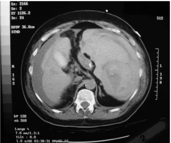
The right femoral arterial sheath was removed on hos-pital day one and an occlusion device deployed at the puncture site. On hospital day two, the patient began to ex-perience abdominal pain associated with nausea. Routine laboratory tests were normal. Hospital day three was mar-ked by worsening nausea and vomiting. The patient also developed a dwindling urine output over the next several hours with a fall in the blood pressure to 80 mm/Hg and a reflex tachycardia of 110 to 120 beats per minute. Physical examination revealed a somnolent patient with a diffusely tender but non-distended abdomen. Bowel sounds were distant and hypoactive. There was voluntary guarding, but no rigidity or rebound. After fluid resuscitation, the hemo-globin was found to be 5.2 gm/dL. The patient was transfu-sed with four units of packed red blood cells. The working diagnosis at this time was a retroperitoneal hemorrhage from the puncture site. After stabilization, a CT scan of the abdomen and pelvis revealed massive intra-abdominal and pelvic free fluid consistent with blood. The spleen was en-larged and fluid noted around the liver (Figure 1).
Operative management
This patient was taken to the operating room for emer-gent laparotomy. After removal of approximately 4 liters of clot, the spleen was examined and found to have a grade III laceration at the hilum. Splenectomy was performed and the patient was transferred to the cardiac care unit where she was extubated on postoperative day zero. The recovery period was marked by development of a large rectus sheath hematoma that was managed conservatively. The patient was discharged on her previous regimen of oral warfarin.
Discussion
Thrombolysis is a complex process whereby clot is or-ganized and then removed by the proteolytic enzyme, plas-min that converts fibrin to fibrin degradation products.11 Thrombolytics such as t-PA are substances that act directly on plasminogen to convert it to plasmin. Because t-PA has specific affinity for fibrin and thus will ideally bind exclu-sively to formed clots, it should act only on the pre-existing fibrin clot for which the patient is being treated and have little effect on peripheral tissues.12This unfortu-nately is not the case in many instances.
Although disagreement exists regarding the classifica-tion of spontaneous splenic rupture, the generally accepted definition is one of disruption of the parenchyma, capsule, or blood supply of a previously normal spleen.13By strict definition, no healthy spleen should rupture. There are myriad reasons that may incite a splenic micro-injury or an injury that would otherwise be subclinical. A multitude of etiologies for spontaneous splenic rupture including coug-hing, sneezing, movement in bed, and even laughing have been proffered in the literature. In general, any condition that produces splenomegaly can predispose a patient to splenic rupture. A literature search revealed only three case of spontaneous splenic rupture resulting from use of t-PA.3,4
The use of t-PA in peripheral arterial occlusion pro-motes clot dissolution. Considering the demonstrated ble-eding complications of t-PA use, the benefit of limb salva-ge salva-generally outweighs the risk of bleeding. Although rare, spontaneous splenic rupture should be kept in the differen-tial diagnosis in a patient undergoing thrombolytic therapy who develops shock with abdominal pain and distention. While aggressive fluid resuscitation and diagnostic
ima-SSR in a patient receiving thrombolytic therapy - Revesz E et al. J Vasc Bras 2009, Vol. 8, N° 3 275
ging are the initial steps in management, early surgical in-tervention may be required to ensure patient survival.
References
Atkinson E. Death from idiopathic rupture of the spleen. Br Med J. 1874;2:403-4.
Lieberman ME, Levitt A. Spontaneous rupture of the spleen. Am J Emerg Med. 1989;7:28-31.
Blakenship JC, Indeck M. Spontaneous splenic rupture after t-PA for acute myocardial infarction [letter]. N Engl J Med. 1991;325:969.
Cheung PK, Arnold JM, McLarty TD. Splenic hemorrhage: a complication of tissue plasminogen activator treatment. Can J Cardiol. 1990;6:183-5.
Weiner RS, Ong LS. Streptokinase and splenic rupture [brief clinical observation]. Am J Med. 1989;86:249.
Gardner-Medwin J, Sayer J, Mahida YR, Spiller RC. Spontane-ous rupture of spleen following streptokinase therapy. Lan-cet. 1989;2:1398.
Eklof B, Jones JE, Lohi A, Staszkiewicz W, Norgren L. Sponta-neous rupture of liver and spleen with severe intra-abdominal bleeding during streptokinase treatment of DVT. Vasa. 1977;6:369-71.
Hibbard LT. Spontaneous rupture of the liver in pregnancy: a re-port of eight cases. Am J Obstet Gynecol. 1976;126:334-8. Toubia NT, Tawk MM, Potts RM, Kinasewitz GT.Cough and
spontaneous rupture of a normal spleen. Chest. 2005;128:1884-6.
Lennard TW, Burgess P. Vomiting and ‘spontaneous’ rupture of the spleen. Br J Clin Pract. 1985;39:407, 410.
Kumar , N, A. Robbins & Cotran pathologic basis of disease. 7th ed. Philadelphia: W.B. Saunders; 2004.
Tissue plasminogen activator for the treatment of acute pulmo-nary embolism. A collaborative study by the PIOPED Inves-tigators. Chest. 1990;97:528-33.
Brunicardi FC, Andersen DK, Billiar TR, et al. Principles of sur-gery. 8th ed. New York: McGraw-Hill; 2005.
Correspondence: Elizabeth Revesz MD Saint Joseph Hospital 60657 - Chicago, IL - USA Tel.: +1 (773) 665.6237 Fax: +1 (773) 665.6232
E-mail: [email protected], [email protected]