Rev Bras Ter Intensiva. 2016;28(2):199-200
To: Severe hypercalcemia as a form of acute
lymphoblastic leukemia presentation in children
LETTER TO THE EDITOR
To the Editor,
Severe hypercalcemia of malignancy in children has been extensively described in the medical literature.(1) However, this complication usually
presents as a late symptom of acute lymphoblastic disease and not as an early manifestation, as described in a case report by Martins et al.(2) Colleti Junior
et al.(3) also observed that hypercalcemia was present as an early symptom of
malignancy, and the serum calcium levels were higher than usual (ionic calcium: 2.95mmol/L; normal value: 1.11-1.40mmol/L). In this case, immediately after diagnosis, in addition to hydration and low doses of loop diuretic, treatment was initiated with pamidronate, a bisphosphonate, and resulted in a rapid decrease in the serum calcium levels. In the case described, it is unclear whether hemoiltration was performed to purify the calcium or to treat kidney failure; the latter does not seem to be the case considering the initial test results. In the irst case, early treatment with bisphosphonates might have avoided an invasive procedure, such as hemoiltration.(3,4) It is important to describe the protein
levels of parathyroid hormone (PTHrP) - often reported as normal - because, in most cases, hypercalcemia caused by malignant disease is associated with the production of PTHrP,(5) and the measurement of this protein is essential for the
diferential etiological diagnosis. In addition, the absence of signs of osteopenia is noteworthy in this case because the high calcium levels are due in part to osteolytic activity, which is blocked by bisphosphonate (otherwise, the calcium levels would not have decreased with the use of zoledronate). Colleti Junior et al.(3) reported that the osteolytic activity is so extensive that classic lesions are
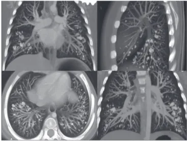
observed in a punch biopsy (Figure 1). Another relevant aspect is the evaluation of tissue calcium deposition after treatment with bisphosphonates. Is there a report of nephrocalcinosis related to calcium deposition? Is there a report of calcium deposition in other tissues? In Colleti Junior et al.,(3) a rare alveolar
lung deposition was shown on chest computed tomography (Figure 2), and the diagnosis of this symptom was essential during patient follow-up. herefore, the side efects of treatment with bisphosphonates should be considered.
Conflicts of interest: None.
Corresponding author:
José Colleti Junior
Unidade de Terapia Intensiva Pediátrica do Hospital Santa Catarina
Avenida Paulista, 200
Zip code: 01310-000 - São Paulo (SP), Brazil. E-mail: [email protected]
Para: Hipercalcemia grave como forma de apresentação de
leucemia linfoblástica aguda em crianças
200 Colleti Junior J, Carvalho WB
Rev Bras Ter Intensiva. 2016;28(2):199-200
Figure 1 - Pelvic X-ray showing osteolytic lesions (arrows). Figure 2 - Computed chest tomography showing alveolar calcification.
Furthermore, we highlight the study of the authors and make a note to pediatricians about this important clinical sign and the need for monitoring treatment-related aspects.
José Colleti Junior Pediatric Intensive Care Unit, Hospital Santa Catarina - São Paulo (SP), Brazil.
Werther Brunow de Carvalho
Department of Pediatrics, Instituto da Criança,
Universidade de São Paulo - São Paulo (SP), Brazil.
REFERENCES
1. Trehan A, Cheetham T, Bailey S. Hypercalcemia in acute lymphoblastic leukemia: an overview. J Pediatric Hematol Oncol. 2009;31(6):424-7.
2. Martins AL, Moniz M, Nunes PS, Abadesso C, Loureiro HC, Duarte X, et al. Severe hypercalcemia as a form of acute lymphoblastic leukemia presentation in children. Rev Bras Ter Intensiva. 2015;27(4):402-5.
3. Colleti Junior J, Carla Armelin Benites E, Spadaccia Dos Santos Fernandes G, Antonio Freddi N, Koga W, Brunow de Carvalho W. Case report: Pulmonary alveolar calcification as a result of severe hypercalcemia due to acute lymphoblatic leukemia. F1000Res. 2015;4:111.
4. Mastrandrea LD, Albini CH. Bisphosphonate treatment of tumor-induced hypercalcemia in a toddler: case report and review of related literature. Endocr Pract. 2006;12(6):670-5.