GASTROENTEROLOGIA EXPERIMENT
AL / EXPERIMENT
AL GASTROENTEROLOGY
HEPATIC HYPERPLASIA AND DAMAGES
INDUCES BY ZEARALENONE
Fusarium
Mycotoxins in BALB/c Mice
Pronobesh CHATOPADHYAY, Anurag PANDEY,
Asshwani K. CHAURASIA, Addesh UPADHYAY,
Sanjev KARMAKAR and Lokendra SINGH
Abstract – Context - Zearalenone is a mycoestrogen and considered a mycotoxin. Objective - To establish whether zearalenone produced hepatotoxicity via oral administration. Methods - Zearalenone was orally administered at a dose of 50 μg, 100 μg and 200 μg ZEN/ body weight/daily, respectively, for 14 days to three groups of BALB/c mice. Diagnostic modalities used to evaluate hepatic damage and impaired hepatic function pre- and post zearalenone administration included hepatic marker enzyme activity, pentobarbital sleeping time, cytochrome P-450 activities and histopathologic evaluation of liver. Results - Signiicant histopathologic changes viz.
sinusoidal congestion, cytoplasmic vacuolization, hepatocellular necrosis and neutrophil iniltration were observed after evaluating of liver section from each group after accumulated zearalenone exposure. Further, zearalenone exposure increased activities of alanine transaminase, aspartate transaminase and lipid peroxides whereas activities of tissue glutathione and cytochrome P450 were
decreased as compared to control mice. Zearalenone also increased the sleeping time and decreased sleeping latency after pentobarbital through intraperitoneal route as compared to control mice which indicates that the impairment of hepatic metabolizing enzymes by zearalenone. Conclusion - Zearalenone is a potential hepatotoxin by oral route.
Headings – Zearalenone. Hyperplasia. Liver, pathology. Mice.
Division of Pharmaceutical Technology, Defence Research Laboratory.
Correspondence: Dr. Pronobesh Chatopadhyay - Post Bag No: 2, Tezpur-784001, Assam, India. E-mail: [email protected]
INTRODUCTION
Zearalenone (6-[10-hydroxy-6-oxo-trans-1-undecenyl]-Bresorcyclic acid lactone) (ZEN), is a non-steroidal mycoestrogen that activates the estrogen receptors (ERs), ESR1 and ESR2, where it acts as an agonist
and partial antagonist to estradiol(5, 8, 10). ZEN also
reported that facilitates carcinogenesis, reproductive
toxicity(10) and suppress immunity(9). ZEN contaminated
of cereals and grains and related products causes food and feed-borne intoxications (myco-toxicoses) in man and livestock. This mycotoxins produces by
various members of the genus Fusarium, including
deoxynivalenol (mostly produced by F. graminearum
and F. sporotrichoides) and zearalenone (produced by F. graminearum and F. culmorum among others) which are major concern in health.
Most of its biological activities of ZEN are attributed to the agonist effect on the ER and certain biological reactions of zearalenone that can not be explained by
its estrogenic activity(5). Estrogens taken by women
for birth control purposes are implicated as a cause for development of both benign and malignant hepatic
neoplasms(11, 12). In addition to endogenous steroidal
estrogens, a variety of compounds with estrogenic
activity also are found in the environment because a number of drugs, insecticides, and natural food products representing many different structures can act like estrogens by products by irst phase metabolic transformation after ingestion. Therefore estrogenic activities of ZEN have possibilities to extend the other biological effects - hepatotoxicity.
The aim of the research was to examine the toxic inluence of ZEN on liver through estimating mycotoxin inluence on markers evaluating hepatic damage.
METHODS
Animals and housing conditions
Study design
Twenty-four Balb/c mice were divided into control group (I) (n = 6) were given 0.9% saline (5 mL/kg, p.o), ZEN treated
groups viz. Groups II, III and IV were given 50 μg,100 μg, 200 μg
ZEN/ body weight /daily, respectively, by oral route for 14 days (n = 6). After 14 days mice were euthanized by ether inhalation and sacriiced. Blood samples were obtained from the right ventricle via a left anterior thoracotomy and separated serum. The serum samples were stored at -20°C until used for ALT and AST assays. A portion of liver lobes were ixed in buffered 10% formalin, embedded in parafin, and used for histopathology by using hematoxylin and eosin (H-E) staining. Another portion of lobe was stored at -20°C for enzymatic assay.
Preparation of mice of liver microsome
Liver microsome was measured as described by previously(18).
Briely, 100 mg liver was excised and homogenized with a
loose-itting Telon pestle in 900 mL of 50 mM L-1 Tris-HC1buffer,
pH 7.4, containing 0.3 M L-1 sucrose, 10 mM L-1 DTT, and
10 mM L-1 EDTA. The homogenate was centrifuged at
20,000 x g for 15 min. The supernatant solution was centrifuged at 10,000 x g for 60 min. The microsomal fraction obtained was suspended in a homogenizing medium without DTT and re-centrifuged at 10,000 x g for 60 min. The resulting
microsomal fraction was suspended in 0.1 ML-1 phosphate
buffer pH 7.4, containing 1 mM L-1 EDTA and volume was
adjusted to 10 mL with phosphate buffer.
Estimation of hepatic marker enzymes
Serum was used for the assay of alanine transaminase (ALT) and aspartate transaminase (AST) using assay kits (Transasia, Mumbai, India) according to the manufacturer’s instructions.
Determination of cytochome P450 (CYP 450)
Hepatic cytochome P450 was measured as described by
previously(16). Briely 1 mL supernatant of liver microsomes
was taken and 10 mg of sodium dithionate was added. Air was bubbled for 10 seconds and then centrifuged for 5 minutes at 3000 x g. Five hundred µl (500 µl) supernatant was mixed with 5 mL Tris HCl buffer (pH 7.4) and absorbance was measured at 450 nm wavelengths using excitation coeficient of 91 mol
L-1cm -1 for A
450.
Light microscopy analysis
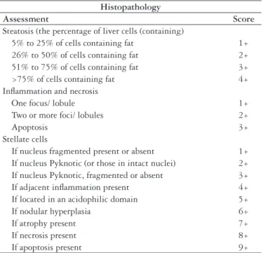
Six µm stains of formalin –ixed and parafin-embedded were taken and stained with H-E. Slide was examined by a pathologist who had no prior knowledge of the treatment groups. Histopathology of liver was evaluated by using a
scoring system (Table 1) as described previously(19).
Two types of eosinophilic hepatocellular changes are presumed to be apoptotic in origin viz. round and detached from surrounding hepatocytes (classical Councilman bodies) and shrunken compared to adjacent hepatocytes, but still irmly attached were counted in 5 to 20 ields from each liver to count at least 100 stellate –Abs. The changes in stellate cells were calculated by using a scoring system.
Determination of lipid peroxides (LPO)
Hepatic LPO was measured as method described(7).
To 100 µl separated microsomes in 0.1(M) phosphate buffer saline, 1 mL of 28% trichloroacetic acid was added and centrifuged at x 2000 g at 4oC for 20 minutes. One milliliter of supernatant was separated and 900 µl of 1% thiobarbituric acid was added and volume was adjusted to 3 mL by using phosphate buffer (pH 7.0), heated in water bath for 60 min and cooled in ice bath. The absorbance was measured at 532 nm. The lipid peroxidation was calculated
on the basis of the molar extinction coeficient (1.56 x 10⁵)
of malondialdehyde.
Tissue glutathione (GSH) assays
The hepatic reduced GSH level was determined by
the method of Ellman(2). Briefly, after 0.2 g liver tissues
were homogenized in 4 mL of 0.02 M EDTA Na2 (using an all glass homogenizer in an ice bath). In 2.5 mL tissue homogenates (aliquots) were mixed with 2.0 mL of distilled water and 0.2 mL of 50% TCA. All tubes were shaken intermittently for 10-15 min and centrifuged for 15 min at approximately 3000 × g. Two milliliter of 0.4 M Tris buffer (pH 8.9) and 0.1 mL of 0.01 M 5,5’-dithiobis- 2-nitrobenzoic acid (DTNB) were added to 2.0 mL of tissue supernatant, and the sample was shaken. The absorbance was read within 5 min of the addition of DTNB at 412 nm against a reagent blank with no homogenate. GSH levels were calculated using standard curve prepared by known amounts of GSH (Aldrich Chemical Co. Ltd, Germany). The concentration of GSH was expressed as mg/g tissue.
Histopathology
Assessment Score
Steatosis (the percentage of liver cells (containing)
5% to 25% of cells containing fat 1+ 26% to 50% of cells containing fat 2+ 51% to 75% of cells containing fat 3+ >75% of cells containing fat 4+ Inlammation and necrosis
One focus/ lobule 1+ Two or more foci/ lobules 2+
Apoptosis 3+
Stellate cells
If nucleus fragmented present or absent 1+ If nucleus Pyknotic (or those in intact nuclei) 2+ If nucleus Pyknotic, fragmented or absent 3+ If adjacent inlammation present 4+ If located in an acidophilic domain 5+ If nodular hyperplasia 6+ If atrophy present 7+ If necrosis present 8+ If apoptosis present 9+
Pentobarbital sleeping time
The pentobarbital sleeping time test was performed using pentobarbital. After 14 days treatment with ZEN animals received pentobarbital (concentration of 3 g/100 mL of solution) through intraperitoneal injection at a dosage of 50 mg/kg of mice body weight (or 0.0017 mL/g of animal weight) and time was measured in minutes, from the loss of position relex to its gaining relexes.
Statistical analysis
Data are expressed as mean ± S.D. The statistical signiicance
between data means was determined by Student’s t-test.
P-values P<0.05 were considered as signiicant
RESULTS
Effect of ZEN on hepatic marker enzymes and cytochrome P450
ZEN increases signiicantly (P<0.01) AST, ALT levels
whereas cytochrome P450 levels decreases as compared to
controlled mice. Activities of ALT and AST were 55.23 ± 7.1
and 52.19 ± 9.40 I.UL-1, respectively, in controlled mice which
increased to 87.96 ± 4.64 and 70.33 ± 3.59.UL-1, respectively,
in ZEN treated (200 µg/kg body weight) mice.
Microsomal, cytochrome P450 levels in the control mice
were 0.66 ± 0.032 nML-1 mg-1 proteins which decreased to
0.29 ± 0.017 nML-1 mg-1 proteins after treatment for 14 days
with ZEN 200 µg/kg body weight (Table 2).
Effect of ZEN on LPO, GST, sleeping time and sleep latency
After treatment with ZEN signiicantly (P<0.01) increased
LPO levels and GST levels were signiicantly decreased
(P<0.01) as compared to control group. Activities of LPO
and GST were 0.7 ± 0.05 nML-1 MDA mg-1 of protein and
52.40 ± 4.90 mM L-1 of 1-chloro 2,4-dinitrobenzene (CDNB)
conjugated min-1 100 mg-1 protein in control groups and after
treatment with ZEN (200 µg/kg body weight) increased to
5.22 ± 0.80 nML-1 MDA mg-1 of protein whereas GST decreased
to 36.20 ± 3.77 mM L-1 of 1-chloro 2,4-dinitrobenzene (CDNB)
conjugated min-1 100 mg-1 protein.
Microsomal, cytochrome P450 levels in the control mice
were 0.66 ± 0.032 nML-1 mg-1 proteins which decreased to
0.29 ± 0.017 nML-1 mg-1 proteins after treatment for 14 days
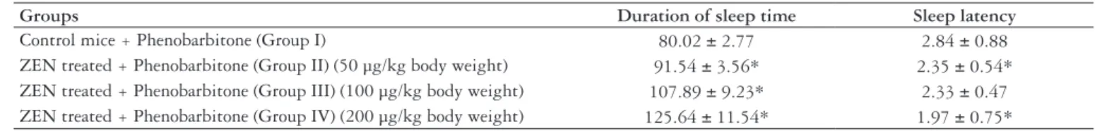
with ZEN 200 µg/kg body weight (Table 3). Pentobarbital
along with ZEN increased signiicantly (P<0.01) sleeping
time as compared to control mice (Table 4).
Effect of ZEN on liver histopathology
Liver histopathology was evaluated based on sinusoidal congestion, cytoplasmic vacuolization, hepatocellular necrosis, and neutrophil iniltration (Table 5). Histopathology examination of liver sections of control group showed normal cellular architecture with distinct hepatic cells, sinusoids spaces and central vein (Figure 1a). ZEN treated group showed that disarrangement and degeneration of normal hepatic cells with intense centri-lobular necrosis extending to mid– zone and sinusoidal hemorrhages and dilation.
Groups aALT aAST bCytochrome P
450
Control mice (Group I) 55.23 ± 7.10 52.19 ± 9.40 0.66 ± 0.032 ZEN treated (Group II) (50 µg/kg body weight) 62.11 ± 5.32* 60.73 ± 4.47* 0.50 ± 0.047 ZEN treated (Group III) (100 µg/kg body weight) 70.33 ± 6.91* 64.77 ± 5.06* 0.33 ± 0.053* ZEN treated (Group IV) (200 µg/kg body weight) 87.36 ± 4.64* 70.33 ± 3.59* 0.29 ± 0.017*
TABLE 2. The effect of ZEN on ALT, AST, cytochrome P450 levels after 14 days treatment
Results are expressed as mean ± SD (n = 6). *Statically different (P<0.01) from control. aExpressed in I.U. L-1, bExpressed as nML-1 protein
Groups aGST aLPO
Control mice (Group I) 52.40 ± 4.90 0.7 ± 0.05 ZEN treated (Group II) (50 µg/kg body weight) 43.27 ± 6.66 2.36 ± 0.77 ZEN treated (Group III) (100 µg/kg body weight) 40.16 ± 2.75 3.56 ± 0.82 ZEN treated (Group IV) (200 µg/kg body weight) 36.20 ± 3.77 5.22 ± 0.80
Results are expressed as mean ± SD (n = 6). *Statically different (P<0.01) from control. aExpressed as nM L-1 of 1-cloro 2,4-dinitrobenzene (CDNB) conjugated min-1 100 mg-1 protein for GST; bExpressed as nML-1 MDA mg-1 of protein
TABLE 3. The effect of ZEN on GST and LPO after 14 days oral administration
Groups Duration of sleep time Sleep latency
Control mice + Phenobarbitone (Group I) 80.02 ± 2.77 2.84 ± 0.88 ZEN treated + Phenobarbitone (Group II) (50 µg/kg body weight) 91.54 ± 3.56* 2.35 ± 0.54* ZEN treated + Phenobarbitone (Group III) (100 µg/kg body weight) 107.89 ± 9.23* 2.33 ± 0.47 ZEN treated + Phenobarbitone (Group IV) (200 µg/kg body weight) 125.64 ± 11.54* 1.97 ± 0.75*
TABLE 4. The effect of ZEN on sleeping time and sleep latency after 14 days oral administration of pentobarbital
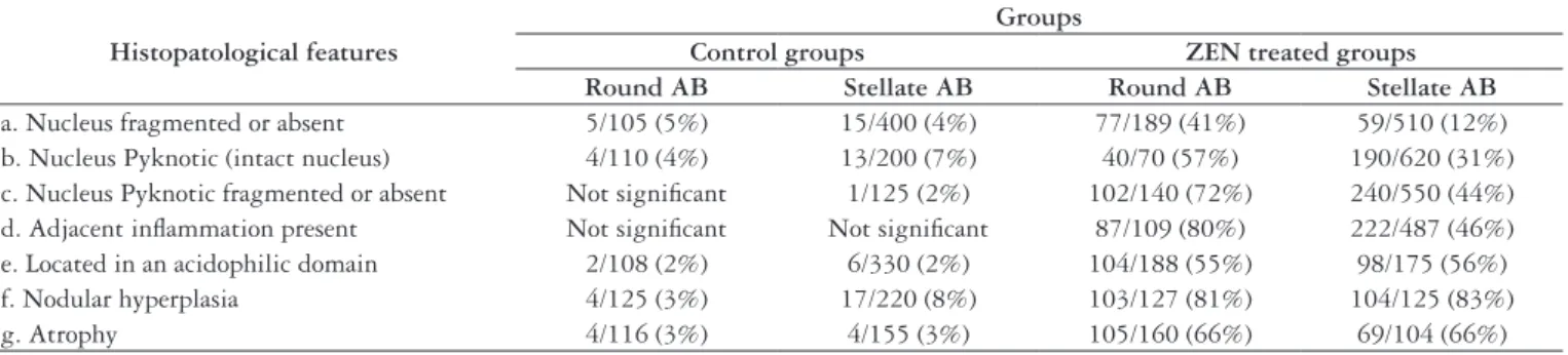
There was chronic inlammatory cells iniltrate in the portal tracks. There was also extensive hepatocellular necrosis, sinusoidal congestion, and neutrophil iniltration. Shrunken acidophilic hepatocytes were both round and detached from surrounding hepatocytes (round – Abs) and stellate – shaped irmly attached to adjacent hepatocytes (stellate – Abs). In 44% of round – Abs, the nucleus was fragmented or absent and among those with an intact nucleus, 80% had nuclear pyknosis stellate. Stellate – Abs often clustered in acidophilic domain usually without signiicant lymphoid iniltrate or necrosis. Round- Abs were usually not clustered and seen more often with an adjust lymphocytes iniltrate. Also apoptotic bodies, in the form of round or stellate – Abs, were seen (Figure 1b).
DISCUSSION
In this present investigation ZEN showed hepatotoxicity in BALB/c mice which characterized by increasing of hepatic marker enzymes (ALT, AST), decreasing of microsomal
cytochrome P450 and histopathology study showed that
ZEN decreased in the number of perfused sinusoids and hepatocellular hypoxia.
Zearalenone is resorcylic acid lactones and functionally is mycoestrogen which found in contaminating grain. Mycoestrogen increased incidence of adenomas (pituitary, liver) was detected in one species after a 2-yr oral carcinogenicity
study(11). In present investigation ZEN shows the potential
hepatotoxicant may due to estrogenic property. Thus, the impaired hepatic function caused after administration of ZEN, that might be a reason for activation of hepatocytes led to increase hepatic marker enzymes, as seen in the present work. The pathophysiology of hepatic injury is complex and it is thought that hepatotoxin activates hepatic cells and subsequently causes free radical- mediated tissue injury and by series of chain reactions produces Lipid
peroxidation (LPO)(17) (Figure 1).
Ayed et al.(1) reported the genotoxicity of ZEN and
concluded that biotransformation of ZEN involved only partial detoxiication and remaining metabolites are relatively
toxic. Another study of Frizzell et al.(3) reported that ZEN
and its metabolites showed potential endocrine disruptors by altering hormone production. Further, ZEN ingestion
of animals from contaminated feed decreased the TNF-α
synthesis and IL-8 synthesis(14). Another study of Marin
et al.(13) reported similar immunosuppressive effects of ZEN
and its derivatives (alpha-ZOL, beta-ZOL, ZAN) in swine. Thus, ZEN showed multiple side effects and our observation reported irst time hepatotoxicity of ZEN.
Formation of LPO leads to many pathological changes
in tissue including liver necrosis(6). The consequences of lipid
peroxidation may be manifested as alternation in membrane integrity of membrane- associated functions in sub-cellular organelles. Reduced glutathione (GSH) is known to function
as an antioxidant and a physiological reservoir for cysteineand
is involved in DNA synthesis, protein synthesis regulation, and detoxiication, etc. Cellular GSH deiciency affects
themitochondrial GSH pool and the cytosolic GSH pool.
MitochondrialGSH is important for the detoxiication of ROS
generated bythe respiratory chain, conjugation of xenobiotics,
maintenanceof thiol-containing proteins, and regulation of
the mitochondrialmembrane potential(15) (Table 5).
FIGURE 1. Showing histopathology of liver sections after staining with H-Eosin (H-E, x.400). (A) Liver section of control mice showing normal architecture of liver with central vein. (B) Liver section of ZEN (50 μg/kg body weight) administered showing sinusoidal congestion and cytoplasmic vacuolization. (C) Liver section of ZEN (100 μg/kg body weight) administered showing hepatocellular necrosis and neutrophil iniltration. (D) Liver section of ZEN (200 μg/kg body weight) administered showing severe nodular hyperplasia, pyknotic nucleus and necrosis
A
B
C
D Histopatological features
Groups
Control groups ZEN treated groups
Round AB Stellate AB Round AB Stellate AB
a. Nucleus fragmented or absent 5/105 (5%) 15/400 (4%) 77/189 (41%) 59/510 (12%) b. Nucleus Pyknotic (intact nucleus) 4/110 (4%) 13/200 (7%) 40/70 (57%) 190/620 (31%) c. Nucleus Pyknotic fragmented or absent Not signiicant 1/125 (2%) 102/140 (72%) 240/550 (44%) d. Adjacent inlammation present Not signiicant Not signiicant 87/109 (80%) 222/487 (46%) e. Located in an acidophilic domain 2/108 (2%) 6/330 (2%) 104/188 (55%) 98/175 (56%) f. Nodular hyperplasia 4/125 (3%) 17/220 (8%) 103/127 (81%) 104/125 (83%) g. Atrophy 4/116 (3%) 4/155 (3%) 105/160 (66%) 69/104 (66%)
In our present study indicates that in parallel increased AST and ALT, increased levels of LPO and decreased GSH activity occurred liver injury by ZEN.
Another point to be considered in this context is that there are increased sleeping times of pentobarbital with combination of ZEN. This result indicates that ZEN impaired metabolism of pentobarbital by hepatic damage with inhibits the activity
of CYP450. Previous study shows that ZEN activates the nuclear
receptor PXR and induces the expression of the drug-metabolizing enzyme CYP3A4, a hepatic monooxygenase involved in the
metabolism of about 60% of clinically used drugs(4). Further, in
our present investigation decreased of CYP450 activities correlates
the increasing sleeping time of pentobarbital. Thus, it is likely
that ZEN inhibits CYP450 activities by hepatotoxicity.
In summary, the present study provides the irst functional anatomical evidence that phenobarbitone sleep time increase may predispose the liver to signiicant oxidative injury and
subsequently hepatic damage, CYP450 deactivation by ZEN.
Although the detailed mechanisms concerning the deactivation
of CYP450 are not fully understood, the current indings present
important insights into the potential detrimental effect of oxidative stress and impaired hepatic function of ZEN.
ACKNOWLEDGEMENTS
This work has been supported by a grant of Defence Research Development Organization (DRDO), Ministry of Defence, Government of India.
Chatopadhyay P, Pandey A, Chaurasia AK, Upadhyay A, Kamarkar S, Singh L. Hiperplasia e danos hepáticos induzidos por micotoxinas do gênero Fusarium-zearalenone em camundongos BAB/c. Arq Gastroenterol. 2012;49(1):77-81.
RESUMO – Contexto - Zearalenone é um micoestrógeno e considerado como micotoxina. Objetivo - Avaliar se o Zearalenone produz hepatotoxicidade por administração via oral. Métodos - Zearalenone foi administrada por via oral em doses de 50 µg, 100 µg e 200 µg/peso corporal/dia/14 dias, respectivamente, para três grupos de camundongos BAB/C. Modalidades diagnósticas usadas para avaliar o dano hepático e comprometimento da função hepática pré- e pós-administração de Zearalenone incluíram atividade enzimática de marcadores hepáticos, tempo de sono por pentobarbital, atividade do citocromo P-450 e avaliação histopatológica hepática. Resultados - Alterações histopatológicas signiicantes como congestão sinusoidal, vacuolização citoplasmática, necrose hepatocelular e iniltração neutrofílica foram observadas após avaliação histológica de cada grupo após exposição acumulada de Zearalenone. Além disto, a exposição à Zearalenone incrementou a atividade das enzimas alanina transaminase e aspartato transaminase e peróxidos lipídicos, ao passo que as atividades teciduais de glutationa e citocromo P-450 diminuiram, quando comparadas com camundongos-controle. Zearalenone também aumentou o tempo de sono e diminuiu a latência do sono após a administração de pentobarbital por via intra-abdominal, quando comparados com camundongos-controle, o que indica o comprometimento das enzimas do metabolismo hepático por ela. Conclusão - Zearalenone é uma potente hepatotoxina quando administrada por via oral.
DESCRITORES – Hiperplasia. Zearalenona. Fígado, patologia. Camundongos.
REFERENCES
1. Ayed Y, Ayed-Boussema I, Ouanes Z, Bacha H. In vitro and in vivo induction of chromosome aberrations by alpha- and beta-zearalenols: comparison with zearalenone. Mutat Res. 2011 [in press].
2. Ellman GL. Tissue sulfhydryl groups. Arch Biochem Biophys. 1959;82:70–7. 3. Frizzell C, Ndossi D, Verhaegen S, Dahl E, Eriksen G, Sorilie M, Ropstad E, Muller
M, Elliott Ct, Connolly L. Endocrine disrupting effects of zearalenone, alpha- and beta-zearalenol at the level of nuclear receptor binding and steroidogenesis. Toxicol Lett. 2011;206:210-7.
4. Guengerich FP. Cytochrome P-450 3A4: regulation and role in drug metabolism. Annu Rev Pharmacol Toxicol. 1999;39:1–17.
5. Hidy PH, Baldwin RS, Greasham RL, Keith CL, McMullen JR. Zearalenone and some derivatives: production and biological activities. Adv Appl Microbiol. 1977;22:59–82.
6. Jaeschke H, Farhood A. Neutrophil and kupffer cell-induced oxidant stress and ischemic–reperfusion injury in rat liver. Am J Physiol. 1991;260:G355-G62. 7. Jordan RA, Schenkman JB. Relationship between malondialdehyde production
and arachidonate consumption during NADPH supported microsomal lipid peroxidation. Biochem Pharmacol. 1982;31:1393-400.
8. Katzenellenbogen BS, Korach KS. A new actor in the estrogen receptor drama-enter ER-beta. Endocrinology. 1997;138:861–2.
9. Kilakivi-Clarke L, Cho E, Onojafe I, Raygada M, Clarke R. Maternal exposure to genistein during pregnancy increases carcinogen–induced mammary tumorgenesis in female rat offspring. Oncol Rep. 1999;6:1089-95.
10. Kuiper GG, Lemmen JG, Carlsson B, Corton JC, Safe SH, van der Saag PT, van
der Burg B, Gustafsson JA. Interaction of estrogenic chemicals and phytoestrogens with estrogen receptor beta. Endocrinology. 1998;139:4252–63.
11. La Vecchia C, Negri E, Parazzini F. Oral contraceptives and primary liver cancer. Br J Cancer. 1989;59:460–1.
12. Mant JW, Vessey MP. Trends in mortality from primary liver cancer in England and Wales 1975-92: inluence of oral contraceptives. Br J Cancer. 1995;72:800–3. 13. Marin DE, Taranu I, Burlacu R, Tudor DS. Effects of zearalenone and its
derivatives on the innate immune response of swine. Toxicon. 2010;56:956-63. 14. Marin DE, Taranu I, Burlacu R, Manda G, Motiu M, Neagoe I, Dragomir C,
Stancu M, Calin L. Effects of zearalenone and its derivatives on porcine immune response. Toxicol In Vitro. 2011;25:1981-8.
15. Marubayashi S, Dohi K, Ochi K, Kawaski T. Role of free radicals in ischemic rat liver cell injury: prevention of damage by alpha-tocopherol administration. Surgery. 1986;99:184-2.
16. Omura T, Sato R. The carbon monoxide-binding pigment of liver microsomes. I. Evidence for its hemoprotein nature. J Biol Chem. 1964;239;2370-8. 17. Salah-Abbès J, Abbès S, Ouanes Z, Houas Z, Abdel-Wahhab MA, Bacha H,
Oueslati R. Tunisian radish extract (Raphanus sativus) enhances the antioxidant status and protects against oxidative stress induced by zearalenone in Balb/c mice. J Appl Toxicol. 2008;28:6-14.
18. Schenkman JB, Cinti DL. Preparation of microsomes with calcium. Methods Enzymol. 1978;52:83-9.
19. Shimamatsu K, Wanless IR. Role of ischemia causing apoptosis, athropy and nodular hyperplasia in human liver. Hepatology. 1997;26:343-50.