AR
TIGO ORIGINAL / ORIGINAL AR
TICLE
INTRODUCTION
Esophageal cancer is an important health problem worldwide, with an increasing incidence and high mor-tality. It has a poor prognosis and is often diagnosed at a late stage. At the time of diagnosis, 60% of the patients are only suitable for palliative therapy(2).
There are two major types of esophageal cancer: adenocarcinoma and squamous cell carcinoma. Squamous cell cancer arises mainly from the upper and middle esophagus. Adenocarcinoma affects the distal esophagus of younger patients and it is usually detected in an earlier stage(6). The known risk factors
PERCUTANEOUS ENDOSCOPIC
GASTROSTOMY FOR NUTRITIONAL
PALLIATION OF UPPER ESOPHAGEAL
CANCER UNSUITABLE FOR
ESOPHAGEAL STENTING
Ana
GRILO
, Carla Adriana
SANTOS
and Jorge
FONSECA
ABSTRACT – Context - Esophageal cancer is often diagnosed at an advanced stage and has a poor prognosis. Most patients with
advanced esophageal cancer have signiicant dysphagia that contributes to weight loss and malnutrition. Esophageal stenting is a widespread palliation approach, but unsuitable for cancers near the upper esophageal sphincter, were stents are poorly tolerated. Generally, guidelines do not support endoscopic gastrostomy in this clinical setting, but it may be the best option for nutritional support. Objective - Retrospective evaluation of patients with dysphagia caused advanced esophageal cancer, no expectation of resuming oral intake and with percutaneous endoscopic gastrostomy for comfort palliative nutrition. Method - We selected adult patients with unresecable esophageal cancer histological conirmed, in whom stenting was impossible due to proximal location, and chemotherapy or radiotherapy were palliative, using gastrostomy for enteral nutrition. Clinical and nutritional data were evaluated, including success of gastrostomy, procedure complications and survival after percutaneous endoscopic gastrostomy, and evolution of body mass index, albumin, transferrin and cholesterol. Results - Seventeen males with stage III or IV squamous cell carcinoma fulilled the inclusion criteria. Mean age was 60.9 years. Most of the patients had toxic habits. All underwent palliative chemothe-rapy or radiothechemothe-rapy. Gastrostomy was successfully performed in all, but nine required prior dilatation. Most had the gastrostomy within 2 months after diagnosis. There was a buried bumper syndrome treated with tube replacement and four minor complications. There were no cases of implantation metastases or procedure related mortality. Two patients were lost and 12 died. Mean survival of deceased patients was 5.9 months. Three patients are alive 6, 14 and 17 months after the gastrostomy procedure, still increasing the mean survival. Mean body mass index and laboratory parameters were roughly stable 1 and 3 months after the gastrostomy procedure. Conclusions - In patients with advanced upper esophageal cancer where only palliative treatment is possible, nutritional support is easily achieved with percutaneous endoscopic gastrostomy, allowing patients to be at homes, surviving a signiicant period of time. Percutaneous endoscopic gastrostomy feeding should be considered as standard deinitive nutritional palliation in patients with upper esophageal cancer, unsuitable for esophageal stenting.
HEADINGS – Gastrostomy. Esophageal neoplasms. Nutritional support.
Declared conflict of interest of all authors: none.
GENE - Grupo de Estudo de Nutrição Entérica - Bloco de Exames Especiais/Serviço de Gastrenterologia Hospital Garcia de Orta, Almada, Portugal. Correspondence: Prof. Jorge Fonseca - Avenida Prof. Torrado da Silva, 2.800 – Almada, Portugal. E-mail: [email protected]
for esophageal cancer are smoking, alcohol consump-tion, chronic gastroesophageal relux disease, Barrett’s esophagus, exposure to nitrosamines, ingestion of lye, Fanconi’s anemia, achalasia, Plummer-Vincent webs, and tylosis(3). Progressive dysphagia and rapid weight loss are the initial symptoms in most patients.
advanced tumors, there are different options of palliative treatment including brachytherapy, chemotherapy and endoscopic palliation techniques such as esophageal dila-tation, intraluminal stents, laser therapy or photodynamic therapy(2).
Most patients with advanced esophageal cancer have signiicant dysphagia that contributes to weight loss and malnutrition, with a negative impact on the disease course and quality of life(5, 9). Therefore, restoring or preserving swallowing and maintaining nutritional status are primary goals of palliation. Esophageal dilatation and stenting are a simple and widespread palliation approach. It allows pa-tients to have an almost normal oral intake. Unfortunately, a large number of cancers arise in upper esophagus, and stents placed near the upper esophageal sphincter, less than 1-2 cm, are poorly tolerated by most patients. For these patients with proximal cancers, gastrostomy may be the best option for nutritional support.
Even when surgery, chemotherapy or radiotherapy are intended to be curative, they frequently compromise oral intake for a large period. Percutaneous endoscopic gastros-tomy (PEG) may be used for transitory nutritional support during this period. Conversely, PEG may be used to provide palliative enteral nutrition in patients with advanced cancer and no expectation of resuming oral intake. As a general rule, guidelines do not support PEG tube placement in evidence of terminal disease, with rapidly progressive tumor, severe malnutrition, and life expectancy less than 1-2 months(8, 14). Nevertheless, in individual cases PEG may be used as an alternative to nasogastric tubes or other nutritional options, in order to prevent patients from starving.
The aim of the present study was the retrospective evalua-tion of the clinical and nutrievalua-tional evoluevalua-tion of patients with upper esophageal cancer using PEG for comfort palliative nutrition.
MATERIALS
From the clinical iles of our enteral nutrition team, we selected adult patients with unresecable esophageal cancer in whom stenting was impossible and chemotherapy or radiotherapy were palliative, without intending a deinitive solution of dysphagia.
Patients were included with:
1. Histological conirmed carcinoma of the proximal esophagus.
2. Unsuitable curative approach due to cancer extension or patient condition.
3. Proximal cancer location preventing esophageal stent placement.
4. Endoscopic gastrostomy as deinitive nutritional pal-liation.
Patients with surgical gastrostomy were excluded. The collected data of the patients included:
1. Clinical data: age and gender, toxic habits, stage of the tumor, success or failure of PEG tube insertion, pro-cedure complications and survival after gastrostomy.
2. Available nutritional and laboratorial markers, at the time of gastrostomy and 1 and 3 months later: body mass index (BMI), serum albumin, serum transferrin and serum total cholesterol.
RESULTS
Seventeen patients fulilled the inclusion criteria. They were all male. Patient’s age ranged from 39 to 78 years (mean age: 60.9 ± 12.8 years). All 17 were diagnosed as having squamous cell carcinoma (SCC). Most of the patients had toxic habits (14 smokers and 11 with major alcoholic intake). All patients presented at an advanced cancer stage, 12 (70%) in stage IV, the remaining 5 (30%) in stage III. All underwent palliative chemotherapy, radiotherapy or booth combined.
PEG Procedure
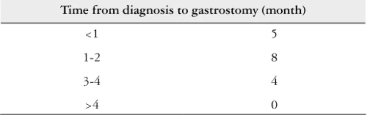
The enteral feeding team discussed the insertion of a PEG tube with the patients and the caregivers. All of them understood the potential and the limitations of the proce-dure. Most of the 17 patients, J (76%) had the gastrostomy within 2 months after the diagnosis (Table 1). The standard pull method was used in all patients. In nine cases (53%), there was an esophageal stenosis requiring prior dilatation. All procedures were performed under conscious sedation, administered according to the patients needs. Gastrostomy was successfully performed in all of them and there were no cases of unsuccessful attempts of PEG tube placement. The procedure was performed during a 24-48 hours hospital stay and all patients were safely discharged to their homes. Follow-up was carried out in an outpatient basis.
TABLE 1. Time from diagnosis to gastrostomy
Time from diagnosis to gastrostomy (month)
<1 5
1-2 8
3-4 4
>4 0
Complications
There was one major complication. It was a buried bumper syndrome (BBS) in a 61 years old patient with a SCC stage IV, treated with PEG tube replacement. Minor com-plications occur in four patients. Three with local infection around the PEG tube, that were treated with dressings and oral antibiotics, and in one case there was a leakage and the PEG tube had to be replaced for a larger one. There were no cases of implantation metastases. There was no procedure related mortality.
Clinical outcome
From the 17 patients, 2 patients were lost for follow-up before the irst month of gastrostomy. Twelve patients died on the course of the advanced disease. Time from PEG tube placement to death ranged from 2 months to 1 year (Table 2). Mean survival time after the procedure of these decease patients was 5.9 months. Other 3 patients are alive and follo-wed by the enteral nutrition team, with 6, 14 and 17 months after gastrostomy, a mean time of 12 months, increasing the survival after the procedure.
TABLE 2 - Time from gastrostomy to death (12 patients)
Time from gastrostomy to death (month)
<1 0
1-3 4
4-6 5
>6 3
Nutritional evolution
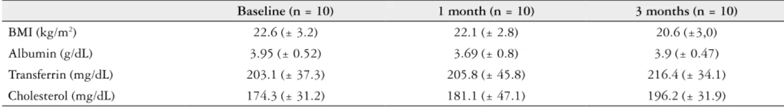
BMI and laboratory data were evaluated at the day of gastrostomy (baseline), 1 and 3 months later. Looking to our 17 patients, the available data demonstrate that, despite of the advance cancers, patients are referred for gastrostomy with reasonable mean BMI (21.3 ± 3.5 kg/m2) and laboratory parameters, including albumin (3.76 ± 0.62 g/dL), transferrin (181.9 ± 45.5 mg/dL) and total cholesterol (162.9 ± 30.9 mg/ dL). Globally, mean BMI and laboratory parameters were roughly stable in the 1 month and 3 months evaluation, but some patients died or were lost for follow-up (Table 3). Look-ing to the 10 patients, with available data at baseline, 1 and 3 months (Table 4), mean BMI and laboratory parameters were also roughly stable in the 1 month and 3 months evaluation.
DISCUSSION
Patients with advanced esophageal cancer have
signii-cant dysphagia, weight loss and malnutrition, that is often worsened during chemotherapy and radiotherapy, by muco-sitis and esophagitis(2, 9). They need an effective nutritional support.
There are many studies where enteral nutrition by PEG compared with nasogastric tube achieves many nutritional advantages. In addition, PEG feeding has a simple manage-ment which do not requires long feeding times for each meal, as needed with a thin diameter of the nasogastric tube. Even more important in this clinical setting of palliation, PEG as a minor interference with the patient’s life, regarding comfort and esthetic aspects(12, 13, 16).
As expected all 17 proximal esophageal carcinomas were squamous cell carcinoma(6). All patients were male. This gen-der speciicity is probably related with tobacco and alcohol consumption of male patients(6, 11, 15).
Gastrostomy should performed be as early as possible. Early insertion may be easier because the stenosis of the esophagus is less pronounced than during the later stages(5). Enteral feeding support must start before the decline of the nutritional status. In our study most of gastrostomies were performed in the irst 2 months after the diagnosis.
There were no technical failures and endoscopic gastros-tomy was performed in all of the patients proposed for the procedure. There were few complications related with the procedure and only one was a major complication, a BBS, an uncommon complication of PEG tube placement, that occurs when the internal bumper of a PEG tube lodges any-where between the gastric wall and the skin. In most cases occurs months after the procedure, but is known that patients like our case, with malignant diseases, poor nutritional con-dition and rapid weight gain after PEG placement, have a greater risk of BBS(10). Probably, the regular follow-up and the care taken in education of patients and caregivers prevented the occurrence of more complications in our patients. It has been described, that patients with head and neck cancer un-dergoing PEG by the pull technique may develop abdominal wall metastases, by cancer seeding(12, 13). This occurs rarely, and there were no cases on our study.
TABLE 4. Evolution of BMI and laboratory markers in patients with data available at the 3 months follow-up
Baseline (n = 10) 1 month (n = 10) 3 months (n = 10) BMI (kg/m2) 22.6 (± 3.2) 22.1 (± 2.8) 20.6 (±3,0)
Albumin (g/dL) 3.95 (± 0.52) 3.69 (± 0.8) 3.9 (± 0.47) Transferrin (mg/dL) 203.1 (± 37.3) 205.8 (± 45.8) 216.4 (± 34.1) Cholesterol (mg/dL) 174.3 (± 31.2) 181.1 (± 47.1) 196.2 (± 31.9) TABLE 3. Evolution of BMI and laboratory markers in all patients included
Baseline (n = 17) 1 month (n = 15) 3 months (n = 10) BMI (kg/m2) 21.3 (± 3.5) 21.0 (± 3.1) 20.6 (± 3.0)
Clinical and nutritional evaluation was performed at the time of gastrostomy, and 1 and 3 months later. As expectable in advanced disease, four patients died before the 3 months follow-up. Two more patients were lost for follow-up. There-fore data from 3 months follow-up refers to a smaller number of patients. Nevertheless, when we compare evolution of the 10 patients with data available at the 3 months follow-up, values are roughly similar.
With our data is evident the stable BMI and serum albumin, and an improvement in transferrin and choles-terol values. The selected laboratory values are known as biomarkers of prognosis and nutritional and inlammatory status(4, 11). In advanced cancer patients it is not expectable that inlammatory status of signiicantly decreases. To the best of our knowledge, the stability or improvement of these laboratory markers, in patients with growing cancers, must relect eficient nutritional support, suggesting that even in this poor clinical setting some improving could be achieved.
Guidelines do not recommend PEG in palliative patients with an unfavorable prognosis or an incurable disease, be-cause in most of them the mean survival time was less than 2 months. Nevertheless, in our experience, decision should always be individualized(1, 8, 14). In the patients we evaluate with unfavorable prognosis and under palliative treatment,
Grilo A, Santos CA, Fonseca J. A gastrostomia percutânea endoscópica na paliação nutricional do câncer do esôfago proximal sem possibilidade de colocação de prótese. Arq Gastroenterol. 2012;49(3):227-31.
RESUMO – Contexto - O câncer do esôfago é frequentemente diagnosticado num estádio avançado, com mau prognóstico. A maioria dos pacientes com
câncer avançado do esôfago sofre de disfagia que contribui para a desnutrição e perda de peso. A colocação de endopróteses é uma forma de paliação muito difundida. Contudo, as próteses muito próximas do esfíncter esofágico superior são mal toleradas pelos doentes, não sendo uma opção adequada se o câncer for muito proximal. Habitualmente, as recomendações para gastrostomia percutânea não incluem a paliação nutricional nestes doentes, mas a gastrostomia percutânea endoscópica pode ser a melhor forma de suporte nutricional no câncer avançado. Objetivo - Avaliação retrospectiva dos doentes com disfagia por câncer avançado do esôfago em que a gastrostomia percutânea endoscópica foi a forma de paliação nutricional, sem expectativa de retomar a ingestão oral. Método - Selecionaram-se doentes adultos com câncer irressecável do esôfago, com conirmação histológica e com localização proximal, impedindo a colocação de prótese, com a radioterapia e quimioterapia paliativas, usando a gastrostomia percutânea endoscópica para a nutrição entérica. Avaliaram-se dados clínicos e laboratoriais, incluindo o sucesso da gastrostomia, complicações e sobrevida após a gastrostomia e evolução do índice de massa corporal, albumina, transferrina e colesterol. Resultados - Foram incluídos 17 homens com carcinoma epidermoide no estádio III ou IV, com média de idade de 60,9 anos. A maioria consumia tabaco e bebidas alcoólicas. Todos foram submetidos a radioterapia ou quimioterapia. A gastrostomia endoscópica foi bem-sucedida em todos, embora nove tenham necessitado de dilatação prévia. A maioria foi gastrostomizada nos 2 meses subsequentes ao diagnóstico. Ocorreu uma “buried bumper syndrome”, resolvida com substituição do tubo e quatro complicações menores. Não houve implantação de metástases, nem mortalidade associada ao procedimento. Dois doentes foram perdidos e 12 morreram. Três doentes estão vivos 6, 14 e 17 meses após a gastrostomia e ainda estão aumentando a sobrevida média. Os valores médios do índice de massa corporal e da avaliação laboratorial mantiveram-se estáveis 1 e 3 meses após a gastrostomia. Conclusão - Em pacientes com câncer avançado do esôfago, em que só a terapêutica paliativa é possível, o suporte nutricional é facilmente obtido com gastrostomia percutânea endoscópica, permitindo aos pacientes permanecer em suas casas por um longo período. A nutrição por gastrostomia percutânea endoscópica deveria ser considerada, por rotina, como a opção deinitiva para paliação nutricional em pacientes com câncer do esôfago proximal em que a colocação de prótese não é possível.
DESCRITORES – Gastrostomia. Neoplasias esofágicas. Apoio nutricional.
insertion of a PEG tube was discussed with the patient and caregivers. The mean survival time after the procedure, in our deceased patients, was 5.9 months, and there are still patients alive, increasing this survival period. This is a period of time clearly in agreement with the general guidelines for PEG(8, 14). During this time we achieved nutritional support to our patients, and contributed for a comfort and a better nutritional support in the last months of life. Patients could be cared at their homes with their families, even keeping some social activities. Our experience demonstrates that an important number of esophageal cancer patients, unsuitable to curative therapy, have a signiicant survival expectance and PEG feeding should be more often proposed to these patients.
CONCLUSION
REFERENCES
1. Angus F, Burakoff R. The percutaneous endoscopic gastrostomy tube: medical and ethical issues in placement. Am J Gastroenterol. 2003;98:272-7.
2. Besharat S, Jabbari A, Semnani S, Keshtkar A, Marjani J. Inoperable esophageal cancer and outcome of palliative care. World J Gastroenterol. 2008;14:3725-8. 3. Enzinger PC, Mayer RJ. Esophageal cancer. N Engl J Med. 2003;349:2241-52. 4. Guerra L, Rosa A, Romani R, Gurski R, Schimer C, Kruel CD. Serum transferrin
and serum prealbumin as markers of response to nutritional support in patients with esophageal cancer. Nutr Hosp. 2009;24:241-2.
5. Hunter J, Lauretano L, Shellito P. Percutaneous endoscopic gastrostomy in head and neck cancer patients. Ann Surg. 1989;210:42-6.
6. Jemal A, Bray F, Center MM, Ferlay J, Ward E, Forman D. Global cancer statistics. CA Cancer J Clin. 2011;61:69–90.
7. Khoshbaten M, Naderpour M, Mohammadi G, Alipoor SH, Estakhri R, Fazeli Z. Epidemiology of esophageal lesions in patients with head and neck squamous cell carcinoma. Asian Pac J Cancer Prev. 2010;11:863-5.
8. Löser C, Aschl G, Hébuterne X, Mathues-Vliegen EM, Muscaritoli M, Niv Y, Rollins H, Singer P, Skelly RH. ESPEN guidelines on artiicial enteral nutrition — percutaneous endoscopic gastrostomy (PEG). Clin Nutr. 2005;24:848–61. 9. Marin FA, Lamônica-Garcia VC, Henry MA, Burini RC. Grade of esophageal
cancer and nutritional status impact on postsurgery outcomes. Arq Gastroenterol. 2010;47:348-53.
10. Meine G, Lukashok H, Mello G, Mansur G, Guimarães D, Carvalho R. Buried bumper syndrome as a complication of percutaneous endoscopic gastrostomy in cancer patients: the Brazilian experience. Dig Endosc. 2007;19:22-5.
11. Raynaud-Simon A, Revel-Delhom C, Hébuterne X; French Nutrition and Health Program; French Health High Authority. Clinical practice guidelines from the French health high authority: nutritional support strategy in protein-energy malnutrition in the elderly. Clin Nutr. 2011;30:312-9.
12. Sironi A, Arcovio C, Bergamini C, Colatruglio S, Gavazzi C. Indications for per-cutaneous luoroscopic gastrostomy in palliative head and neck cancer patients. Nutr Ther Metab. 2010;28:143-7.
13. Stockeld D, Fagerberg J, Granström L, Backman L. Percutaneous endoscopic gastrostomy for nutrition in patients with oesophageal cancer. Eur J Surg 2001;167:839-44.
14. Stroud M, Duncan H, Nightingale J; British Society of Gastroenterology: guidelines for enteral feeding in adult hospital patients. Gut. 2003;52(Suppl VII):VII1-VII12 15. Thuler FP, Forones NM, Ferrari AP. Neoplasia avançada de esófago – diagnóstico
ainda muito tardio. Arq Gastroenterol. 2006;43:206-11.
16. Zuercher B, Grosjean P, Monnier P. Percutaneous endoscopic gastrostomy in head and neck cancer patients: indications, techniques, complications and results. Eur Arch Otorhinolaryngol. 2011;268:623-9.