AR
TIGO ORIGINAL / ORIGINAL AR
TICLE
INTRODUCTION
Fibrosis is an important process that results from hepatic injury and chronic disease process. Both of these causes can have common etiologies that can contribute to the onset of cirrhosis(2).
Cirrhosis is considered to be the most advanced stage of tissue ibrosis and is characterized by the disruption of hepatic parenchyma, the appearance of the septa and ibrotic nodules, changes in hepatic blood low and a risk of liver failure(10).
Cirrhosis can be caused by altered vascular tis-sue, which causes a shift in the supply of blood and portal blood low directly to the liver (central vein). The altered vascular tissue compromises the hepatic sinusoids and hepatocytes. Some of the circulatory changes found in cirrhosis are splanchnic vasodilata-tion, vasoconstriction and renal hypoperfusion, salt and water retention and increased cardiac output, which are all closely linked to vascular disease of the liver that may cause portal hypertension(8, 25).
Portopulmonary hypertension (PPH) is a relative-ly rare complication occurring in cirrhosis, and its prevalence is high in patients with refractory ascites. PPH is mostly likely caused by excessive pulmonary vasoconstriction and factors such as transforming
LUNG AND LIVER CHANGES DUE
TO THE INDUCTION OF CIRRHOSIS
IN TWO EXPERIMENTAL MODELS
Renata Salatti
FERRARI
1, 3, Maurício
TIEPPO
1, 2, Darlan Pase da
ROSA
1, 2, 3,
Luiz Alberto
FORGIARINI JR
1, Alexandre Simões
DIAS
1, 3and Norma Possa
MARRONI
1, 2, 3ABSTRACT - Context - To evaluate lung and liver changes in two experimental models using intraperitoneal carbon tetrachloride (CCl4) and bile duct ligation (BDL). Methods - Twenty-four male Wistar rats were divided into a control group (CO) and an experimental group (EX). We evaluated the liver transaminases (AST, ALT, AP), arterial blood gases (PaO2, PCO2 and SpO2) and lipid peroxidation by TBARS (substances that react to thiobarbituric acid) and chemiluminescence. We also evaluated the antioxidant enzyme superoxide dismutase (SOD) and histology of lung tissue and liver. Results - There were signiicant differences in AST, ALT, ALP and PaO2 between CO group and EX group (P<0.05). The levels of TBARS, chemiluminescence and activity of enzyme superoxide dismutase were in-creased to different degrees in the CCl4 groups: CO and in the BDL -EX (P<0.05, respectively). In the lung histology, an increase in the wall thickness of the pulmonary artery and a diameter reduction in the CCl4 animal model were observed: comparing CO group with EX group, we observed a reduction in thickness and an increase in the diameter of the artery wall lung. Conclusion - Both experimental models have caused liver damage and alterations in the artery wall that are associated with major changes in pulmonary gas exchange. HEADINGS - Liver diseases. Lung diseases. Liver cirrhosis experimental. Carbon tetrachloride, diagnostic use. Common bile duct,
physiopathology. Diseases models, animal.
Declared conflict of interest of all authors: none
1 Research performed at: Laboratório de Hepatologia e Gastroenterologia, Hospital das Clínicas de Porto Alegre (HCPA); 2Universidade Luterana do Brasil (ULBRA); 3 Universidade Federal do Rio Grande do Sul (UFRGS), Porto Alegre, RS, Brasil. Financial Support: Fundo de incentivo à pesquisa e eventos do Hospital de Clínicas de
Porto Alegre. FIPE / HCPA.
Correspondence: Dr. Renata Salatti Ferrari - Street Evaristo da Veiga, 196/04 – 90620-230 - Porto Alegre, RS, Brasil. E-mail: [email protected]
growth factor beta1 (TGF-β1)(4). However, we do
not have a perfect experimental model for the study of PPH, making the discovery of effective substances that can reduce and even prevent the progression of the disease impossible. Developing an experimental model is be very useful, as such a model could lead to substantial cost savings in treatment.
Carbon tetrachloride (CCl4) can be applied both intraperitoneally and by inhalation. CCl4 induces ibrosis liver cirrhosis and may be used for the study of PPH. The signals observed in the CCl4 liver injury model are similar to those found in cirrotic patients(19).
The CCl4 trichloromethyl radical is converted to (•CCL3) and trichloromethyl peroxide (°OOCCl3). The CCl4 trichloromethyl radical has been described as causing hepatotoxic effects such as ibrosis, steatosis, necrosis and hepatocellular carcinoma(9, 22).
Bile duct ligation is another experimental model related to secondary biliary cirrhosis because it causes cell proliferation, hepatocellular necrosis, apoptosis, activation of stellate cells, and inally the formation of liver ibrosis and cirrhosis. This model has been described by our group as causing Experi-mental Hepatopulmonary Syndrome(26, 29).
hepatic injury that cause liver cirrhosis, a CCl4 model and a BDL model, to evaluate which model can best reproduce characteristics found in PPH.
METHODS
Animals and experimental groups
We used 24 male Wistar rats (average weight 250 g), purchased from the Center for Reproduction and Labo-ratory Animal Experimentation (CREAL), Federal Uni-versity of Rio Grande do Sul (UFRGS). The animals were housed in plastic cages (47×34×18 cm) and maintained in light/dark cycles of 12/12 hs with a controlled temperature (20-25 °C) and ad libitum access to food and water. All of the procedures followed the parameters established by the Ethics and Research of Hospital Clinics of Porto Alegre, and all of the animals received care according to the “Principles of Laboratory Animal Care” formulated by the National Society for Medical Research and the “Guide for the Care and Use of Laboratory Animals” published by the National Institutes of Health (NIH publication 86-23, revised 1985).
The animals were randomly divided into four groups, an experimental and a control group (CO), for each of the two models used in the study, intraperitoneal Carbon tetrachlo-ride (CCl4) and bile duct ligation (BDL):
• CCl4-CO: animals that received only phenobarbital in drinking water;
• CCl4-EX: animals that received phenobarbital in drink-ing water one week before startdrink-ing the injections of CCl4 inductions + ip;
• BDL-CO: animals that underwent laparotomy and only manipulation of the duct biliar.
• BDL-EX:animals subjected to BDL.
Experimental procedures
To develop the CCl4 model, we used the standard rec-ommended by Pavanato et al.(21). Phenobarbital was added
to the drinking water of animals (0.3 mL/dL), serving as an enzyme inducer to potentiate the effect of CCl4(6).
To develop the BDL model, we followed the standard described by Kontouras et al.(14) where, after the experimental
procedures, the animals were caged individually for a period of 28 days.
When sacriicing animals from either group, the animals were anesthetized with xylazine (50 mg/kg body weight) and ketamine (100 mg/kg body weight), both administered intraperitoneally. Post mortem, blood samples were collected via retro-orbital plexus for liver function analysis(12).
After the abdominal region was shaved, a laparotomy was performed to collect blood from the abdominal aorta for arterial blood gas analysis. After collection of blood samples, the animals were sacriiced with an “overdose” of anesthetics.
Histology
For histological analysis of the lung, a fragment of the
right lower lobe was collected for histological analysis, and a fragment of the liver was removed from the right hepatic lobe. The tissues were cut and ixed by immersion in 10% buffered formalin for 24 hours. The remainders of lung and liver were immediately frozen in liquid nitrogen and stored at -80°C for later analysis.
Integrity liver enzymes
Blood samples taken from the retro-orbital plexus were used to assess the levels of aspartate aminotransferase (AST), alanine aminotransferase (ALT), and alkaline phosphatase (AP) and were expressed in IU/L and measured with routine laboratory methods in the Hospital Clinics of Porto Alegre.
Determination of oxidative stress
The liver tissue and lung of each animal were homoge-nized in phosphate buffer solution (140 mM KCl, 20 mM phosphate, pH 7.4) and centrifuged at 3,000 rpm for 10 minutes. Lipid peroxidation was determined by levels of thiobarbituric acid reactive substances (thiobarbituric acid reactive substances - TBARS) mg/prot. read at 535 nm. For the determination of chemiluminescence (CL), 0.5 mL of homogenate was added to 120 mM KCl, 30 mM phosphate buffer, pH 7.4, and 3 mM tert-butyl hydroperoxide at 30°C and analyzed for chemiluminescence in a counter liquid scintillation expressed as cps/mg/prot(12, 18).
Superoxide dismutase enzyme activity
The activity of the antioxidant enzyme superoxide dis-mutase (SOD) was determined by quantifying the oxidation of adrenaline to adrenochrome according to Misra and Fridovich and was expressed in U/mg/prot(18).
Statistical analysis
The results were expressed as mean ± standard error. Data were compared by analysis of variance (ANOVA), and if the analysis indicated a statistically signiicant difference, we used a post hoc Student Newman-Keuls test. The statistical signiicance level was set at P<0.05. The Statistical Package
for Social Sciences, version 13.0 (SPSS Inc., Chicago, IL, USA), was used.
RESULTS
Activity of transaminases
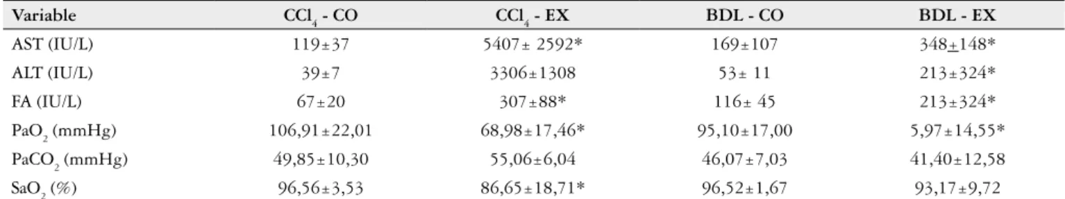
Table 1 indicates a signiicant difference between the CCl4 and CCl4-CO-EX groups and between BDL and BDL-CO-EX in serum AST, ALT and AP, which were higher in the experimental groups compared to the controls (P<0.05).
Arterial blood gas analysis
respective controls (P<0.05). Only the variable PCO
2 showed
no signiicant difference between the CO and EX animals in the BDL model.
Markers of Oxidative Stress and Antioxidant Activity
Analysis of lipid peroxidation in lung tissue (Table 2) it appears that TBARS as well as high CL in two models are used in the study (P<0.05). Regarding the antioxidant enzyme SOD, a signiicant increase (P<0.05) in both
exper-imental models was observed compared to control animals.
Histology
On the photomicrograph of the lung tissue (Figure 1), we observed an increase of 47.87 mM in wall thickness in TABLE 1. Activity of transaminases and arterial blood gas analysis
Variable CCl4 - CO CCl4 - EX BDL - CO BDL - EX
AST (IU/L) 119±37 5407± 2592* 169±107 348+148*
ALT (IU/L) 39±7 3306±1308 53± 11 213±324*
FA (IU/L) 67±20 307±88* 116± 45 213±324*
PaO2 (mmHg) 106,91±22,01 68,98±17,46* 95,10±17,00 5,97±14,55*
PaCO2 (mmHg) 49,85±10,30 55,06±6,04 46,07±7,03 41,40±12,58
SaO2 (%) 96,56±3,53 86,65±18,71* 96,52±1,67 93,17±9,72
CCl4:Carbon tetrachloride; BDL:bile duct ligation; CO:control; EX:experimental; AST:aspartate aminotransferase; ALT:alanina aminotransferase; AP:alkaline phosphatase; PaO2:pressure oxygen; PaCO2:cabon dioxide partial pressure; SaO2: oxygen saturation. Results expressed as mean + standard deviation. * P<0,05 corresponding control group
TABLE 2. Evaluation of lipid peroxidation in the lung tissue (TBARS e QL) and antioxidant enzyme SOD
Model TBARS (nmol/mg prot)
QL (cps/mg prot)
SOD
(IU/mg prot)
CCl4
CO 0,56±0,22 16764,20±515,48 11,24±4,99
EX 1,48±0,52* 17797,20±531,65 27,27±1,23*
BDL
CO 0,835±0,16 16008,41±1171,45 6,66±1,34
EX 1,91±0,91* 20250,36±827,82* 16,06±2,67* TBARS: thiobarbituric acid reactive substances; QL: chemiluminescence; SOD: superoxide dismutase; CO: control; EX: experimental. Results expressed as mean + standard deviation. * P<0,05 corresponding control group
Figure 1. Histology of the pulmonary tissue
Photomicrograph of the lung tissue in the models experimentals. a) CCl4-CO; b) CCl4-EX; c) BDL-CO; d) BDL-EX
CO
CO
CCL
4
the pulmonary artery model CCl4 as compared to the CO. However, when analyzing the histology of the pulmonary tissue of the animal model BDL, we observed an increase of 69.67 mM in vascular diameter when compared to con-trols. The photomicrograph of the liver (Figure 2) revealed the presence of necrotic foci, ibrotic nodules, lymphocytic iniltration, steatosis and cellular changes, suggesting that cirrhosis was present in the two models.
DISCUSSION
In an attempt to develop a speciic model for the study of PPH, we used two experimental models developed in our laboratory that cause cirrhosis.
The CCl4 has been used to reproduce liver cirrhosis due to its potent hepatotoxic effect in which a single dose of CCl4 is associated with steatosis and necrosis but a prolonged administration leads to liver ibrosis, cirrhosis and hepa-tocellular carcinoma. CCl4 acts directly on hepatocytes by changing the permeability of the plasma and mitochondrial membranes. This model has been widely used to elucidate the pathogenesis of cirrhosis(11).
Secondary biliary cirrhosis is a chronic and diffuse liver disease that alters intrahepatic or extrahepatic biliary function. The BDL model is used as an animal model for secondary biliary cirrhosis and cirrhosis caused by progres-sive and fatal damage to the liver. This model simulates the effects of the disease present in humans, causing changes in the inlammatory reaction by leaking bile and the subsequent disorganization of parenchymal inflammation, collagen deposition and formation of ibrosis(7, 13, 20).
Studies by Kontouras et al.(14) showed that 15 days after
bile duct ligation (BDL), the metabolic and biochemical changes were not fully established. Thus, in our experiment, the animals were sacriiced only after 28 days of BDL, when studies conirmed liver cirrhosis(17, 29).
In this study, both the BDL and the CCl4 caused liver damage and elevated serum levels of AST, ALT and ALP, which are commonly known to be markers of liver injury. These changes in liver enzyme levels indicate a loss of tissue integrity with consequent apoptosis and necrosis of hepato-cytes. These enzymes are eventually released into circulation
after cellular damage, and this inding has been reported in several studies using models of BDL and CCl4(3, 27, 31).
Patients who have secondary biliary cirrhosis exhibit hyperbilirubinemia due to biliary obstruction resulting from liver damage and present results of liver function tests in higher ranges for FA and aminotransferases. High levels of these enzymes are largely caused by the necrosis of tissues that are rich in aminotransferases(1, 7).
In the presence of liver cirrhosis, there is an increase in lipid peroxidation due to the formation of reactive oxygen species and lipid peroxidation. Some authors report that these phenomena determine the changes in the structures and cell membranes by two mechanisms, covalent binding to macromolecules and cell action on lipids, and that these two mechanisms are the result of the metabolism of CCl4 by cytochrome P-450, which produces two radicals which are highly toxic and CCl3 CCl3O2••(28) Additionally, reactive
oxygen species (ROS) are involved in the BDL model. Cholestasis in the BDL model reduces the excretion of bile salts, causing the retention of hydrophobic bile salts with-in hepatocytes and apoptosis and necrosis, the destruction of liver parenchyma, which contributes to a redox imbalance, and the formation of ROS. In addition, ROS cause oxidation of cellular proteins and extensive damage to mitochondrial DNA and mitochondrial synthesis by liver damage(8, 24, 34).
When we evaluated lipid peroxidation in the two models of cirrhosis, animals in the EX group had signiicantly higher values compared to those in the CO group, and the level of TBARS and QL were elevated in the lung and liver in both groups. This overproduction of ROS might occur in several pathophysiological situations and may explain the inding that in our study, the levels of TBARS and QL were high(22, 23).
Physiological defense mechanisms are effective in pre-venting or counteracting the damage caused by free radicals, which can include a set of endogenous antioxidant enzymes such as superoxide dismutase (SOD), catalase (CAT), and glutathione peroxidase (GPx). The two models used in this study showed an increase in lipid peroxidation in lung tissue and are likely to increase in response to an increase of ROS in the SOD, which plays an important role in the balance of the redox cell, as the catalyst free radicals are generated in an attempt to protect the tissue against lesions(1, 2, 7).
Figure 2. Histology of the liver tissue.
Photomicrograph of the liver tissue in the models experimentals. BDL- and CCl4-EX
BDL
CCI
Arterial blood gas analysis is an indicator used to facili-tate the evaluation of trade gasosas(20). In our study, there was
a signiicant difference between the experimental and control groups with respect to arterial blood gasses. We found that PaO2 and SpO2 were signiicantly lower in the CCl4 group. In the BDL model, the values of SpO2 were also reduced, although this did not reach statistical signiicance.
Approximately 85% of PPH patients show an increase in pulmonary vascular resistance in the course of the disease, of which, 15% experience increases in their pulmonary pressures that are caused by just a state of high lux(15).
The relationship between arterial hypoxemia and PPH has received little attention because PPH is a clinical problem that involves an altered hemodynamic status. PPH patients exhibit arterial hypoxemia and arterial oxygenation that is signiicantly worse when comparing liver transplantation candidates with normal vascular resistance as determined by echocardiography Doppler(16).
In the BDL model, hypoxemia may be associated with bacterial translocation occurring in 45%-75% of cirrhotic animals whereby the induced hepatocellular damage prevents adequate blood iltration and promotes the development of portosystemic shunts; this phenomenon dramatically decreases the phagocytic ability of the liver, and it also allows the entry of bacteria and endotoxins into the lung circulation(23).
Furthermore, studies have reported arterial hypoxemia in BDL animals, which contributes to a reduction in the values of PaO2 due to increased volume of lung shunt(32).
The differences found in this study between the BDL and CCl4 gas analyses showed that regardless of etiology, animals with cirrhosis are hypoxemic because the values of PaO2 and SpO2 were lower in both experimental models. This hypox-emia might be triggered by the activation of macrophages as well as the presence of cytokines and nitric oxide in the extracellular medium. Nitric oxide is a potent vasodilator that promotes dilation of intrapulmonary vessels, leading to hypoxemia. Nitric oxide may also be involved in the mech-anisms that cause an imbalance between the antioxidants and oxidants(23).
Regarding photomicrographs of the lung tissue, the BDL
model caused an increase in diameter of arterioles of the lung tissue. This inding corroborates studies reporting the increase of animals subjected to BDL(29). This result may be
due to the increased intrapulmonary shunt caused by liver injury leading to an accumulation of blood in the body, which in turn dilates the arterioles of the lung tissue and releases nitric oxide. In the CCl4 model, the observed reduction of these structures can be explained by the fact that cirrhotic CCl4 animals had increased levels of ET-1 and collagen depo-sition, promoting the reduction in thickness of lung tissue.
The histology of liver tissue revealed damage to liver cells and conirmed the changes observed in biochemical analyses. The presence of necrotic foci, ibrotic nodules, the iniltration of lymphocytes, and the fatty changes in liver cells are typical characteristic after the induction of cirrhosis(5, 33).
The two experimental models used in this study reinforce and conirm the onset of liver damage and cirrhosis of the liver with consequent changes in gas exchange. The BDL model is most often used to evaluate the Hepatopulmonary Syndrome because it shows changes that are typical charac-teristic of the disease. The CCl4 model seems to be promising for the study of portopulmonary hypertension (PPH) as well as cirrhosis and changes the thickness and diameter of the pulmonary artery wall. More studies are needed to assess and investigate the speciic mechanisms involved in these changes.
CONCLUSION
The model of hepatic cirrhosis caused by CCl4 modiies the gas exchange, increases the thickness of the pulmonary artery and reduces its diameter. The CCl4 model is a promis-ing model for the study of PPH. The BDL model also causes alterations in the lung and liver, reinforcing that is a reliable model of HPS.
ACKNOWLEDGEMENTS
Hospital Clinics of Porto Alegre – HCPA.
Fund research incentive events of Hospital Clinics of Porto Alegre - FIPE/HCPA.
Ferrari RS, Tieppo M, Rosa DP, Forgiarini Jr LA, Dias AS, Marroni NP. Alterações hepáticas e pulmonares decorrentes da indução de cirrose em dois modelos experimentais. Arq Gastroenterol. 2013,50(3):208-13.
RESUMO - Objetivo - Avaliar as alterações pulmonares e hepáticas em dois modelos experimentais de cirrose hepática pelo uso de tetracloreto de carbono intraperitoneal (CCl4) e ligadura de ducto biliar. Métodos - Vinte e quatro ratos machos Wistar foram divididos em grupo controle (CO) e experimental (EX). Foram avaliadas as transaminases hepáticas (AST, ALT, FA), gasometria arterial (PaO2, PCO2 e SatO2) e a lipoperoxidação através de TBARS (substâncias que reagem ao ácido tiobarbitúrico) e por quimiluminescência. Também foi avaliada a atividade antioxidante da enzima superóxido dismutase e a histologia do tecido pulmonar e hepático. Resultados - Nas enzimas hepáticas (AST, ALT e FA), bem como na PaO2 foram observadas diferenças signiicativas (P<0,05) entre os grupos CO vs EX em ambos modelos. Os níveis de TBARS, quimiluminescência e a atividade da enzima superóxido dismutase encontram-se aumentados nos grupos CCl4 e ligadura de ducto biliar: CO vs EX (P<0,05). Na análise histológica do pulmão observamos um aumento na espessura da parede da artéria pulmonar e uma redução no diâmetro no modelo CCl4: CO vs EX, e no modelo de ligadura de ducto biliar podemos observar uma redução da espessura e aumento no diâmetro da parede da artéria pulmonar. Conclusão - Ambos os modelos experimentais provocaram dano hepático, além de causar alterações na parede da artéria pulmonar contribuindo na redução das trocas gasosas. DESCRITORES - Hepatopatias. Pneumopatias. Cirrose hepática experimental. Tetracloreto de Carbono, uso diagnóstico. Ducto colédoco, isiopatologia.
REFERENCES
1. Ahmed AF, Mahmoud MF, Ouf MA, El-Fathaah EA. Aminoguanidine potenti-ates the hepatoprotective effect of silymarin in CCL4 treated rats. Ann Hepatol. 2011;10:207-15.
2. Ali SI, Said MM, Hassan EK. Prophylactic and curative effects of purslane on bile duct ligation-induced hepatic ibrosis in albino rats. Ann Hepatol. 2011;10:340-346.
3. Amália PM, Possa MN, Augusto MC, Francisca LS. Quercetin prevents oxidative stress in cirrhotic rats. Dig Dis Sci. 2007;52:2616–21.
4. Blendis L, Wong F. Portopulmonary hypertension: an increasingly important complication of cirrhosis. Gastroenterology. 2003;125:622-4.
5. Bona S, Filippin LI, Di Naso FC, de David C, Valiatti B, Isoppo Schaun MI, Xavier RM, Marroni NP. Effect of Antioxidant Treatment on Fibrogenesis in Rats with Carbon Tetrachloride-Induced Cirrhosis. ISRN Gastroenterol. doi: 10.5402/2012/762920.
6. Buege JA, Aust SD. Microsomal lipid peroxidation. Methods Enzymol. 1978;52:302-10.
7. Cuevas MJ, Tieppo J, Marroni NP, Tuñón MJ, González-Gallego J. Suppression of amphiregulin/epidermal growth factor receptor signals contributes to the protective effects of quercetin in cirrhotic rats. J Nutr. 2011;141:1299-305. 8. Fallon MB. Mechanisms of Pulmonary Vascular Complications of Liver Disease
Hepatopulmonary Syndrome. J Clin Gastroenterol. 2005;39:S138-42. 9. Fang HL, Lin WC. Lipid peroxidation products do not activate hepatic stellate
cells. Toxicology. 2008;253:36-45.
10. Friedman SL. Mechanisms of hepatic fibrogenesis. Gastroenterology. 2008;134:1655-69.
11. Fujii T, Fuchs BC, Yamada S, Lauwers GY, Kulu Y, Goodwin JM, Lanuti M, Tanabe KK. Mousecmodel of carbon tetrachloride induced liver ibrosis: His-topathological changes and expression of CD133 and epidermal growth factor. BMC Gastroenterology. 2010;10:79.
12. Gonzalez Flecha B, Llesuy S, Boveris A. Hydroperoxide-initiated chemilumines-cence: an assay for oxidative stress in biopsies of heart, liver, and muscle. Free Radic Biol Med. 1991;10:93-100.
13. Gonzalez-Gallego JAE. El hígado. Fisiopatologia de las Hepatopatias. Madrid, Spain: McGraw-Hill-Interamericana; (Fundamentos de isiopatologia, edited by E. A. M. Cordero).1998.
14. Kountouras J, Billing BH, Scheuer PJ. Prolonged bile duct obstruction: a new experimental model for cirrhoses in the rat. Br J Exp Pathol. 1984;65:305-11. 15. Krowka MJ, Cortese DA. Hepatopulmonary syndrome: an evolving perspective
in the era of liver transplantation. Hepatology. 1990;11:138-42. 16. Krowka MJ. Hepatopulmonary syndromes. Gut. 2000;46:1-4.
17. Miltersteiner A, Milterstainer D, Pereira-Filho N, Frota AR, Ely PB, Zettler CG, Marroni CA, Marroni NP. Uso da quercetina a longo prazo em ratos cirróticos. Acta Cir Bras. 2003;18:232-7.
18. Misra HP, Fridovich I. The role of superoxide anion in the autoxidation of epineph-rine and a simple assay for superoxide dismutase. J Biol Chem. 1972;247:3170-5.
19. Miyazaki T, Karube M, Matsuzaki Y, Ikegami T, Doy M, Tanaka N, Bouscarel B. Taurine inhibits oxidative damage and prevents ibrosis in carbon tetrachlo-ride-induced hepatic ibrosis. J Hepatol. 2005;43:117-25.
20. Pastor A, Collado PS, Almar M, Gonzalez-Gallego J. Antioxidant enzyme status in biliary obstructed rats: effects of N-acetylcysteine. Journal of Hepatology. 1997;27:363–70.
21. Pavanato A, Tuñón MJ, Sánchez-Campos S, Marroni CA, Llesuy S, González-Gal-lego J, Marroni N. Effects of quercetin on liver damage in rats with carbon tetrachloride-induced cirrhosis. Dig Dis Sci. 2003;48:824-9.
22. Pérez Tamayo R. Is cirrhosis of the liver experimentally produced by CCl4 and adequate model of human cirrhosis? Hepatology. 1983;3:112-20.
23. Rabiller A, Nunes H, Lebrec D, Tazi KA, Wartski M, Dulmet E, Libert JM, Mougeot C, Moreau R, Mazmanian M, Humbert M, Hervé P. Prevention of gram-negative translocation reduces the severity of hepatopulmonary syndrome. Am J Respir Crit Care Med. 2002;166:514-7.
24. Sangameswaran B, Reddy TC, Jayakar B. Hepatoprotective Effect of Leaf Extracts of Andrographis lineata Nees on Liver Damage Caused by Carbon Tetrachloride in Rats. Phytother. 2008;22:124-6.
25. Schuppan D, Afdhal NH. Liver Cirrhosis. Lancet. 2008;371:838-51.
26. Silva Jr OC, Mazzetto SA, Souza MEJ, Picinato M, Sankarankutty AK.
Ob-strução biliar extra hepática. Modelos experimentais de pesquisa em cirurgia. São Paulo: Robe,1998.
27. Tieppo J, Vercelino R, Dias AS, Marroni CA, Marroni N. Common bile duct ligation as a model of hepatopulmonary syndrome and oxidative stress. Arq Gastroenterol. 2005;42:244-8.
28. Tieppo J, Vercelino R, Dias AS, Silva Vaz MF, Silveira TR, Marroni CA, Marroni NP, Henriques JAP, Picada JN. Evaluation of the protective effects of quercetin in the hepatopulmonary syndrome. Food and Chemical Toxicology. 2007;7:1140–6. 29. Vercelino R, Tieppo J, Forgiarini Junior LA, Dias AS, Marroni CA, Marroni NP. [Experimental models for assessment of pulmonary alterations in hepatopulmo-nary syndrome]. J Bras Pneumol. 2008;34:453-60.
30. Vieira EK, Bona S, Di Naso FC, Porawski M, Marroni NP. Quercetin Treatment Ameliorates Systemic Oxidative Stress in Cirrhotic Rats. ISRN Gastroenterology. 2011;6.
31. Vieira EK, Bona S, Di Naso FC, Porawski M, Tieppo J, Marroni NP. Quercetin Treatment Ameliorates Systemic Oxidative Stress in Cirrhotic Rats. ISRN Gas-troenterol. 2011;2011:1-6. doi: 10.5402/2011/604071.
32. Wang G, Shen H, Rajaraman G, Roberts MS, Gong Y, Jiang P, Burczynski F. Expression and antioxidant function of liver fatty acid binding protein in normal and bile-duct ligated rats. Eur J Pharmacol. 2007;560:61-8.
33. Yuan LP, Chen FH, Ling L, Bo H, Chen ZW, Li F, Zhong MM, Xia LJ. Protective effects of total lavonoids of Bidens bipinnata L. against carbon tetrachloride-in-duced liver ibrosis in rats. J Pharm Pharmacol. 2008;60:1393-402.
34. Zhang XJ, Katsuta Y, Akimoto T, Ohsuga M, Aramaki T, Takano T. Intrapul-monary vascular dilatation and nitric oxide in hypoxemic rats with chronic bile duct ligation. J Hepatol. 2003;39:724-30.