Effects of bromopride on the healing of left colon anastomoses
Effects of bromopride on the healing of left colon anastomoses
Effects of bromopride on the healing of left colon anastomoses
Effects of bromopride on the healing of left colon anastomoses
Effects of bromopride on the healing of left colon anastomoses
of rats
of rats
of rats
of rats
of rats
Efeitos da bromoprida na cicatrização de anastomoses no cólon esquerdo de
Efeitos da bromoprida na cicatrização de anastomoses no cólon esquerdo de
Efeitos da bromoprida na cicatrização de anastomoses no cólon esquerdo de
Efeitos da bromoprida na cicatrização de anastomoses no cólon esquerdo de
Efeitos da bromoprida na cicatrização de anastomoses no cólon esquerdo de
ratos
ratos
ratos
ratos
ratos
SILVANA MARQUESE SILVA1; VÂNIA MARIA MORAES FERREIRA2; FABIANA PIRANI CARNEIRO3; OMAR FERES4; PAULO GONÇALVESDE OLIVEIRA,
TCBC-DF5; JOÃO BATISTADE SOUSA, TCBC-DF5
A B S T R A C T A B S T R A C T A B S T R A C T A B S T R A C T A B S T R A C T
Objective Objective Objective Objective
Objective: To evaluate the effects of bromopride on the formation of adhesions and anastomotic healing in the left colon of rats. Methods
Methods Methods Methods
Methods: We divided 40 rats into two groups of 20 animals, administration of bromopride (study E) or saline (control group-C). Each group was divided into subgroups containing 10 animals each for euthanasia in the third (C3 and E3) or the seventh (E7 and C7) postoperative days. The rats were submitted to section of the left colon and end-to-end anastomosis. On the day of reoperation, we evaluated the total amount of adhesions and removed a colonic segment containing the anastomosis for histopathological analysis, assessment of rupture strength and hydroxyproline concentration. ResultsResultsResultsResultsResults: There was no difference between groups in relation to clinical outcome. Two animals in the study group had blocked anastomotic leakage. The animals that received bromopride had the number of intracavitary adhesions and adhesions to the anastomosis similar to the control group. The anastomoses from the group E3 animals showed lower resistance to rupture the one from the C3 group (p = 0.04). This effect did not occur on the seventh postoperative day (p = 0.37). There was no significant difference between groups in relation to histopathology and hydroxyproline concentration in the anastomoses. ConclusionConclusionConclusionConclusionConclusion: The use of bromopride was associated with decreased tensile strength of left colon anastomosis in rats in the third postoperative day.
Key words: Key words: Key words: Key words:
Key words: Anastomosis, surgical. Colon. Gastrointestinal motility. Tissue adhesions. Rats.
Work performed at the Laboratory of Experimental Surgery, Surgical Clinics, Faculty of Medicine, Universidade de Brasilia – UNB-DF-Brazil.
1. PhD Graduate, Post-Graduation Program in Medical Sciences, Faculty of Medicine, Universidade de Brasilia-DF-BR; 2. Associate Professor, School of Pharmaceutical Sciences, Faculty of Health Sciences at the Universidade de Brasilia-DF-BR; 3. Associate Professor, Pathology, Faculty of Medicine, Universidade de Brasilia-DF-BR; 4. PhD, Assistant Professor, Coloproctology, Department of Surgery and Anatomy, FMRP-USP; 5. Associate Professor, Surgical Clinics, Faculty of Medicine, Universidade de Brasilia - DF-BR.
INTRODUCTION
INTRODUCTION
INTRODUCTION
INTRODUCTION
INTRODUCTION
T
he anastomotic leakage is a potential complication of colorectal surgery and has variable incidence, occurring in up to 1.8 to 12% of cases1. It is associated with increasedmorbimortality, hospital stay and total treatment costs2.
Multiple local and systemic factors affect the healing process of anastomoses in its various phases3, among them we can
mention blood supply, oxygenation, tension in the anastomotic line, surgical technique and material, the magnitude of inflammation, age and nutritional status of the patient, drug use and intraperintoneal infection3.
Adhesions are a consequence of the normal healing process 4. After any trauma to the peritoneal surface,
mesothelial cells form connective tissue, containing blood vessels, collagen, lymphocytes, fibroblasts, macrophages, plasma cells and mast cells4. Almost all patients develop
adhesions after laparotomy with penetration of the peritoneal cavity. They can be formed between any organs,
but adhesions between the omentum and the internal aspect of the wound are the most common.
Given the physiological similarities between the healing of intestinal anastomoses and adhesion formation, it appears that agents that affect the formation of adhesions should also modulate the healing process.
The formation of adhesions showed a beneficial effect during the critical period of healing of the anastomosis. This effect may be explained by the ability of these adhesions, especially the omentum’s, to supply oxygen and nutrients to the area under repair by continuity or as a result of the development of microscopic vascular connections 5.
It has been described an increased risk of anastomotic leakage with the prevention of adhesion formation by hyaluronic acid derivatives 6.
postoperative period to aid gastric emptying and speed the resolution of ileus.
The bromopride (4-amino-5-bromo-N-[2-(diethylamino)-ethyl]-2-metoxy benzamide) is a prokinetic agent that has antiemetic action and stimulates the motility of the gastrointestinal tract8, being used in the treatment
of ileus and control of vomiting in the postoperative period. There are no reports on the effect of bromopride on the healing of colonic anastomoses. The determination of factors detrimental to healing is of great clinical relevance, since it can contribute in reducing the risk of dehiscence and, consequently, morbidity and mortality associated with these operations.
The present study was to evaluate the effects of bromopride on the process of adhesion formation and anastomotic healing in left colon of rats.
METHODS
METHODS
METHODS
METHODS
METHODS
The study was conducted at the Laboratory of Experimental Surgery, Surgical Clinics, Faculty of Medicine, Universidade de Brasilia – UNB. The research project was approved by the Ethics Committee on Animal Use (CEUA) of the Institute of Biological Sciences, Universidade de Brasilia (UnBDOC No. 67336/2009), and followed the recommendations of the Colégio Brasileiro de Experimen-tação em Animais (COBEA).
The study included 40 rats, Rattus norvegicus, Wistar strain, male and healthy, aged between 90 and 120 days old and weighing between 371 and 592g. During the preoperative period, they were kept for two weeks in cages with five animals each, under 12 hours of artificial light and 12 hours of darkness. They received standard diet and water ad libitum. There was no preoperative fasting.
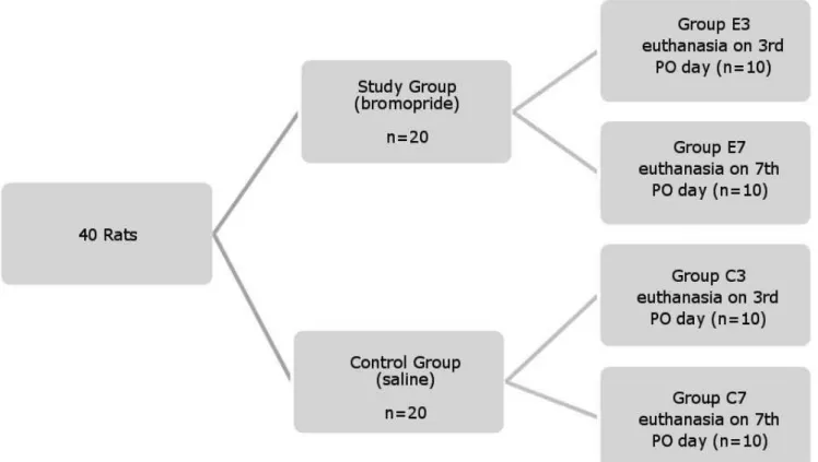
The rats were randomly distributed into two groups of 20 animals each for administration of bromopride in the postoperative period (study group E) or saline (control -C). Subsequently, we performed a new randomization into subgroups containing 10 animals each for euthanasia in the third or seventh postoperative day (Figure 1).
General anesthesia was performed with xylazine hydrochloride 10mg/Kg and ketamine hydrochloride 75mg/ Kg intramuscularly. All surgical procedures were performed by one surgeon. A laparotomy with 4cm extension was initiated at 1cm above the external genitalia of animals. The distal colon was exposed and a section of 0.5cm segment of the left colon was made about 2.5 to 3.5cm above the peritoneal reflection, followed by an end-to-end, transmural, single plane anastomosis of the segment, with running 6.0 polypropylene suture with cylindrical needle. The synthesis of the abdominal wall was performed in two planes of running 3.0 silk stitches.
A f t e r t h e p r o c e d u r e , b r o m o p r i d e w a s administered to the corresponding groups in the daily
Figure 1 Figure 1Figure 1 Figure 1
dose of 1mg/100g of weight, subcutaneously, every 12 hours, until the day of euthanasia. The control group received identical volumes of saline solution at 0.9%, a l s o e v e r y 1 2 h o u r s , b y t h e s a m e r o u t e o f administration.
In the postoperative period we evaluated clinical patterns of apathy, erection of hair, diarrhea, bloating, and wound complications, such as bruising and signs of surgical site infection.
The reoperation was performed on the day determined by the draw for each subgroup. After exposure of the abdominal cavity we searched for signs of peritonitis, abscess, or anastomotic leakage. The total amount of intra-abdominal adhesion was evaluated by the Nair score9: 0
-no adhesions; 1 - presence of one band between organs or between an organ and the abdominal wall; 2 - presence of two bands between organs or between organs and the abdominal wall; 3 - presence of more than two bands between organs or between organs and the abdominal wall, or all bowel loops forming a mass not adhering to the abdominal wall; 4 - presence of viscera directly attached to the abdominal wall, no matter the number or extension the adhesions.
A colonic segment of 4cm long containing the anastomosis in its central portion was removed together with the structures attached to it. After this procedure, we evaluated the type of structure adherent to the anastomosis. The amount of these adhesions was graded according to the proportion of the anastomosis covered by them.
The specimen was opened at the antimesenteric border and divided into three longitudinal segments. The central segment was sent for analysis of tensile strength by means of a digital test apparatus called Versa Test (Mecmesin Versa Test, United Kingdom) coupled to a AGF digital dynamometer (Mecmesin Versa Test, United Kingdom). The rectangular piece of the specimen was fixed at its two ends by the upper clamp of the dynamometer and the lower clamp of the Versa Test, with the anastomotic region equidistant and parallel to the clamps 10. The speed
used during the test was 30mm/min. The rupture strength was expressed in Newtons (N).
The right lateral segment was sent for histopathological analysis. The specimens fixed in formalin were processed and stained with hematoxylin-eosin. An experienced pathologist, blinded as for the groups of animals that the specimens came from, examined them by light microscopy. We evaluated the following parameters: congestion, edema, hemorrhage, ulceration, necrosis, mononuclear and polymorphonuclear infiltration, neovascularization, granulation, fibrosis, and fibroblasts (amount, disposition and maturity). Each of these parameters was graded as absent (-), mild (+), moderate (++), marked (+++), intense (++++).
The left lateral segment was used to measure the concentration of hydroxyproline according to Stegemann and Stalder technique, modified by Medugorac 11,12.
Statistical analysis was performed using the SPSS®. The Student’s t test was used for analysis of pre-and postoperative weight, tensile strength pre-and hydroxyproline. The evaluation of clinical parameters, the amount of adhesions and histological analysis was performed using Fisher’s exact test. The significance value of p <0.05.
RESULTS
RESULTS
RESULTS
RESULTS
RESULTS
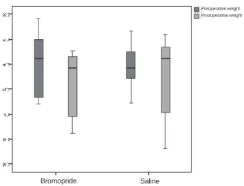
There was no difference between groups regarding weight pre-and postoperatively. The animals in Group E3 showed a slight increase of weight in the postoperative period when compared to the preoperative period. The other groups (E7, C7 and C3) showed a decrease of weight after surgery (Figures 2 and 3).
The clinical course of the animals was similar in both groups regarding the parameters apathy, hair erection and diarrhea. No animal showed abdominal distention, hematoma, or surgical site infection. There were no deaths in either group.
No animal showed signs of peritonitis at the evaluation performed in the abdominal surgical reintervention, but one animal from Group E3 had an intraabdominal abscess. One animal in a group of E3 and one in group E7 had anastomotic leakage blocked by adjacent organs, noticed only after the opening of the surgical specimen. No animal in the control group had anastomotic leakage.
Animals that had received bromopride present with Nair score values similar to the control group, both on the third day and on the seventh day after surgery (Table 1).
The evaluation of the anastomosis percentage covered by adhesions was similar between groups, both on the third and seventh postoperative days (Table 2).
Figure 2 Figure 2 Figure 2 Figure 2
Figure 2 - Weight of animals euthanized on the third day after surgery.
Bromopride Saline
The anastomoses of group E3 animals had lower rupture strength when compared to animals in group C3, this being statistically significant (Table 3). There was no statistical difference in the rupture strength of anastomoses on the seventh day after surgery (Table 3). There was also no statistical difference between groups in relation to the concentration of hydroxyproline (Table 3) or histopathological evaluation.
DISCUSSION
DISCUSSION
DISCUSSION
DISCUSSION
DISCUSSION
Bromopride is a prokinetic agent used in the postoperative period for treatment of gastroesophageal reflux, nausea, vomiting and gastrointestinal motility disorders. It is a substituted benzamide, such as metoclopramide. Its main action is related to the blockade of the dopamine receptor-2 (D2) in the central nervous system and gastrointestinal tract.
Figure 3 -Figure 3 -Figure 3 Figure 3
-Figure 3 - Weight of animals euthanized on the seventh day after surgery.
Bromopride Saline
Preoperative weight Postoperative weight
T TT
TTable 1 able 1 able 1 able 1 able 1 - Intra-abdominal adhesions found during the intraoperative assessment.
Score of Nair(Adhesions) Score of Nair(Adhesions)Score of Nair(Adhesions) Score of Nair(Adhesions)
Score of Nair(Adhesions) Number of animalsNumber of animalsNumber of animalsNumber of animalsNumber of animals 3
3 3 3
3rdrdrdrdrd postoperative day postoperative day postoperative day postoperative day postoperative day 77777ththththth postoperative day postoperative day postoperative day postoperative day postoperative day
(p = 0.628) (p = 0.628) (p = 0.628) (p = 0.628)
(p = 0.628) (p = 0.243)(p = 0.243)(p = 0.243)(p = 0.243)(p = 0.243) B r o m o p r i d e
B r o m o p r i d e B r o m o p r i d e B r o m o p r i d e
B r o m o p r i d e S a l i n eS a l i n eS a l i n eS a l i n eS a l i n e B r o m o p r i d eB r o m o p r i d eB r o m o p r i d eB r o m o p r i d eB r o m o p r i d e S a l i n eS a l i n eS a l i n eS a l i n eS a l i n e
0- Absent 6 8 2 6
1- One band 4 2 7 3
2- Two bands 0 0 1 1
Total animals 10 10 10 10
Table 2 -Table 2 -Table 2 Table 2
-Table 2 - Percentage of anastomosis covered by adhesions.
% of anatomose covered % of anatomose covered% of anatomose covered % of anatomose covered
% of anatomose covered Number of animalsNumber of animalsNumber of animalsNumber of animalsNumber of animals by adhesions
by adhesionsby adhesions by adhesions
by adhesions 33333rdrdrdrdrd postoperative day postoperative day postoperative day postoperative day postoperative day 77777ththththth postoperative day postoperative day postoperative day postoperative day postoperative day
(p = 0.141) (p = 0.141) (p = 0.141) (p = 0.141)
(p = 0.141) (p = 0.359) (p = 0.359) (p = 0.359) (p = 0.359) (p = 0.359) B r o m o p r i d e
B r o m o p r i d e B r o m o p r i d e B r o m o p r i d e
B r o m o p r i d e S a l i n eS a l i n eS a l i n eS a l i n eS a l i n e B r o m o p r i d eB r o m o p r i d eB r o m o p r i d eB r o m o p r i d eB r o m o p r i d e S a l i n eS a l i n eS a l i n eS a l i n eS a l i n e
up to 25% 0 0 0 2
between 25 and 50% 0 0 0 1
between 50 and 75% 1 1 1 0
more than 75% but less than 100% 5 1 1 2
100% 4 8 8 5
Total animals 10 10 10 10
Table 3 -Table 3 -Table 3 Table 3
-Table 3 - Averages of the values of rupture strength and concentration of hydroxyproline.
3 3 3 3
3rdrdrdrdrd postoperative day postoperative day postoperative day postoperative day postoperative day 77777ththththth postoperative day postoperative day postoperative day postoperative day postoperative day
B r o m o p r i d e B r o m o p r i d eB r o m o p r i d e B r o m o p r i d e
B r o m o p r i d e S a l i n eS a l i n eS a l i n eS a l i n eS a l i n e ppppp B r o m o p r i d eB r o m o p r i d eB r o m o p r i d eB r o m o p r i d eB r o m o p r i d e S a l i n eS a l i n eS a l i n eS a l i n eS a l i n e PPPPP
Rupture strength (mmHg) 0,051 0,235 0,04 0,342 0,241 0,37
Dopamine antagonists decrease the activity of the vomiting center and the activation of visceral nerves13.
They promote inhibition of the gastrointestinal tract, with relaxation or inhibition of smooth muscle contraction from the esophagus to the colon 14. Thus, inhibition of dopamine
receptors results in a prokinetic effect. Specific dopamine receptors have been described in the gastrointestinal tract (particularly in the stomach and the exocrine pancreas) and in the renal, mesenteric, coronary and cerebral vasculature 15. The D2 receptors are located both pre and
post junctional and exert negative modulation on the release of acetylcholine on intrinsic cholinergic nerve terminals 16.
Similar to other benzamide derivatives, stimulation of the gastrointestinal tract by bromopride also appears to be mediated, at least in part, by its indirect cholinergic activity, partially dependent on its anticholinesterase properties.
Garcia-Olmo et al.17 conducted a study to
deter-mine the effects of pharmacological manipulation of gastrointestinal motility in the resistance of colic anastomoses. Seventy-two rats undergoing colic anastomoses were randomized into three groups and subcutaneously given 1ml of saline, metoclopramide (1.2mg/100g body weight) or hyoscine (2mg/100g body weight). The animals were killed on the fourth day after surgery. The authors concluded that the use of metoclopramide in the early postoperative period was associated with an increase in the number of colonic anastomosis dehiscence and, in the surviving animals, with a significant decrease in the resistance of the anastomosis. Hyoscine, an inhibitor of gastrointestinal motility, did not improve healing.
In the present study, the use of bromopride was associated with a statistically significant decrease of the values of rupture strength of anastomoses evaluated on the third day after surgery when compared to the control group. These results are consistent with the findings described in the study previously cited17.
However, there was no statistical difference in the evaluation of the anastomoses on the seventh day after surgery. Similar values of tensile strength were also reported on the seventh day after surgery between control animals and rats treated by another prokinetic agent, cisapride 7.
The stimulation of gastrointestinal motility by prokinetic agents can cause significant reduction of peritoneal adhesions 18. This effect could be deleterious,
interfering with the healing of the anastomosis by direct mechanical action or by reducing the formation of adhesions to the anastomosis 5.
A previous study found a strong relationship between the resistance of the anastomosis and the
proportion covered by the adjacent organs. It was shown that the group of animals subjected to the administration of prokinetic agents had a greater amount of intra-abmoninais adhesions, but fewer adhesions actually beneficial, those between adjacent organs and anastomosis in the fourth postoperative day17. The
present study showed similar results. Despite the lack of statistical significance, the animals that received bromopride showed higher values of both Nair score in the third and seventh days. However, there were fewer adhesions to the anastomosis on the third day after surgery, at which time there was a reduction of values of tensile strength. This effect was reversed on the seventh day.
These findings could be explained, at least in part, by the physiology of wound healing. The process of healing begins with hemostasis. Inflammatory response follows, then the formation of connective tissue and wound remodeling10,19. During this crucial stage, macrophages and
polymorphonuclear cells migrate from the circulation to the wound site in response to increased levels of cytokines 20.
This phase is followed by fibroblast proliferation, collagen synthesis, connective tissue and its parenchymal component remodeling and the acquisition of resistance of the wound
21. Thus, resistance depends on the initial anastomotic suture,
since, until the fourth day, the anastomosis is filled with loose and disorganized collagen fibers. Perhaps this resistance may also be dependent on blockade by adjacent organs, which would act as a protective factor for the anastomosis.
The peak of collagen synthesis by fibroblasts is reached between the fifth and seventh day and anastomotic strength is mainly dependent on these new arranged fibers22.
In this study, the formation of collagen was assessed by measurement of hydroxyproline. On the seventh day after surgery, the levels were higher in the control group when compared with the group receiving bromopride. This was accompanied by higher values of tensile strength in this group, although the differences were not statistically significant.
Importantly, despite the differences between the groups, there was no major interference in clinical parameters. The animals had similar postoperative clinical course and there were no cases of peritonitis or deaths in either group. In addition, the anastomotic dehiscences that occurred in the study group were blocked by adjacent organs and were found only after the opening of the surgical specimen. Therefore, these animals showed no clinical consequences of this complication.
R E S U M O R E S U M O R E S U M O R E S U M O R E S U M O
Objetivo: Objetivo: Objetivo: Objetivo:
Objetivo: Avaliar os efeitos da bromoprida sobre a formação de aderências e a cicatrização de anastomoses de cólon esquerdo de ratos. Métodos: Métodos: Métodos: Métodos: Métodos: Foram incluídos 40 ratos, divididos em dois grupos contendo 20 animais, para administração de bromoprida (grupo de estudo- E) ou solução fisiológica (grupo controle- C). Cada grupo foi dividido em subgrupos contendo 10 animais cada, para eutanásia no terceiro (E3 e C3) ou no sétimo dia (E7 e C7) de pós-operatório. Os ratos foram submetidos à secção do cólon esquerdo e anastomose término-terminal. No dia da relaparotomia, foi avaliada a quantidade total de aderências e removido um segmento colônico contendo a anastomose para análise histopatológica, da força de ruptura e da concentração de hidroxiprolina. Resulta-Resulta-Resulta-Resulta- Resulta-dos:
dos: dos: dos:
dos: Não houve diferença entre os grupos em relação à evolução clínica. Dois animais do grupo de estudo apresentaram deiscência de anastomose bloqueada. Os animais que receberam bromoprida apresentaram número de aderências intracavitárias e aderências à anastomose semelhantes ao grupo controle. As anastomoses dos animais do grupo E3 apresentaram menor resistência de ruptura do que as do grupo C3 (p=0,04). Este efeito não ocorreu no sétimo dia de pós-operatório (p=0,37). Não houve diferença significativa entre os grupos em relação à histopatologia ou concentração de hidroxiprolina das anastomoses. Conclusão: Conclusão: Conclusão: Conclusão: Conclusão: O uso da bromoprida está associado à diminuição da resistência tênsil de anastomoses do cólon esquerdo de ratos no terceiro dia de pós-operatório.
Descritores: Descritores: Descritores: Descritores:
Descritores: Anastomose cirúrgica. Colo. Motilidade gastrointestinal. Aderências teciduais. Ratos.
REFERENCES
REFERENCES
REFERENCES
REFERENCES
REFERENCES
1. Fielding LP, Stewart-Brown S, Blesovsky L, Kearney G. Anastomotic integrity after operations for large-bowel cancer: a multicentre study. Br Med J 1980; 281(6237):411-4.
2. Goligher JC, Graham NG, De Dombal FT. Anastomotic dehiscence after anterior resection of rectum and sigmoid. Br J Surg 1970;57(2):109-18.
3. Thornton FJ, Barbul A. Healing in the gastrointestinal tract. Surg Clin North Am 1997; 77(3):549-73.
4. Liakakos T, Thomakos N, Fine PM, Dervenis C, Young RL. Peritoneal adhesions: etiology, pathophysiology, and clinical significance. Recent advances in prevention and management. Dig Surg 2001; 18(4):260-73.
5. Garcia-Olmo D, Lucas FJ, Paya J. Relationship between peritoneal adhesion phenomena and the experimental resistance of colonic anastomoses: influence of omentoplasty. Eur Surg Res 1996; 28(4):315-22.
6. Beck DE, Cohen Z, Fleshman JW, Kaufman HS, van Goor H, Wolff BG: Adhesion Study Group Steering Committee. A prospective, randomized, multicenter, controlled study of the safety of Seprafilm adhesion barrier in abdominopelvic surgery of the intestine. Dis Colon Rectum 2003; 46(10):1310-9.
7. Springall RG, Spitz L. The prevention of post-operative adhesions using a gastrointestinal prokinetic agent. J Ped Surg 1989; 24(6):530-3.
8. Longo WE, Vernava AM 3rd. Prokinetic agents for lower gastrointestinal motility disorders. Dis Colon Rectum 1993; 36(7):696-708.
9. Nair SK, Bhat IK, Aurora AL. Role of proteolytic enzyme in the prevention of postoperative intraperitoneal adhesions. Arch Surg 1974; 108(6):849-53.
10. Lopes JV, Freitas LAM, Marques RD, Bocca AL, Sousa JB, Oliveira PG. Análise da força tênsil na cicatrização da parede abdominal de ratos tratados com infliximabe. Acta Cir Bras 2008; 23(5):441-6.
11. Stegemann H, Stalder K. Determination of hydroxyproline. Clin Chim Acta 1967; 18(2):267-73.
12. Medugorac I. Collagen content in different areas of normal and hypertrophied rat myocardium. Cardiovasc Res 1980; 14(9):551-4.
13. Pinder RM, Brogden RN, Sawyer PR, Speight TM, Avery GS. Metoclopramide: a review of its pharmacological properties and clinical use. Drugs 1976; 12(2):81-131.
14. Willems JL, Buylaert WA, Lefebvre RA, Bogaert MG. Neuronal dopamine receptors on autonomic ganglia and sympathetic nerves and dopamine receptors in the gastrointestinal system. Pharmacol Rev 1985; 37(2):165-216.
15. Thorner MO. Dopamine is na important neurotransmitter in the autonomic nervous system. Lancet 1975;1(7908):662-5. 16. Tonini M. Recent advances in the pharmacology of gastrointestinal
prokinetics. Pharmacol Res 1996; 33(4-5):217-26.
17. Garcia-Olmo D, Payá J, Lucas FJ, Garcia-Olmo DC. The effects of the pharmacological manipulation of postoperative intestinal motility on colonic anastomoses. An experimental study in a rat model. Int J Colorectal Dis 1997; 12(2):73-7.
18. Sparnon AL, Spitz L. Pharmacological manipulation of postoperative intestinal adhesions. Aust N Z J Surg 1989; 59(9):725-9. 19. Sousa JB, Oliveira PG. Cuidados com a ferida operatória -
infec-ção. Clin bras cir 1999; 5(2):215-37.
20. Martin CW, Muir IF. The role of lymphocytes in wound healing. Br J Plast Surg 1990; 43(6):655-62.
21. Goldman R. Growth factors and chronic wound healing: past, present, and future. Adv Skin Wound Care 2004; 17(1):24-35. 22. Haciyanli M, Fuzun M, Unek T, Tokgoz Z. Does the administration
route of leucovorin have any influence on the impairment of colonic healing caused by intraperitoneal 5-fluorouracil treatment ? Eur Surg Res 2001; 33(2):80-5.
Received on 16/02/2011
Accepted for publication 07/04/2011 Conflict of interest: none
Source of funding: none
How to cite this article: How to cite this article:How to cite this article: How to cite this article:How to cite this article:
Silva SM, Ferreira VMM, Carneiro FP, Feres O, Oliveira PG, Sousa JB. Effects of bromopride on the healing of left colon anastomoses of rats. Rev Col Bras Cir. [periódico na Internet] 2011; 38(6). Disponível em URL: http://www.scielo.br/rcbc
Correspondence address: Correspondence address:Correspondence address: Correspondence address:Correspondence address: Silvana Marques e Silva