Original papers
DOI: 10.11152/mu-981
Can fetal adrenal artery Doppler velocimetry predict delivery date
in pregnant women with spontaneous preterm birth?
Francisco Herlânio Costa Carvalho
1, Aline Pinto Lemos
1, Francisco Edson De Lucena Feitosa
1,
Helvécio Neves Feitosa
1, Lucas Costa Carvalho Augusto
1, Rosa Maria Salani Mota
1,
Edward Araujo Júnior
21Fetal Medicine Service, Maternidade-Escola Assis Chateaubriand,Federal University of Ceará (UFC), Fortaleza-CE, 2Department of Obstetrics, Paulista School of Medicine–Federal University of São Paulo (EPM-UNIFESP), São
Paulo-SP, Brazil
Received 10.10.2016 Accepted 04.01.2017 Med Ultrason
2017, Vol. 19, No 3, 295-301
Corresponding author: Prof. Edward ARAUJO JÚNIOR, Ph.D Department of Obstetrics, Paulista School of Medicine–Federal University of São Paulo (EPM-UNIFESP)
Rua Belchior de Azevedo, 156 apto. 111 Torre Vitoria, São Paulo–SP, Brazil CEP 05089-030
Phone/Fax: +55-11-37965944 E-mail: araujojred@terra.com.br Introduction
Health care practitioners have had limited ability to identify pregnant women likely to experience preterm birth. The main preventive interventions for preterm birth, such as vaginal progesterone, are restricted to high-risk pregnancies including women with previous preterm births or those who have a short uterine cervix. However, the majority of preterm births occur in low-risk
pregnan-cies, and all available methods should be used to identify these cases [1].
The identification of several biochemical and bio -physical markers has been proposed as a means to iden-tify pregnancies at high risk for spontaneous preterm birth because most high-risk cases are asymptomatic [2]. At present, cervical length (CL) measurement is an ex-cellent ultrasound marker for predicting preterm birth in both high- and low-risk pregnancies [2-4].
In 2007, the first literature description regarding the
relevance of fetal adrenal gland volume for predicting preterm births was published. An increase in fetal adre-nal gland volume was found to have an accuracy of 98% when used to predict delivery within 5 days [5]. Turan et al [6] demonstrated that fetal adrenal gland volume was a better predictor of preterm birth than CL measurement. In that study, in addition to the increase in total fetal ad-renal gland volume, the central zone showed a dispropor-tional increase.
Abstract
Aim: To assess the accuracy of delivery date predictions made using fetal adrenal artery Doppler velocimetry in pregnant women with spontaneous preterm birth (PB) and to compare these predictions with cervical length (CL) measurements. Mate-rial and methods: A prospective study was performed with 51 pregnant women whose gestational lengths were between 24 and 36 weeks. The main outcome was the time between the Doppler velocimetry examination and delivery, categorized as delivery within 7 days or 7 days later after the examination. A receiver operating characteristics curve was performed to define the cutoffs among deliveries within 7 days for fetal adrenal artery Doppler velocimetry parameters and CL measurements.
Results: The incidence of delivery within 7 days was 37.3%, with a statistically significant difference for the pulsatility index
(PI; p=0.045) and resistance index (RI; p=0.030) of the fetal adrenal artery. The best cutoff values of PI and RI for predicting deliveries within 7 days were 1.65 and 0.78, respectively. The sensitivity and specificity of PI, RI, and CL (20 mm) were 73.7% (95% CI: 51.9–95.5) and 56.3% (95% CI: 38.1–74.4); 68.4% (95% CI: 45.4–91.4) and 62.5% (95% CI: 44.8–80.2); and 76.5% (95% CI: 54.0–99.0) and 78.1% (95%: CI 71.1–97.7), respectively. Conclusion: Fetal adrenal artery Doppler velocimetry can predict delivery within 7 days among pregnant women in cases of spontaneous PB and this prediction is similar to the predic-tions made using CL measurements.
To the best of our knowledge, no studies in literature have examined the use of fetal adrenal artery Doppler velocimetry for the prediction of preterm birth. There-fore, the objective this study was to assess the capacity of fetal adrenal artery Doppler velocimetry to predict delivery within 7 days for pregnant women with intact membranes in cases of spontaneous preterm birth and to compare these predictions with CL measurements.
Materials and methods
The prospective study was conducted between April 2014 and March 2015. Approval by the Local Ethics Committee (nº 640.692) was obtained and the pregnant women provided their informed consent. Singleton preg-nant women who were admitted to the hospital with signs or symptoms of spontaneous preterm birth and with ges-tations between 26 weeks and 34 weeks were invited to participate in the study. Gestational age was determined
by the last menstrual period and confirmed by an ul -trasound examination performed until the 20th week of
pregnancy.
The following signs and symptoms were considered to diagnose spontaneous preterm birth: abdominal pain
with uterine contractions, modifications of the uterine
cervix, and the need for tocolytic medication. The assess-ment of uterine contractions was routinely performed during a 20 minute period but we took into considera-tion any number of regular uterine contracconsidera-tions that were capable to produce cervical changes and necessitate to-colytic medication for inclusion in this study. Cervical changes were assessed by vaginal examination at 1–2 hour intervals. All pregnant women with risk of imped-ing spontaneous preterm birth, except for pregnant wom-en who were at least 34 weeks of gestation, received oral nifedipine (20 mg) as tocolytic medication for at least 24 h and intramuscular dexamethasone for fetal lung matu-ration (12 mg, two doses, 24 h intervals). If contractions persisted after 30 min, a second dose of nifedipine (20 mg) was administered orally followed by another 20 mg every 8 h for 48 h. The exclusion criteria were as fol-lows: premature rupture of membranes, pregestational and gestational diabetes mellitus, chronic arterial hy-pertension, pre-eclampsia, endocrine diseases, amniotic
fluid disorders (amniotic fluid index <5 cm or >25 cm),
fetal malformations detected during the ultrasound
ex-amination and/or confirmed in the postnatal exam, fetal
distress detected by cardiotocography, suspicion of fetal
growth restriction (estimated fetal weight <10th
percen-tile for gestational age [7] and/or umbilical artery
pul-satility index >95th percentile or cerebroplacental ratio <5th percentile at Doppler velocimetry [8]), maternal use
of vaginal progesterone to prevent spontaneous preterm birth or spontaneous preterm birth treatment during the actual pregnancy, and newborn size that was too small or too large for gestational age according to the Alexander curve [9]. It was a study protocol unpublished in the lit-erature and because of lack of all the changes that may occur in the fetal adrenal gland with growth impairment (large or small for gestational age), we decided to select a very homogeneous population.
The ultrasound exams were performed within 24 h of hospital admission using a Voluson S6 apparatus (Gen-eral Electric Medical Systems, Milwaukee, WI, USA) equipped with a convex probe (4C-RS). The sonographic evaluation of the CL measurement was performed trans-vaginally. Fetal adrenal artery Doppler velocimetry as-sessment was performed according to the procedure described by Fujita et al [10] using the transabdominal route. With the mothers in the semi-Fowler position, an axial view of the fetal abdomen was obtained at the level of the adrenal glands. Color and/or power Doppler ve-locimetry were applied to identify the middle adrenal ar-tery, which is an aortic branch, at the level of the adrenal gland hilum. To the posterior side, the gate of the spectral Doppler velocimetry was positioned closest to the mid-point of the fetal adrenal artery in the absence of uterine contractions or respiratory or body movements of the fetus. The following Doppler velocimetry settings were
standard in all exams: filter 100 Hz, sample size 2 mm, and insonation angle ≤30°. When at least three typical
waveforms were obtained, the pulsatility index (PI), re-sistance index (RI), and systole/diastole (S/D) ratio were measured by ultrasound apparatus automatically in one waveform. We also assessed the presence or absence of
end-diastolic flow (fig 1).
All ultrasound exams were performed by a single
cer-tified examiner (FHCC) with 15 years of experience in
obstetrical ultrasound. This examiner performed a learn-ing curve with 20 adrenal artery Doppler velocimetry exams prior to the study to increase the intra-observer reproducibility of measurements.
the Maternidade-Escola Assis Chateaubriand – Federal University of Ceará, the data were obtained by medical records. When the birth did not occur in the institution, the data were obtained by phone calls with the patients.
The following variables were measured: maternal data (age, parity, history of prior preterm birth, gestation-al age at ultrasound examination); Doppler examination data (PI, RI, S/D ratio, and presence or absence of
end-diastolic flow), perinatal outcome data (time between the
Doppler velocimetry examination and delivery, gesta-tional age at delivery, delivery route, birth weight and Apgar score at 1st and 5th minute).
The outcome variable was the time period between the Doppler velocimetry examination and delivery,
which was classified into two groups: delivery within 7
days and delivery after 7 days. We chose dichotomizing similarly to Turan et al [6].
Statistical analysis
The sample size was calculated considering the high prevalence of suspected diagnosis of preterm birth at Maternidade-Escola Assis Chateaubriand – Federal Uni-versity of Ceará and to differentiate true from false pre-term birth by ultrasound and fetal adrenal artery Doppler
velocimetry, since we did not use fibronectin or other
biochemical markers to evaluate patients with suspicion of preterm birth. We decided to calculate the sample size considering the risk of delivery within 7 days in
sympto-matic pregnant women (36.5%) [6], a significance level
of 5% (alpha error 0.05 - two-sided), 90% power (beta error of 10%), and bias of 20% (alternative p=0.175).
A minimum of 52 subjects would be necessary for this study.
Data were transferred to an Excel spreadsheet (Mi-crosoft Corp., Redmond, WA, USA) and analyzed using the PASW Statistics program (version 20.0; SPSS Inc., Chicago, IL, USA). Fisher’s exact test to nominal varia-bles was applied, and either the Mann–Whitney U test or Student t-test to continuous variables. A receiver operat-ing characteristics (ROC) curve was performed to predict the best cutoffs regarding delivery within 7 days to fetal adrenal Doppler velocimetry artery and CL measure-ment. A point on the ROC curve near the top left corner was selected, with smaller proportions of false positives and false negatives. We prioritized getting better
speci-ficity rather than sensitivity because the aim was not a
screening test, but to obtain a test to support the diagno-sis of true preterm labor applicable in clinical practice.
Sensitivity, specificity, positive predictive value, false
positive, negative predictive value, false negative, posi-tive likelihood ratio, and negaposi-tive likelihood ratio were calculated. McNemar’s test was applied to compare the
sensitivity and specificity regarding delivery within 7
days between the obtained cutoffs for adrenal artery PI and RI values, and CL measurement (20 mm). The rela-tive risk of these cutoffs to delivery within 7 days as well as the prediction capacity for variables combination were calculated, which turned out to be good predictors (both below the cutoff points) of delivery occurring within 7 days. A P value of less 0.05 was considered an indicative
of statistical significance.
Results
We initially selected 64 pregnant women but 13 were excluded for the following reasons: one fetus had hydro-nephrosis, one newborn was small for gestational age, one pregnant woman had pre-gestational diabetes mel-litus, one pregnant woman had gestational diabetes, two pregnant women had chronic arterial hypertension, Dop-pler velocimetry of the adrenal artery was not assessed in
two cases due to technical difficulties (one case of mater -nal obesity and one case of excessive fetal movement),
and five pregnant women missed the follow-up.
Therefore, data from 51 pregnant women were used for the statistical analysis. A total of 19 (37.3%) mothers had their deliveries within 7 days from hospital admis-sion.
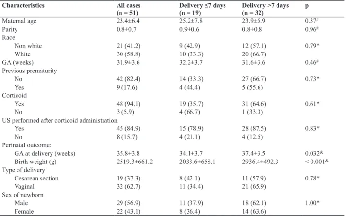
Table I shows the maternal characteristics and peri-natal outcomes for both groups. The assessment of fetal mean adrenal artery Doppler velocimetry was possible for 51 out of 56 patients (51 regularly included patients and 5 missing follow-ups) with a 91.1% success rate.
Table II shows the comparison of Doppler velocimetry parameters between both groups.
The ROC curve for the mean fetal adrenal artery PI for a 37.3% prevalence of delivery within 7 days had an area under the ROC curve of 0.669 (95% CI: 0.523– 0.816, p=0.045) with an optimal selected cutoff of 1.65. The ROC curve for the mean RI of the fetal mean adrenal artery had an area under the ROC curve of 0.679 (95% CI: 0.528–0.831, p=0.034), with an optimal selected
cut-off of 0.78. Table III shows the sensitivity, specificity,
positive predictive value, false positive, negative predic-tive value, false negapredic-tive, posipredic-tive likelihood ratio, and
negative likelihood ratio for PI (cutoff 1.65), RI (cutoff at 0.78), and CL measurement (cutoff at 20 mm).
The area under the ROC curve for CL measurement
was 0.807 (95% CI: 0.663-0.951, p<0.001) and the S/D
ratio of the fetal adrenal artery was 0.548 (95% CI 0.411-0.692, p=0.39). The relative risk of delivery to occur over
the next 7 days for CL measurement <20 mm was 5.6 (95% CI: 2.2-14.6), RI <1.65 was 2.3 (95% CI: 1.0-5.4), and PI <0.78 was 2.3 95% CI: 1.0-4.9).
There were no significant differences in sensitiv
-ity or specific-ity for the detection of delivery within 7
days, using the MacNemar test. Sensitivity of the mean
Table I. Distribution of maternal and perinatal characteristics according to the outcome.
Characteristics All cases
(n = 51)
Delivery ≤7 days (n = 19)
Delivery >7 days (n = 32)
p
Maternal age 23.4±6.4 25.2±7.8 23.9±5.9 0.37#
Parity 0.8±0.7 0.9±0.6 0.8±0.8 0.96#
Race
Non white 21 (41.2) 9 (42.9) 12 (57.1) 0.79*
White 30 (58.8) 10 (33.3) 20 (66.7)
GA (weeks) 31.9±3.6 32.2±3.7 31.6±3.6 0.46#
Previous prematurity
No 42 (82.4) 14 (33.3) 27 (66.7) 0.73*
Yes 9 (17.6) 4 (44.4) 5 (55.6)
Corticoid
Yes 48 (94.1) 19 (35.7) 31 (64.6) 0.61*
No 3 (5.9) 4 (66.7) 1 (33.3)
US performed after corticoid administration
Yes 45 (84.9) 15 (78.9) 28 (87.5) 0.83*
No 8 (15.7) 4 (21.1) 4 (12.5)
Perinatal outcome:
GA at delivery (weeks) 35.8±3.8 34.1±3.7 37.4±3.5 0.032&
Birth weight (g) 2519.3±661.2 2033.6±658.1 2936.4±492.3 < 0.001&
Type of delivery
Cesarean section 19 (37.3) 8 (42.1) 11 (57.9) 0.78*
Vaginal 32 (62.7) 11 (34.4) 21 (65.9)
Sex of newborn
Male 29 (56.9) 11 (37.9) 18 (62.1) 1.00*
Female 22 (43.1) 8 (36.4) 14 (63.6)
The results are expressed as n(%) or mean± standard deviation, US: ultrasound, GA: gestational age, *Fisher’s exact test, #Student’s t test, &Mann–Whitney U test
Table II. Distribution of fetal adrenal artery Doppler parameters according to the outcome.
Doppler parameter All cases Delivery ≤7 days Delivery >7 days p
End-diastolic peak
Positive 34 (66.7) 16 (47.1) 18 (52.9) 0.07*
Absent 17 (33.3) 3 (17.6) 14 (82.4)
RI 0.80±0.17 0.74±0.15 0.84±0.17 0.030&
PI 1.71±0.85 1.36±0.35 1.91±0.99 0.045&
Systole/diastole ratio 5.30±7.85 4.04±2.20 6.30±4.46 0.56&
adrenal artery PI (73.7%) versus the CL (76.5%) meas-urement was p=1.00 and versus the mean adrenal artery
RI (68.4%) was p=1.000. The specificity of the mean
adrenal artery PI (73.5%) compared to the CL (78.1%) measurement was p=0.21 and that compared to the mean adrenal artery RI (62.5%) provided a value of p=1.00.
The ability of delivery prediction occurrence within 7 days by the combination of variables below the cut-offs was calculated. For the combination of cervical biometry with RI (both below the cutoff points),
sensi-tivity was 94.4% (95% CI: 82.7-100.0), specificity was
48.4% (95% CI: 29.8–67.0), accuracy was 31.1%, posi-tive likelihood ratio was 1.83, and negaposi-tive likelihood ratio was 0.12. For the combination of cervical biom-etry with PI (both below the cutoff points), the
sensitiv-ity was 88.9% (95% CI: 72.8–100.0), specificsensitiv-ity was
45.2% (95% CI: 26.6–63.7), accuracy was 29.0%, posi-tive likelihood ratio was 1.62, and negaposi-tive likelihood ratio was 0.25.
Discussions
This study is the first one, to the best of our knowl -edge, to test the hypothesis that the fetal adrenal artery reduces its resistance in cases of imminent preterm birth. We observed that both PI and RI were good predictors of delivery within 7 days for pregnant women in cases of spontaneous preterm birth, suggesting that Doppler ve-locimetry indices could be incorporated into the obstet-rics practice together with biophysical (CL measurement [2,4] and fetal adrenal biometry [5,6]) and biochemical markers [12] to obtain a greater predictive power for pre-term birth in symptomatic pregnant women.
Previous observations have shown vasodilation in the adrenal artery of fetuses with chronic hypoxemia [13-15]. These studies describe the presence of vasodilata-tion in the fetal adrenal artery as being associated with prematurity, higher Cesarean section rates, fetal distress
detected by cardiotocography, and longer time periods spent in neonatal intensive care units.
The only reference range of the fetal mean adrenal artery PI was described by Mari et al [15]. They assessed 131 singleton pregnancies and observed that PI values decreased with increasing gestational age. The authors
did not observe statistically significant differences be -tween the PI of the mean adrenal artery and the PI of the inferior adrenal artery or between the right or left adre-nal arteries. The success rate of obtaining the Doppler velocimetry waveforms was 84% and the intra-observer reproducibility was 7.4%. Dudiel et al. [13] had a suc-cess rate of 81% for obtaining the Doppler waveform parameters, with failures due to fetal movements. In our study, we obtained a 96% success rate, which could be hypothesized as a consequence of using a higher-quality ultrasound apparatus today.
Fujita et al [10] described a success rate of 90% in obtaining Doppler velocimetry waveforms in the fetal mean adrenal artery and 50% in the fetal superior adrenal
artery. The fetal inferior adrenal artery was not identified
in 10 attempts. These authors stated that RI values in-creased until the 31st week of gestation and progressively
decreased after this age. Fetal adrenal artery Doppler velocimetry showed the ability to differentiate among fetuses with high risks for hypoxemia and those with ad-verse perinatal outcomes [14,15].
It has been shown that the volume of the fetal adrenal gland enlarges in the last 6 weeks before term or pre-term delivery, especially with disproportionate increase between the central and the peripheral areas 5 to 7 days before preterm birth. Fetal adrenal gland synthesizes steroid precursors which are transformed into estrogen by the placenta. It has been suggested that this increase of fetal adrenal gland, particularly its central zone may have an important role in the endocrine regulation of parturi-tion. [6,16-18]. Turan et al [6] considered reasonable to propose that a disproportionate increase in the size of the
Table III. Comparative analysis between the different cutoffs of fetal adrenal artery Doppler parameters and cervical length measure-ment.
Variable Se
(95% CI)
Sp (95% CI)
PPV NPV Accuracy LR+ LR−
Positive end-diastolic peak 61.1% (41.2-82.3)
73.5% (55.3-94.9)
56.3% 77.2% 69.1% 2.309 0.529
PI ≤1.65 73.7%
(51.9-95.5)
56.3% (38.1-74.4)
48.4% 79.3% 62.5% 1.684 0.468
RI ≤0.78 68.4%
(45.4-91.4)
62.5% (44.8-80.2)
50.4% 78.0% 64.6% 1.825 0.505
Cervical length <20 mm 76.5% (54-99)
78.1% (71.1-97.7)
66.1% 85.6% 77.5% 3.493 0.301
fetal adrenal gland would allow noninvasive recognition of premature activation of the delivery cascade.
Therefore, higher vascularization would be necessary to increase the volume of this gland. The formulated hy-pothesis was based on the fact that there was an increase of the gland in consequence of the increased vascularity. In other words, the reduced resistance in the fetal adrenal
artery Doppler velocimetry should reflect the increase in
the vascularization concomitant with the increase in the volume.
According to Ishimoto et al [17], the central zone of the fetal adrenal gland produces large amounts of andro-gens, which are transformed into estrogens in the placen-ta. Therefore, the human fetus presents with large adrenal glands, and after delivery, these glands involute quickly, decreasing by 50%, because of decreased androgen se-cretion. The growth of the fetal adrenal gland involves proliferation, hypertrophy, apoptosis, and cellular migra-tion [9]. The biometry of the central zone, because of its disproportionate increase relative to the total gland, has been found to be a good predictor of imminent preterm
delivery in pregnant women [6]. A specific study on the
vascularization of the central zone could perhaps contrib-ute to a better understanding of the function of the fetal adrenal gland in determining delivery.
In our study, all of the pregnant women had intact membranes. Turan et al [5,6,19] assessed fetal adrenal gland volume in a heterogeneous population of pregnant women, including 34% with premature rupture of mem-branes [5]. The mechanisms involved in preterm birth in the presence of premature membrane rupture are
associ-ated with inflammatory and infectious factors, and it is
not known what effect these factors could have on the volume and vascularization of the fetal adrenal gland
[20]. Moreover, scientific evidence shows that antibiot -ic prophylaxis increases the latency period in pregnant women with premature rupturing of the membranes [21].
In our protocol, we recorded whether steroids admin-istration occurred before or after the fetal adrenal artery
Doppler assessment. There were no statistically signifi -cant differences between the groups suggesting similar corticoid effects on delivery in both groups.
Statistical analysis to associate the risk of delivery within 7 days in this population were performed using
the same data used to define the cutoff points (values of
PI and RI for predicting delivery within 7 days were 1.65 and 0.78, respectively). Therefore, these estimates may be biased and should be considered as a study limitation. These cutoff points should be tested in other populations and institutions.
Considering the need for specific operator training
and time for scanning as well as cost and considering
that at the end the predictive value of fetal adrenal ar-tery Doppler velocimetry is similar to CL measurement, it becomes apparent that the clinical usefulness of this method in order to be proposed or included in obstetrical practice is very limited if not recommendable.
Conclusions
In conclusion, fetal adrenal artery Doppler
velocime-try was identified as a good predictor of delivery in preg -nant women who had spontaneous preterm births with intact membranes. The predictive value of this method was similar to that of CL measurements. However, fu-ture studies with larger population size are warranted to
demonstrate the real significance of fetal adrenal artery
Doppler velocimetry in predicting preterm births.
Conflict of interest: none
References
1. Tedesco RP, Passini R, Cecatti JG, Camargo RS, Paca-gnella RC, Sousa MH. Estimation of preterm birth rate, associated factors and maternal morbidity from a Demo-graphic and Health Survey in Brazil. Mater Child Health J 2013;17:1638-1647.
2. Di Renzo GC, Roura LC, Facchinetti F, et al. Guidelines for the management of spontaneous preterm labor: identi-fication of spontaneous preterm labor, diagnosis of preterm premature rupture of membranes, and preventive tools for preterm birth. J Mater Fetal Neonatal Med 2011;24:659-667.
3. Orzechowski KM, Boelig R, Nicholas SS, Baxter J, Berghella V. Is universal cervical length screening indicat-ed in women with prior term birth? Am J Obstet Gynecol 2015;212:234.e1-5.
4. Lim K, Butt K, Crane JM. SOGC Clinical Practice Guide-line. Ultrasonographic cervical length assessment in pre-dicting preterm birth in singleton pregnancies. J Obstet Gynaecol Can 2011;33:486-499.
5. Turan OM, Turan S, Fuani EF, Buhimschi IA, Copel JA, Buhimschi CS. Fetal adrenal gland volume. A novel meth-od for identify women at risk for impending preterm birth. Obstet Gynecol 2007;109:855-862.
6. Turan OM, Turan S, Funai EF, et al. Ultrasound measure-ment of fetal adrenal gland enlargemeasure-ment: an accurate predic-tor of preterm birth. Am J Obstet Gynecol 2011;204:311e1-10.
7. Hadlock FP, Harrist RB, Martinez-Poyer J. In utero analy-sis of fetal growth: a sonographic weight standard. Radiol-ogy 1991;181:129-133.
9. Alexander GR, Himes JH, Kaufman RB, Mor J, Kogan M. A United States national reference for fetal growth. Obstet Gynecol 1996;87:163-168.
10. Fujita Y, Satoh S, Nakano H. Doppler velocimetry in the adrenal artery in human fetuses. Early Human Dev 2001;65:47-55.
11. Rouse DJ, Hirtz DG, Thom EA, et al. A randomized trial of magnesium sulfate for the prevention of cerebral palsy. New Engl J Med 2008;359:895-905.
12. Chauhan SP, Berghella V, Sanderson M, Magann EF, Mor-rison JC. American College of Obstetricians and Gynecolo-gists practice bulletins: an overview. Am J Obstet Gynecol 2006;194:1564-1572.
13. Dubiel M, Breborowicz GH, Marsal K, Gudmundsson S. Fetal adrenal and middle cerebral artery Doppler velocime-try velocimevelocime-try in high-risk pregnancy. Ultrasound Obstet Gynecol 2000;16:414-418.
14. Mari G, Uerpairojkit B, Abuhamad AZ, Copel JA. Ad-renal artery velocity waveforms in the appropriate and small-for-gestational-age fetus. Ultrasound Obstet Gynecol 1996;8:82-86.
15. Mari G, Abuhamad AZ, Uerpairojkit, B, Martinez E, Copel JA. Blood flow velocity waveforms of the abdominal ar -teries in appropriate-and small-for-gestational-age fetuses. Ultrasound Obstet Gynecol 1995;6:15-18.
16. Guler A, Pehlivan H, Cakmak B. Assessment of fetal adre-nal gland enlargement in term and preterm labor cases. Int J Res Med Sci 2015;3:1035-1040.
17. Ishimoto H, Jaffe RB. Development and function of the human fetal adrenal cortex: a key component in the feto-placental unit. Endocr Rev 2011;32:317-355.
18. Challis JR, Bloomfield FH, Bocking AD, et al. Fetal signals and parturition. J Obstet Gynaecol Res 2005;31:492-499. 19. Turan OM, Turan S, Buhimschi IA, et al. Comparative
analysis of 2-D versus 3-D ultrasound estimation of the fe-tal adrenal gland volume and prediction of preterm birth. Am J Perinatol 2012;29:673-680.
20. ACOG. Practice bulletins No. 139: premature rupture of membranes. Obstet Gynecol 2013;122:918-930.