Declaration of Interest: The authors certify that they have no commercial or associative interest that represents a conflict of interest in connection with the manuscript.
Corresponding Author: Raquel Pippi Antoniazzi
E-mail: [email protected]
https://doi.org/10.1590/1807-3107BOR-2017.vol31.0042
Submitted: Aug 05, 2015
Accepted for publication: Mar 23, 2017 Last revision: Apr 17, 2017
Association between crack cocaine use
and reduced salivary flow
Abstract: Crack cocaine use appears to have an impact on oral conditions. However, changes in the salivary low among crack users have not been fully clariied. The aim of this study was to compare stimulated salivary low and the occurrence of hyposalivation between crack users and non-users. A cross-sectional study was conducted involving 40 crack users and 40 controls matched for sex, age, and smoking habits. Interviews were conducted to acquire data on the perception of dry mouth (xerostomia) and drug use. Stimulated salivary low was determined using the spitting method. A signiicant reduction in stimulated salivary low was found among crack users in comparison to non-users (1.02 vs. 1.59 ml/min). A total of 42.5% and 15% of crack users had very low and low stimulated salivary low, respectively. Moreover, 65% of users reported xerostomia in comparison to 37.5% non-users (p < 0.012). No signiicant association was found between xerostomia and hyposalivation (p = 0.384). A multivariate analysis revealed that individuals older than 26 years of age, those with a low household income, and crack users (prevalence ratio: 2.59) had a signiicant association with the occurrence of hyposalivation. A signiicant association was found between the use of crack and reduced salivary low. The use of crack was associated with the occurrence of hyposalivation in the multivariate analysis.
Keywords:Crack Cocaine; Drug Users; Saliva; Xerostomia.
Introduction
Crack cocaine use is characterized as a public health problem mainly due to the systemic1,2 and behavioral consequences.3 In the oral cavity,
crack appears to generate heat, vasoconstriction, harmful effects to the immune response, and higher prevalence of periodontitis.4,5,6,7 However, the occurrence of changes in salivary low among crack users has not been fully clariied.
Saliva has different functions in the oral cavity, such as physical–mechanical cleaning, tissue protection, lubrication, hydration, permeability barrier formation, changes in microbiota, the regulation of the calcium/phosphate balance, antacid action, the digestion of the food bolus, and swallowing.8 Salivary low is essential for these actions9 and a reduction in salivary low
can lead to an increase in the occurrence of dental caries, oral infections, gastrointestinal complications, and a reduction in the quality of life.10,11
Raquel Pippi ANTONIAZZI (a)
Amanda Rodrigues SARI(b)
Maísa CASARIN(c)
Cristina Machado Bragança de MORAES(a)
Carlos Alberto FELDENS(d)
(a) Centro Universitário Franciscano,
Santa Maria, RS, Brazil;
(b) Universidade Federal do Rio Grande
do Sul - UFRGS, Faculty of Dentistry, Porto Alegre, Brazil.
(c) Universidade Federal de Santa Maria - UFSM,
Faculty of Dentistry, Santa Maria, RS, Brazil.
(d) Universidade Luterana do Brasil,
Hyposalivation is deined as an objective measure of a reduction in salivary flow and is associated with xerostomia, which is the subjective perception of dry mouth.12 Hyposalivation can be caused by
different factors, such as systemic diseases, the use of medications, radiotherapy of the head and neck,12
and the use of illicit drugs.13,14
Cannabis, ecstasy, and methamphetamine are illicit drugs already associated with dry mouth.13,14,15
Despite the sympathomimetic effects of cocaine, there is little evidence of dry mouth found in the drug dependents.16 A study of crack users showed a reduced pH and normal salivary low compared to non-users.17
The effect of crack in salivary flow is poorly known. This information is important for planning prevention strategies as well as improving healthcare and recovery of dependent individuals. Thus, the aim of the present study was to compare stimulated salivary low between crack users and non-users.
Methodology
Participants and study design
A cross-sectional study with a control group was conducted in the city of Santa Maria, state of Rio Grande do Sul, Brazil, in two facilities for the treatment of chemical dependency. The data were collected between August 2012 and December 2013. This study received approval from the human research ethics committee of the Franciscan University Center (Brazil) under process number 02451812.6.0000.5306. All participants signed a statement of informed consent.
Eligible individuals exposed to crack were in treatment for chemical dependency, used the drug for at least 1 year, and were free of neurological or psychological disorders. Further details on the methods used in this study can be found in a previous publication.7 Each crack user was paired with a
control who had never used any street drugs and was matched for sex, age (± 3 years), and exposure to tobacco (smoker or never smoked). This group was recruited from public schools and screening (previously submitted for dental examination) at the dentistry course of the Franciscan University Center in Santa Maria, state of Rio Grande do Sul, Brazil.
The sample was calculated based on the difference in mean and standard deviation of salivary pH between crack users and controls (7.11 ± 0.21 and 7.26 ± 0.25, respectively),17 at a 5% significance level and 95% conidence interval. Considering a proportion of 1:1, a minimum of 38 participants was needed for each group.
Data collection
A face-to-face questionnaire was administered on the use of drugs (crack, cocaine, heroin, marijuana, alcohol, tobacco, and others). This questionnaire has been tested and adapted for use on the Brazilian population.18 Demographic, socioeconomic, medical,
and dental data were also collected.
Exposure and adjustment variables
The variables were categorized into the following: age in years [≤ or >26 years (median of controls)]; sex (female or male); self-reported skin color (white or non-white); schooling in completed years of study; household income based on the Brazilian monthly minimum salary [BMMS = R$750, approximately equivalent to US$ 300 during the study (median of controls: ≤ 1 / > 1 BMMS)]; tobacco use [non-smoker (≤ 1 pack in lifetime) and smoker (current smoker)]; crack cocaine use [(never used/used for at least one year) or (adapted18)]; xerostomia [does your mouth feel dry?
Stimulated salivary flow
The amount of saliva was determined by the chewing of a mechanical stimulus (2 x 2 cm strip of rubber). The participants were instructed not to eat, drink (except water), or smoke 1 h prior to the saliva collection. Salivary low was determined using the spitting collection method22 between 8:30 am and 12 pm. The participant was seated in a comfortable chair and instructed not to speak or interrupt the collection process. Otherwise, the collection would be initiated again. Chewing was performed with a sterile strip of rubber of a standardized size and the collection duration lasted for 5 min. Prior to the collection, the participants were instructed to maintain a regular and calm chewing pace. The collection of saliva after the irst 2 min was discarded to avoid the possible presence of food scraps that could inluence the weight of the saliva. After every 60 s, the patients expelled the saliva into a previously deined and weighed recipient. The researcher timed the collection with a chronometer. The amount of saliva was determined based on weight, which was measured using a precision scale (Balança Eletrônica, Gehaka BG 200). Saliva was expressed as ml/min, which is similar to g/min and was used to determine the velocity of salivary secretion. Salivary low was determined by a trained examiner. Stimulated salivary low was categorized as normal (> 1 ml/min), low (0.7–1.0 ml/min) or very low (< 0.7 ml/min).23,24 Hyposalivation was considered when a participant had low salivary low (≤ 1 ml/min).
Data analysis
The data were expressed as mean, standard deviation, and median values. The Shapiro–Wilk test was used to determine the distribution (normal or non-normal) of the data. Simulated salivary low, hyposalivation, and xerostomia were compared between crack users and non-users using the Wilcoxon and McNemar tests, with a 5% level of significance. Poisson regression models with robust variables were run to evaluate co-variables (age, sex, skin color, crack use, smoking, alcohol use, xerostomia, missing teeth, and the DMFT index) associated with hyposalivation. Variables with a high p-value were removed from the model one by one until only those with a p-value < 0.20 remained. The data were analyzed with the aid of the Statistical Package for the Social Sciences (SPSS, version 22.0, Chicago, IL, USA).
Results
In total, 74 crack users were eligible for the study. Of these, 21 were excluded for being pregnant, HIV positive and/or having used crack for less than 1 year, 8 were considered unable to answer the questions during the interview, 3 refused to participate, and 2 were not present during the three attempts to obtain the collections. The total sample was composed of 83 individuals. Additionally, 2 crack users and 1 non-user were excluded due to problems during the collection, transportation, and weighing of the saliva samples. Thus, the data from 80 individuals (40 in each group) were analyzed. Both groups were predominantly male (80%), 85% were smokers and 15% were non-smokers, and age ranged from 14 to 46 years (Table 1). Exposed individuals used crack for 4.6 ± 3.72 years, with a mean of 20.72 ± 26.28 rocks per day. Mean duration in the rehabilitation facility was 79.90 ± 84.64 days, but 35% had been interned for less than 21 days. A majority of crack users had previously or concomitantly taken other street drugs, such as marijuana (75%), cocaine (87.5%), and inhalants (37.5%).
Table 1. Distribution of demographic variables of participants (n = 40).
Variable Crack users Crack non-users
n (%) n (%)
Matched characteristics
Age (Years)
Mean ± SD 27.32 ± 7.27 26.43 ± 6.99
Sex
Female 8 (20) 8 (20)
Male 32 (80) 32 (80)
Tobacco use
Non-smoker 6 (15) 6 (15)
Smoker 34 (85) 34 (85)
Unmatched characteristics
Ethnicity
White 31 (77.50) 28 (70)
Non-white 9 (22.50) 12 (30)
Schooling (years)
Mean ± SD 9.81 ± 2.13 8.54 ± 2.70
Household income
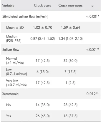
Crack users exhibited a signiicant reduction in stimulated salivary flow compared to non-users (1.02 vs. 1.59 ml/min, respectively) (p < 0.001). A total of 42.5% and 15% of crack users had very low and low stimulated salivary low, respectively (p < 0.001). Moreover, 65% of crack users perceived dry mouth in comparison to 37.5% on non-users (p < 0.012) (Table 2). However, no signiicant association was found between xerostomia and hyposalivation (p = 0.384) (Figure 1).
The crude model revealed that the prevalence of hyposalivation was 2.88-fold greater among crack users. After adjustment for possible confounding variables, the prevalence of hyposalivation was 2.59-fold greater among crack users (prevalence ratio, 2.59 and 95% conidence interval, 1.37–4.90). Moreover, the multivariate analysis demonstrated that the prevalence of the outcome was also signiicantly higher among individuals older than 26 years and those with a household income less than the BMMS (Table 3).
Discussion
In the present study, salivary low was compared between crack users and non-users and a signiicant reduction in stimulated salivary low along with a greater occurrence of hyposalivation was found among the crack users.
The occurrence of xerostomia in users of illicit drugs has been reported in previous studies, which is in agreement with the present indings.13,14,15,25 However,
only one study compared the salivary characteristics between crack users and controls that detected the changes in salivary pH but not in salivary low or buffering capacity (p > 0.05).17 Likewise, nearly all
crack users took other legal and street drugs prior or concomitantly to crack, such as marijuana, cocaine, and tobacco, and alcohol. The control group was composed nearly completely of smokers. These factors may have contributed to the reduction in salivary low in both groups.14
A reduction in normal salivary flow may or may not be accompanied by xerostomia, which is characterized by a subjective sensation of dry mouth.19,26 In the present study, xerostomia was not signiicantly associated with a reduction in salivary
Table 2. Comparison of stimulated salivary flow and xerostomia between crack users and non-users (n = 40).
Variable Crack users Crack non-users p
Stimulated salivar flow (ml/min) < 0.001*
Mean ± SD 1.02 ± 0.70 1.59 ± 0.64
Median
(P25–P75) 0.87 (0.46–1.52) 1.34 (1.07–2.10)
Salivar flow < 0.001**
Normal
(>1 ml/min) 17 (42.5) 32 (80.0)
Low
(0.7–1 ml/min) 6 (15.0) 7 (17.5)
Very low
(<0.7 ml/min) 17 (42.5) 1 (2.5)
Xerostomia 0.012**
No 14 (35.0) 25 (62.5)
Yes 26 (65.0) 15 (37.5)
*Wilcoxon test; **McNemar test.
Figure 1. Association between xerostomia and stimulated salivary flow in crack users and non-users (§ Mean ± standard deviation).
0.88±0.41 1.10±0.82
1.55±0.59 1.60±0.68
0 0.5 1 1.5 2 2.5
Crack users Crack nonusers
Stimulate flow rate (mL/min)
low (hyposalivation), which is in agreement with data described in the literature.9
Different factors are associated with the reduction in salivary low, such as smoking, alcohol, the use of street drugs, beverages with caffeine, and systemic diseases (especially Sjögren’s syndrome).14,27 However, reduced salivary low is more commonly associated with the use of medications, such as diuretics, laxatives, antacids, anorectics, anti-hypertension agents, antidepressants, antipsychotics, sedatives, antihistamines, anticholinergics, and medications for the treatment of Parkinson’s Disease.14,28,29
Individuals who take two or more medications have a 5.5-fold greater chance of having low salivary low (< 0.7 ml/min) in comparison to individuals who take no medication.19 Among the crack users in the present
study, 52.5% were taking some type of medication at the time of saliva collection, such as anxiolytics, anticonvulsants, antihistamines, antipsychotics, and antidepressants. However, no association was found between the use of medication and hyposalivation. This may be explained by the fact that 73.4% of crack
users were taking medications only at the beginning of the treatment period (detoxiication period in a maximum of 21 days).
In the adjusted model, an age over 26 years, income, and crack use remained associated with hyposalivation. A meta-analysis showed that the aging process is associated with reduced salivary low.30 Drug use may be associated with systemic and local factors. With regard to systemic factors, some drugs may increase sympathetic activity in the central nervous system, reducing salivary secretion through the stimulation of alpha receptors.25 Considering local
factors, drug use seems to generate vasoconstriction,4,5
which is similar to what occurs among smokers and is also associated with a reduction in salivary control.31
The gold standard for determining the limit between normal and abnormal salivary low is not yet clear in the literature. However, normal salivary low is considered to be between 0.7 and 1.5 ml/min.32 Some authors
consider hyposalivation to be at a stimulated salivary low with a rate lower than 0.7 ml/min,24 whereas
others consider 0.5 ml/min to be the cutoff point.33
Table 3. Association between hyposalivation and demographic, behavioral, and clinical variables.
Variable n Hyposalivation n (%) RP* (95% CI) crude p RP* (95%CI) Adjusted p
Age (years)
≤ 26 41 10 (24.4) 1.0 0.011 1.0 0.001
> 26 39 21 (53.8) 2.21 (1.20–4.07) 2.52 (1.50–4.24)
Household income#
≤ 1 BMW 38 20 (52.6) 1.0 0.015 1.0 0.024
> 1 BMW 42 11 (26.2) 0.45 (0.24–0.86) 0.52 (0.29–0.92)
Crack users
No 40 8 (20.0) 1.0 0.002 1.0 0.003
Yes 40 23 (57.5) 2.88 (1.46–5.65) 2.59 (1.37–4.90)
Missing teeth
≤ 2 teeth 65 23 (35.4) 1.0 0.163 - -
> 2 teeth 15 8 (53.3) 1.51 (0.85–2.68)
-Periodontitis
No 52 15 (28.8) 1.0 0.012 - -
Yes 28 16 (57.1) 1.98 (1.16–3.38) -
-DMFT
≤ 5 39 9 (23.1) 1.0 0.010 - -
> 5 41 22 (53.7) 2.33 (1.23–4.41) - -
Ericsson and Hardwick23 propose three categories for mean stimulated salivary low: > 0.7 ml/min, very low; 0.7–1.0 ml/min, low; and 1 ml/min, normal. However, such limits are not well established. In the present investigation, hyposalivation was deined as below ≤ 1 ml/min due to the small prevalence of very low stimulated salivary low (< 0.7 ml/min) in the control group. Moreover, our interest was to evaluate factors that can exert an impact on salivary low rather than the impact of hyposalivation on oral conditions. Most studies addressing reduced salivary flow use the spitting collection method established by Navazesh34. Objective measures of saliva quantity and quality are obtained through salivary stimulation22 and different
methods are used to stimulate salivary secretion, including the chewing of gum, parafin wax, rubber strips, and citric acid.35
There is evidence that a very low salivary low signiicantly increases the risk of developing dental caries36, which underscores the importance of the recognition of reduced salivary low by dentists. Because a permanent manner for increasing saliva is not yet known,37 preventive measures regarding
dental caries should be performed.
The present study has limitations that should be addressed. The two study groups were composed of convenience samples; therefore, the indings do not represent the general population. Moreover, reference
centers may concentrate individuals with a greater occurrence and severity of the disease, which could exert an inluence on the differences detected between the groups. The two groups were matched for variables that could confound the outcome (sex, age, and tobacco use), and most of the controls were recruited from public schools, thereby exhibiting a similar socioeconomic status to that of the crack users, and did not seek any reference treatment center. The cross-sectional design does not allow the establishment of a relationship of causality. Another limitation was the fact that no investigation was made regarding saliva quality, such as pH and buffering capacity.
Conclusion
Crack users have a signiicant reduction in salivary flow and a greater occurrence of hyposalivation in comparison to non-users, independent of socio-demographic, behavioral, or clinical factors.
Acknowledgments
The authors are grateful to the volunteers who participated in this study. This study was supported by the State of Rio Grande do Sul Research Foundation (FAPERGS-12/1655-7), Brazil. The authors declare no conlicts of interest.
1. Ribeiro M, Dunn J, Laranjeira R, Sesso R. High mortality among young crack cocaine users in Brazil: a 5-year follow-up study. Addiction. 2004;99(9):1133-5. https://doi.org/10.1111/j.1360-0443.2004.00804.x 2. Morano JP, Gibson BA, Altice FL. The burgeoning
HIV/HCV syndemic in the urban Northeast: HCV, HIV, and HIV/HCV coinfection in an urban setting. PLoS One. 2013; 14;8(5):e64321. https://doi.org/10.1371/journal.pone.0064321 3. Narvaez JC, Jansen K, Pinheiro RT, Kapczinski F, Silva RA,
Pechansky F et al. Psychiatric and substance-use comorbidities associated with lifetime crack cocaine use in young adults in the general population. Compr Psychiatry. 2014;55(6):1369-76. https://doi.org/10.1016/j.comppsych.2014.04.021 4. Mitchell-Lewis DA, Phelan JA, Kelly RB, Bradley JJ,
Lamster IB. Identifying oral lesions associated with crack cocaine use. J Am Dent Assoc. 1994;125(8):1104-8. https://doi.org/10.14219/jada.archive.1994.0122
References
5. Baldwin GC, Buckley DM, Roth MD, Kleerup EC, Tashkin DP. Acute activation of circulating polymorphonuclear neutrophils following in vivo administration of cocaine: a potential etiology for pulmonary injury. Chest. 1997;111(3):698-705. https://doi.org/10.1378/chest.111.3.698
6. Woyceichoski IE, Arruda EP, Resende LG, Machado MA, Grégio AM, Azevedo LR et al. Cytomorphometric analysis of crack cocaine effects on the oral mucosa. Oral Surg Oral Med Oral Pathol Oral Radiol Endod. 2008;105(6):745-9. https://doi.org/10.1016/j.tripleo.2007.09.011
7. Antoniazzi RP, Zanatta FB, Rösing CK, Feldens CA.
Association among periodontitis and the use of crack cocaine and other illicit drugs. J Periodontol. 2016;87(12):1396-405. https://doi.org/10.1902/jop.2016.150732
9. Ship JA, Fox PC, Baum BJ. How much saliva is enough? “Normal” function defined. J Am Dent Assoc.1991;122(3):63-9. https://doi.org/10.14219/jada.archive.1991.0098
10. Pedersen AM, Bardow A, Jensen SB, Nauntofte B. Saliva and gastrointestinal functions of taste, mastication, swallowing and digestion. Oral Dis. 2002;8(3):117-29. https://doi.org/10.1034/j.1601-0825.2002.02851.x 11. Thomson WM, Lawrence HP, Broadbent JM, Poulton R. The
impact of xerostomia on oral-health-related quality of life among younger adults. Health Qual Life Outcomes. 2006;8;4:86. https://doi.org/10.1186/1477-7525-4-86
12. Orellana MF, Lagravère MO, Boychuk DG, Major PW, Flores-Mir C, Ortho C. Prevalence of xerostomia in population-based samples: a systematic review. J Public Health Dent. 2006;66(2):152-8. https://doi.org/10.1111/j.1752-7325.2006.tb02572.x 13. Duxbury AJ. Ecstasy: dental implications. Br Dental J.
1993;175(1):38. https://doi.org/10.1038/sj.bdj.4808200 14. Scully C. Drug effects on salivary glands: dry mouth.
Oral Dis. 2003;9(4):165-76.
https://doi.org/10.1034/j.1601-0825.2003.03967.x 15. Rommel N, Rohleder NH, Wagenpfeil S, Haertel-Petri R,
Kesting MR. Evaluation of methamphetamine-associated socioeconomic status and addictive behaviors, and their impact on oral health. Addict Behav. 2015;50:182-7. https://doi.org/10.1016/j.addbeh.2015.06.040
16. Parry J, Porter S, Scully C, Flint S, Parry MG. Mucosal lesions due to oral cocaine use. Br Dent J. 1996;22;180(12):462-4. 17. Woyceichoski IE, Costa CH, Araújo CM, Brancher JA,
Resende LG, Vieira I et al. Salivary buffer capacity, pH, and stimulated flow rate of crack cocaine users. J Investig Clin Dent. 2013;4(3):160-3.
https://doi.org/10.1111/j.2041-1626.2012.00126.x
18. Smart RG, Hughes DPH, Johnston LD, Anumonye A, Khant U, Medina-Mora ME. Methodology for students drug-use surveys. Geneva: World Health Organization; 1980. 19. Flink H, Bergdahl M, Tegelberg A, Rosenblad A,
Lagerlöf F. Prevalence of hyposalivation in relation to general health, body mass index and remaining teeth in different age groups of adults. Community Dent Oral Epidemiol. 2008;36(6):523-31. https://doi.org/10.1111/j.1600-0528.2008.00432.x 20. World Health Organization. Oral health surveys: basic
methods. Geneva: World Health Organization; 1997. 21. Borrell LN, Burt BA, Taylor GW. Prevalence and trends in periodontitis in the USA: the [corrected] NHANES, 1988 to 2000. J Dent Res.2005;84(10):924-30. https://doi.org/10.1177/154405910508401010 22. Navazesh M, Christensen CM. A comparison of whole
mouth resting and stimulated salivary measurement procedures. J Dent Res. 1982; 61(10):1158-62. https://doi.org/10.1177/00220345820610100901
23. Ericsson Y, Hardwick L. Individual diagnosis, prognosis and counselling for caries prevention. Caries Res. 1978;12 Suppl 1:94-102. https://doi.org/10.1159/000260369
24. Nederfors T. Xerostomia and hyposalivation. Adv Dent Res. 2000;14(1):48-56. https://doi.org/10.1177/08959374000140010701 25. Curtis EK. Meth mouth: a review of methamphetamine abuse
and its oral manifestations. Gen Dent. 2006;54(2):125-9. 26. Wang SL, Zou ZJ, Wu QG, Sun KH. Sialographic changes
related to clinical and pathologic findings in chronic obstructive parotitis. Int J Oral Maxillofac Surg. 1992;21(6):364-8. https://doi.org/10.1016/S0901-5027(05)80764-7
27. Antoniazzi RP, Miranda LA, Zanatta FB, Islabão AG, Gustafsson A, Chiapinotto GA et al. Periodontal conditions of individuals with Sjögren’s syndrome. J Periodontol. 2009;80(3):429-35. https://doi.org/10.1902/jop.2009.080350
28. Field EA, Longman LP, Bucknall R, Kaye SB, Higham SM, Edgar WM. The establishment of a xerostomia clinic: a prospective study. Br J Oral Maxillofac Surg. 1997;35(2):96-103.
https://doi.org/10.1016/S0266-4356(97)90683-5 29. Torres SR, Peixoto CB, Caldas DM, Silva EB, Akiti T,
Nucci M et al. Relationship between salivary flow rates and Candida counts in subjects with xerostomia. Oral Surg Oral Med Oral Pathol Oral Radiol Endod. 2002;93(2):149-54. https://doi.org/10.1067/moe.2002.119738
30. Affoo RH, Foley N, Garrick R, Siqueira WL, Martin RE. Meta-analysis of salivary flow rates in young and older adults. J Am Geriatr Soc. 2015;63(10):2142-51. https://doi.org/10.1111/jgs.13652 31. Rad M, Kakoie S, Niliye Brojeni F, Pourdamghan N. Effect of
long-term smoking on whole-mouth salivary flow rate and oral health. J Dent Res Dent Clin Dent Prospects. 2010;4(4):110-4. https://doi.org/10.5681/joddd.2010.028
32. Närhi TO, Meurman JH, Ainamo A. Xerostomia and hyposalivation: causes, consequences and treatment in the elderly. Drugs Aging. 1999;15(2):103-16. https://doi.org/10.2165/00002512-199915020-00004 33. Longman LP, Higham SM, Rai K, Edgar WM, Field EA.
Salivary gland hypofunction in elderly patients attending a xerostomia clinic. Gerodontology. 1995;12(2):67-72. https://doi.org/10.1111/j.1741-2358.1995.tb00133.x 34. Navazesh M1. Methods for collecting saliva. Ann N Y Acad
Sci. 1993;20;694(694):72-7.
https://doi.org/10.1111/j.1749-6632.1993.tb18343.x 35. Chambers MS, Posner M, Jones CU, Biel MA, Hodge KM,
Vitti R et al. Cevimeline for the treatment of postirradiation xerostomia in patients with head and neck cancer. Int J Radiat Oncol Biol Phys. 2007;15;68(4):1102-9. https://doi.org/10.1016/j.ijrobp.2007.01.019
36. Leone CW, Oppenheim FG. Physical and chemical aspects of saliva as indicators of risk for dental caries in humans. J Dent Educ. 2001;65(10):1054-62.