In their original work, Dyke, Davidoff and Mas-son1 described the radiological changes occurring in
the skull of nine patients with the clinical
diagno-LA
TERALIZATION OF EPILEPTIFORM DISCHARGES
IN PATIENTS WITH EPILEPSY AND PRECOCIOUS
DESTRUCTIVE BRAIN INSULTS
Ricar
do A. Teixeira
1, Li M. Li
1, Sergio L.M. Santos
2,
Bárbara J. Amorim
2, Elba C.S.C. Etchebehere
2,
Verônica A. Zanardi
2, Carlos A.M. Guerreiro
1, Fernando Cendes
1ABSTRACT - Unilateral destructive brain lesions of early development can result in compensatory thicken-ing of the ipsilateral cranial vault. The aim of this study was to determine the frequency of these bone changes among patients with epilepsy and precocious destructive lesions, and whether a relationship exists between these changes and epileptiform discharges lateralization. Fifty-one patients had their ictal / inter-ictal scalp EEG and skull thickness symmetry on MRI analyzed. Patients were divided into three main groups according to the topographic distribution of the lesion on the MRI: hemispheric (H) (n=9); main
arterial territory (AT) (n=25); arterial borderzone (Bdz) (n=17). The EEG background activity was abnormal in 26 patients and were more frequent among patients of group H(p= 0.044). Thickening of the skull was
more frequent among patients of group H(p= 0.004). Five patients (9.8%) showed discordant lateraliza-tion between epileptiform discharges and structural lesion (four of them with an abnormal background, and only two of them with skull changes). In one of these patients, ictal SPECT provided strong evidence for scalp EEG false lateralization. The findings suggest that compensatory skull thickening in patients with precocious destructive brain insults are more frequent among patients with unilateral and large lesions. However, EEG lateralization discordance among these patients seems to be more related to EEG background abnormalities and extent of cerebral damage than to skull changes.
KEY WORDS: hemiatrophy, infarct, MRI, EEG, Dyke-Davidoff-Masson syndrome.
Lateralização de descargas epileptiformes em pacientes com epilepsia e lesões cerebrais destru-tivas precoces
RESUMO - Lesões cerebrais destrutivas unilaterais ocorridas em fase precoce do desenvolvimento podem resultar em espessamento compensatório da calota craniana ipsilateral. O objetivo deste estudo foi deter-minar a freqüência destas alterações ósseas em pacientes com epilepsia e lesões destrutivas precoces e avaliar se há associação entre estas alterações e lateralização de descargas epileptiformes. Foram analisados EEGs interictais / ictais e espessura do crânio pela RM de 51 pacientes. Os pacientes foram divididos em três gru-pos de acordo com a distribuição topográfica da lesão à RM: hemisférico (H) (n=9); território arterial (AT)
(n=25); fronteira arterial (Bdz) (n=17). A atividade de base no EEG foi anormal em 26 pacientes e foi mais freqüente entre os pacientes do grupo H(p=0,044). Espessamento unilateral da calota craniana foi mais
freqüente entre os pacientes do grupo H(p=0,004). Cinco pacientes (9,8%) apresentaram discordância late-ralizatória entre as descargas epileptiformes e lesão estrutural (quatro deles com atividade de base anor-mal, e apenas dois deles com espessamento da calota craniana). Em um destes pacientes, o SPECT ictal reve-lou forte evidência de falsa lateralização pelo EEG. Os achados sugerem que o espessamento compensatório da calota craniana é mais freqüente entre pacientes com lesões unilaterais e extensas. No entanto, a late-ralização de descargas epileptiformes parece estar mais relacionada ao grau de alteração da atividade de base e extensão da lesão cerebral do que às alterações ósseas.
PALAVRAS-CHAVE: hemiatrofia, infarto, RM, EEG, Dyke-Davidoff-Masson syndrome.
sis of “infantile hemiplegia”: thickening of the cra-nial vault on the same side of the cerebral lesion, overdevelopment of the frontal and ethmoid
sinus-Departments of Neurology1and Radiology2, State University of Campinas (UNICAMP), Campinas SP, Brazil. Dr. Ricardo A. Teixeira
was supported by a grant from Fundação de Amparo à Pesquisa do Estado de São Paulo (FAPESP), # 98/13101-8.
Received 27 June 2003. Accepted 16 September 2003.
es and of the air cells of the petrous pyramid of the temporal bone. These compensatory bone changes are the result of precocious unilateral loss of cerebral volume, due to a variety of congenital and acquired pathological processes. The clinical presentation consists of variable degrees of hemi-plegia, hemiatrophy, mental retardation and sei-zures. The combination of those clinical and imag-ing features became known as the Dyke-Davidoff-Masson syndrome2,3. Sammaritano et al.2emphasized
the importance of a critical interpretation of the surface EEG in patients with epilepsy and large uni-lateral cerebral lesions. Patients with large hemi-spheric lesions can present bilateral synchronous or independent interictal epileptic discharges that in some instances “predominate” in the hemisphe-re contralateral to the lesion. The EEG background activity of the intact hemisphere will be higher in amplitude and can overshadow the low voltage activity of the lesioned hemisphere, leading to a false lateralization.
Besides the large extension of the lesion, thi-ckening of the adjacent skull could also contrib-ute to the false lateralization described by Samma-ritano et al.2in patients with destructive lesions of
early development. However, as far as we know, no study has investigated the significance of these skull abnormalities in lateralization of epileptic discharges. There is evidence that magnetic reso-nance imaging (MRI) can demonstrate the com-pensatory bone changes in Dyke-Davidoff-Masson syndrome as easily as computed tomography (CT) and can afford additional details of brain lesion that CT can not2-4.
This study was designed to access the lateraliza-tion of epileptogenic discharges and the frequen-cy of compensatory skull changes in patients with different patterns of precocious destructive lesions. We also tested the hypothesis that lateralization of epileptogenic discharges varies according to the presence of skull thickening.
METHOD
We studied 51 consecutive adult patients (22 women) seen at the Epilepsy Clinic of UNICAMP from March 1999 to April 2001 with the diagnosis of epilepsy secon-dary to a destructive brain lesion of early development (mean age = 31.8 years; range = 15-55 years). Informed consent was obtained from all subjects. This study was approved by the Ethics Committee of the Faculty of Medical Sciences of UNICAMP. Further details about clinical and MRI findings about these patients were des-cribed in a recent publication5.
Detailed histories of prenatal, neonatal and early childhood events were systematically reviewed through the medical records and direct interview with the par-ents. All patients had disease onset before the fifth year of age. We excluded patients with foreign tissue lesions detected on MRI as well as those with history of major traumatic brain injury or with signs of progressive dis-ease.
Previous electroencephalographic data were revie-wed and all patients had serial routine EEG studies using the 10-20 system with additional anterior temporal and zygomatic electrodes. Eleven patients had long-term video-EEG monitoring with scalp electrodes for record-ing of their habitual seizures.
MRI was performed in a 2.0 T scanner (Elscint Prestige, Haifa, Israel). Our epilepsy protocol consists of: a) sagital T1 spin-echo, 6 mm thick (TR= 430, TE= 12) for optimal orientation of the subsequent images; b) coronal T1 inver-sion recovery (IR), 3 mm thick (flip angle = 200o; TR = 2700,
TE= 14, TI = 840, matrix =130x256, FOV =16x18 cm); c) coro-nal T2-weighted “fast spin-echo” (FSE), 3-4mm thick (flip angle = 120o, TR = 4800, TE = 129, matrix = 252x320, FOV
= 18x18 cm); d) axial images parallel to the long axis of the hippocampus; T1 gradient echo (GRE), 3mm thick (flip angle = 70o, TR = 200, TE = 5, matrix =180x232, FOV
= 22x22 cm); e) axial T2 FSE, 4mm thick (flip angle =120o,
TR = 6800, TE = 129, matrix = 252x328, FOV = 21x23 cm); f) volumetric (3D) T1 GRE, acquired in the sagital plane for multiplanar reconstruction, 1-1.5mm thick (flip angle = 35o, TR = 22, TE=9, matrix = 256x220, FOV = 23x25 cm).
Visual analysis of MRI and multiplanar reconstruction were systematically performed in a workstation (O2 Silicon Graphic) using Omnipro software (Elscint Prestige, Haifa, Israel). Qualitative analysis of the skull thickness symmetry was made by one of us (SLMS) who was blind-ed to the clinical and EEG data. Patients were dividblind-ed in three different groups according to the topographi-cal distribution of the lesion: Hemispheric lesions (H); pa-tients in this group had an homogeneous atrophy of an entire hemisphere without loss of tissue continuity. Ar te-rial territory lesions (AT);this group had their lesions li-mited to a main arterial territory, constituted by a cav-itation or a localized retraction of the cerebral tissue with an heterogeneous appearance suggestive of a substan-tial gliotic scar. Arterial borderzone lesions (Bdz); these
patients had their lesions consisting of atrophy between the main cerebral arterial territories with an ulegyric aspect6,7. Eleven patients showed more than one of
the-se lesional patterns and they were classified according to the most exuberant pattern.
a fan-beam collimator. Sixty images were acquired, at 6o intervals for a total of 360o. Images were normalized, a Metz filter and attenuation correction were applied. The images were reconstructed in the transaxial, coro-nal, and sagittal planes.
We used the Pearson’s chi-square (χ2) or Fisher’s exact test for comparing proportions among groups. We con-sidered the significance level of 0.05.
RESULTS
Fifty-one patients were distributed in the three different groups as follows: H (n=9), AT (n=25), Bdz
(n=17). The EEG and MR findings are summarized in Table 1.
EEG analysis
The background activity was abnormal in 26 patients (51%) including voltage asymmetry (n=15), continuous slowing over one hemisphere (n=9) and diffuse slowing (n=3). These abnormalities were more frequent among patients of group H(77,7%) than groups Bdz(29.4%) and AT(56%) (χ2 [2]=6.257, p = 0.044).
Epileptiform activity was present in routine EEGs of 38 patients (74.5%); in 33 of them it was lateralized and concordant with the lesion, in one it was not lateralized, and in four it was consistent-ly discordant. Three out of these four patients (Patients 3, 6 and 49) presented an abnormal back-ground activity with voltage reduction ipsilateral to the lesion and contralateral to the epileptiform activity. In patients from Bdzgroup who had bilat-eral lesions, EEG was considered concordant when it was ipsilateral to the most robust lesion.
Ictal surface EEG was recorded in eleven pa-tients. In seven patients (Patients 24, 27, 33, 44, 46, 48 and 51) ictal onset was ipsilateral to the lesion. Two patients had nonlocalizing ictal EEG (Patients 32 and 42). We observed in further two patients (Patients 3 and 39) a bitemporal seizure onset with electrical activity of higher voltage over the hemi-sphere contralateral to the lesion. Both present-ed an abnormal background activity with voltage reduction ipsilateral to the lesion.
MRI analysis
Bone thickening of the skull adjacent to the le -sion was observed more frequently among patients of group H(7/9) than groups AT(11/25) and Bdz
(2/17) (χ2 [2]=11.29, p = 0.004).
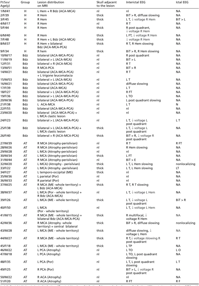
Patients from group Hconstituted a continu -um of hemiatrophy severity. The two patients who presented a symmetric skull thickness could be
ca-tegorized as the less severe pole of this spectrum of hemiatrophy (Fig 1).
Among patients from group Bdz, fourteen (82%) exhibited bilateral lesions. Twelve (70%) had the lesion distributed posteriorly on the watershed between the three main arteries. Five patients had their lesions more anteriorly, on the watershed bet-ween the anterior cerebral artery (ACA) and mid-dle cerebral artery (MCA). The two patients who exhibited skull thickening (Patients 25 and 26) were those who had the more extensive lesions from the group, one of them associated to a clastic lesion on the territory of the MCA (Patient 26).
Nineteen patients from group AT (76%) pr e-sented lesions on the territory of the MCA, four on the posterior cerebral artery (PCA) and two on the ACA. Among patients with MCA lesions, those with lesions of the whole territory had skull thick-ening more frequently (7/9) than those with lesions over MCA divisions (4/10), but this difference did not reach significance (χ2 [1]=2.77, p = 0.096). Patients with lesions on PCA and ACA did not show skull thickening. There was no difference in fre-quency of skull thickening between patients with cystic (5/14) and atrophic lesions (6/11) (χ2 [1]=0.89, p = 0.346). However, we could observe that there were no skull changes among patients with large cystic lesions communicating to the lateral ventri-cle and extending up to the convexity as a single cavity (porencephaly) (Patients 35,36,38,40,48 and 49). Conversely, patients with multicystic encephalo-malacia had intact ventricular walls (Patients 34, 39,41,43,44 and 45), and all showed skull thicken-ing (Fig 2).
Patients with EEG and MRI discordance
Five patients out of 51 (9.8%) showed discor -dant lateralization between epileptiform discharges and structural lesion.
Table 1. EEG and MRI features
Pt/Sex/ Group Lesion distribution Skull adjacent Interictal EEG Ictal EEG Age(y) on MRI to the lesion
1/M/41 H L Hem + R Bdz (ACA-MCA) nl nl NA 2/F/39 H R Hem thick BiT > R, diffuse slowing NA 3/F/45 H R Hem thick L T, ↓voltage R Hem BiT > L 4/M/17 H R Hem nl R T NA 5/F/44 H R Hem thick R post quadrant, NA
↓ voltage R Hem
6/M/40 H R Hem thick L FT, ↓voltage R Hem NA 7/F/48 H R Hem + L Bdz (ACA-MCA) thick ↓voltage R Hem NA 8/M/37 H R Hem + bilateral thick R T, R Hem slowing NA
Bdz (ACA-MCA-PCA)
9/F/34 H R hem thick BiT > R, R Hem slowing NA 10/M/17 Bdz bilateral (ACA-MCA-PCA) nl R post quadrant NA 11/M/19 Bdz bilateral > L (ACA-MCA) nl BiT > L NA 12/F/31 Bdz bilateral > R (ACA-MCA) nl R T NA 13/M/51 Bdz R MCA-PCA nl BiT > R NA 14/M/21 Bdz bilateral (ACA-MCA-PCA) nl R T NA
+ L trigone leucomalacia
15/M/53 Bdz bilateral > L (ACA-MCA) nl L T NA 16/M/21 Bdz bilateral (ACA-MCA-PCA) nl diffuse slowing NA 17/F/39 Bdz bilateral (ACA-MCA) nl L T NA 18/F/27 Bdz bilateral > L (ACA-MCA-PCA) nl R FT NA 19/F/36 Bdz bilateral > L (ACA-MCA-PCA) nl L TO NA 20/M/36 Bdz bilateral (ACA-MCA-PCA) nl L post quadrant slowing NA 21/F/38 Bdz L ACA-MCA nl L T N 22/F/55 Bdz bilateral (ACA-MCA-PCA) nl nl NA 23/M/20 Bdz bilateral (ACA-MCA-PCA) + nl nl NA
L MCA clastic lesion
24/F/23 Bdz bilateral > L (ACA-MCA-PCA) nl L T, ↓voltage L L T post quadrant
25/F/38 Bdz bilateral > L (ACA-MCA-PCA) + thick L T, ↓voltage L NA L MCA clastic lesion post quadrant
26/F/40 Bdz bilateral > R (ACA-MCA-PCA) thick BiT > R, ↓voltage R NA post quadrant
27/M/39 AT R MCA (Atrophy-perisilvian) nl R T R FT 28/M/26 AT R MCA (Atrophy-perisilvian) nl R Hem slowing NA 29/M/28 AT L MCA (Atrophy-perisilvian) nl nl NA 30/M/34 AT L MCA (Atrophy-perisilvian) thick L FT NA 31/M/44 AT R MCA (Atrophy-perisilvian) nl BiT > E NA
32/M/20 AT L MCA) (Atrophy - perisilvian) thick L T, L Hem slowing nonlocalizing 33/F/22 AT L MCA (Atrophy -perisilvian) thick L T, L Hem slowing LT
34/F/27 AT L temporo-occipital (ME) thick nl NA 35/M/36 AT L parietal (Por) nl nl NA 36/M/33 AT R parietal (Por) nl nl NA 37/M/25 AT R MCA (ME -whole territory) + thick R T, R T slowing NA
L Bdz (ACA-MCA)
38/M/37 AT L MCA (Por - whole territory) + nl L T, ↓voltage L Hem NA R Bdz (ACA-MCA)
39/F/26 AT L MCA (ME - whole territory) thick L T, ↓voltage L BiT > R post quadrant
40/F/50 AT L MCA nl L T, ↓voltage L Hem NA (Por - whole territory)
41/M/15 AT R MCA (ME - whole territory) + thick R multifocal, ↓ NA
bilateral Bdz (ACA-MCA-PCA) voltage R Hem
42/M/36 AT R MCA (Atrophy -whole thick BiF > R, diffuse slowing nonlocalizing territory) + central bilateral
43/M/28 AT L MCA (ME - whole territory) thick diffuse slowing, ↓ NA
voltage L Hem
44/M/27 AT R MCA (ME - whole territory) thick R T,↓voltage /slowing R R T post quadrant
45/F/18 AT L MCA (ME - whole territory) thick L TP NA 46/M/22 AT L PCA (Atrophy) nl L TO L O 47/M/18 AT L PCA (Atrophy) nl L TO, L post quadrant NA
slowing
48/F/35 AT L PCA (Por) nl L T, L post quadrant L T slowing
49/F/25 AT R PCA (Por) nl BiT > L, ↓voltage R NA post quadrant
50/M/22 AT R ACA (Atrophy) nl nl NA 51/F/20 AT R ACA (Atrophy) nl R FT R F
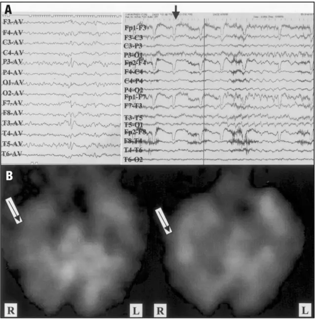
hand dystonia. The ictal EEG discharges first began to appear approximately 5 seconds after clinical onset, involving both hemispheres but with une-quivocal left side amplitude predominance. In one of the seizures, radioligand for SPECT images was injected 4 seconds after clinical onset and one se-cond before EEG discharges. Interestingly, the ictal brain SPECT images showed marked hyperperfu-sion in the right temporal region (Fig 3). This same region was hypoperfused in the interictal study indi-cating that this region was the epileptic focus. The MRI showed a right hemiatrophy associated with a marked right hippocampal atrophy with ipsilat-eral compensatory skull thickening. She was sub-mitted to a right standard anterior temporal lobe removal including amygdala and anterior portion of the hippocampus, and remains seizure free after a period of 12 months.
Patient 6. A 44-year-old man with a previous his-tory of neonatal seizure had normal development until the 6thyear of age when he presented an
epi-sode of status epilepticus, followed by a per ma-nent left hemiparesis and recurrent seizures. His habitual seizures were partially controlled with antiepileptic drugs (AED) and were of two kinds: sensitive partial seizures involving the left arm and complex partial seizures. Interictal EEG showed Fig 1. Illustration of different grades of hemiatrophy severity and their relationship to skull changes. A) Patient 1. Slight to moderate left hemiatrophy and no skull changes; B) Patient 7. Severe right hemiatrophy associated to adjacent thickening of the cranial vault.
Fig 2. Examples of two kinds of cystic lesions showing differ-ent effects over the adjacdiffer-ent skull. A) Patidiffer-ent 38. A 37-year-old man with a congenital right hemiparesis and a left poren-cephaly over the territory of the middle cerebral artery (MCA). The lesion is in broad communication with the lateral ventri-cle extending until the convexity and no compensatory bone thickening can be detected; B) Patient 37. A 25-year-old man with a congenital left hemiparesis and a multicystic encephalo-malacia over the right MCA. Adjacent compensatory bone thickening can be observed, probably due to impairment of pres-sure against the convexity exerted by the gliotic “cells”. In this way, the equalization of pressure is brought in part by thick-ening of the skull.
Patient 31.A 44-year-old man with a discrete right hemiparesis and recurrent seizures since the age of 13. He had monthly motor partial seizures involving the left hand and occasionally a complex partial seizure. Interictal EEG showed bitemporal epileptic discharges, synchronic or independently, and it was substantially more common on left side. The MRI showed a right perisylvian atrophy and no skull changes.
without loss of tissue continuity9-10. Our
classifica-tion follows this neuropathological perspective. On the opposite end of the spectrum of lesions ana-lyzed in this study, there are borderzone lesions that are usually subtle, bilateral and rarely associated with skull abnormalities.
An interesting finding among patients from group AT was that cystic lesions in communication with the lateral ventricle and extending until the convexity as a single cavity (porencephaly) were not associated with skull changes. Conversely, all pa-tients with multicystic encephalomalacia present-ed compensatory bone thickening. This suggests that the pressure that these two patterns of cys-tic lesions exert against the skull is quite different. A multicystic lesion would have a reduced poten-tial for expansion since the gliotic tissue works as “containers”, and in this way the equalization of pressure is brought in part by thickening of the skull. This is in agreement with Laplace’s law which states that the force exerted by a fluid-filled cavity against its surface is directly proportional to the diameter of the cavity.
In our study, lateralization of epileptiform dis-charges was discordant to the lesion in nearly 10% of the patients. In a study of twenty-five children with post-hemiconvulsive hemiplegia, nine (36%) showed interictal epileptiform discharges exclusive-ly over the non-affected hemisphere11. Ito et al.12
des-cribed two cases with discordance of epileptiform discharges on EEG and CT among 41 children with congenital hemiplegia. A study before the advent of CT in children with congenital hemiplegia evi-denced a lateralization discordance between EEG and presumed affected hemisphere in 24% of pa-tients13. The study of Sammaritano et al.2was very
elucidative as it could be elegantly demonstrated by invasive EEG recordings that in patients with lar-ge destructive lesions, a lateralization discordance between lesion and scalp EEG can be false due to a limitation of the method (extracranial EEG).
In our study, only two out of five patients with lateralization discordance showed skull changes. In contrast, four out of five patients presented EEG background abnormalities. This suggests that changes of EEG background are more relevant than skull changes for this lateralization discor-dance, but this needs to be confirmed by a larger series.
It is likely that the dorsal hippocampal comis-sure14,15 may also have a relevant role in
inter-drant and remarkable epileptic discharges over the left temporal region. Ictal EEG recordings sho-wed a bitemporal electrographic ictal onset with right side amplitude predominance, in a total of four seizures recorded. The MRI showed a multi-cystic encephalomalacia over the whole territory of the left MCA with ipsilateral hippocampal atro-phy and no skull changes. A left standard anterior temporal lobe removal including amygdala and an-terior portion of the hippocampus was performed and the patient remained seizure-free for a peri-od of 26 months and now presents rare seizures (Engel’s class II)8.
Patient 49. A 25-year-old woman with a normal development until the age of 11 years when she began to present recurrent seizures described as a rising epigastric sensation followed by a complex partial seizure. One seizure was witnessed by one of us, when a dystonic posture of the left hand could be observed. The seizures were refractory to AED and occurred weekly. The MRI revealed a cystic le-sion in communication with the lateral ventricle over the territory of the right PCA, an ipsilateral hippocampal atrophy and no skull changes. Interictal EEGs demonstrated reduced voltage over the right posterior quadrant and bitemporal epilep-tic discharges with predominance of voltage and frequency over the left side. A right standard ante-rior temporal lobe removal including amygdala and anterior portion of the hippocampus was per-formed and the patient remains only with rare simple partial seizures (epigastric sensation) after a follow-up of two years and eight months.
DISCUSSION
We studied a group of epileptic patients with heterogeneous neuropathologic conditions that have in common the finding of precocious brain tissue destruction. We grouped these patients ba-sed on the topographical distribution of their le-sions on MRI and in this way we created three near-ly homogenous groups 5.The frequency of
compen-satory skull thickening was greater among patients with hemiatrophy (group H). This is in agreement with previous studies that show precocious unilat-eral and large lesions associated to Dyke-Davidoff-Masson syndrome1,3,4. In fact, the term
hemiatro-phy is often used in the neurological and radiolog-ical literature to describe any large destructive unilateral lesion (e.g, MCA infarct)3,4, but the
tive brain insults are more frequent among patients with unilateral and large lesions. However, EEG lat-eralization discordance among these patients seems to be more related to EEG background abnormal-ities and the extent of cerebral damage than to skull changes.
REFERENCES
1. Dyke CG, Davidoff LM, Masson CB. Cerebral hemiatrophy with homo-lateral hypertrophy of the skull and sinuses. Surg Gynecol Obstet 1933;57:588-600.
2. Sammaritano M, de Lotbinière A, Andermann F, et al. False lateraliza-tion by surface EEG of seizure onset in patients with temporal lobe epilepsy and gross focal cerebral lesions. Ann Neurol 1987;21:361-369. 3. Zeiss J, Brinker RA. MR imaging of cerebral hemiatrophy. J Comput
Assist Tomogr 1988;12:640-643.
4. Sener RN, Jinkins JR. MR of craniocerebral hemiatrophy. Clin Imaging 1992;16:93-97.
5. Teixeira RA, Li LM, Santos SL, et al. Early developmental destructive brain lesions and their relationship to epilepsy and hippocampal dam-age. Brain Dev 2003; “in press”.
6. Kuzniecky RI, Jackson GD, Magnetic resonance in epilepsy. New York: Raven Press, 1995:142-144.
7. Barkovich AJ. Pediatric neuroimaging, 2nd ed. Philadelphia: Lippincott-Raven, 1996.
8. Engel JJ, Van Ness PC, Rassmussen TB, Ojemann LM. Outcome with respect to epileptic seizures. In Engel JJ (ed). Surgical treatment of the epilep-sies, 2nd edition. New York: Raven Press, 1993: 609-621.
9. Alpers BJ, Dear RB. Hemiatrophy of the brain. J Nerv Ment Dis 1939;89:653-671.
10. Friede RL. Developmental neuropathology. New York: Springer-Verlag,1989.
11. Kataoka K, Okuno T, Mikawa H, Hojo H. Cranial computed tomograph-ic and electroencephalographtomograph-ic abnormalities in children with post-hemi-convulsive hemiplegia. Eur Neurol 1988;28:279-284.
12. Ito M, Okuno T, Takao T, et al. Electroencephalographic and cranial com-puted tomographic findings in children with hemiplegic cerebral pal-sy. Eur Neurol 1981;20:312-318.
13. Dyken ML, White PT, Nelson G. Electroencephalographic lateraliza-tion in chronic infantile hemiplegia. Electroenceph Clin Neurophysiol 1964;17:693-695.
14. Palmini AL, Gloor P, Jones-Gotman M. Pure amnestic seizures in tem-poral lobe epilepsy. Brain 1992;115:749-769.
15. Gloor P, Salanova V, Olivier A, Quesney LF. The human dorsal hippocam-pal comissure. Brain 1993;116:1249-1273.
hemispheric synchrony among patients with large destructive hemispheric lesions, since the neocor-tical pathways are extensively damaged. Seizure spread to the contralateral temporal hippocampus and neocortex would occur prior to involvement of adjacent neocortical structures of the ipsilater-al hemisphere. However, the SPECT study from Pa-tient 3 does not support this hypothesis, since the-re was a clear and pthe-recocious activation of the damaged temporal lobe.
In this study, interictal epileptiform discharges among patients with EEG-lesion discordance were present in three different patterns: a) restricted to the non damaged hemisphere; b) bilateral but predominating on the non-damaged hemisphere; c) bilateral predominating on the damaged hemi-sphere. Different magnitudes of hemispheric injury and skull thickness could explain these lateralizing variations. These interictal EEG lateralizing varia-tions were also present in the study of Sammaritano et al.2.
It is worth to emphasize the decisive role of the SPECT study on surgical indication of patient 3. Along with clinical lateralizing data, the method can be very useful for noninvasive lateralization of ictal onset in patients with large destructive lesions with discordant EEG features, and further studies will be helpful for better delineation of its useful-ness at these circumstances.