Department of Neurology, Hospital das Clínicas da Faculdade de Medicina da Universidade de São Paulo (FMUSP), São Paulo SP, Brazil.
Received 28 February 2003, received in final form 30 July 2003. Accepted 9 September 2003.
Dra. Letícia Brito Sampaio - Department of Neurology, Hospital das Clínicas FMUSP - Rua Dr. Eneas de Carvalho Aguiar 255 - 05403-000 São Paulo SP - Brasil. E-mail: [email protected]
THE ROLE OF MIRROR FOCUS IN THE SURGICAL
OUTCOME OF PATIENTS WITH INDOLENT
TEMPORAL LOBE TUMORS
Leticia Sampaio, Elza Mar
cia Yacubian, Maria Luiza Manreza
ABSTRACT - Purpose: To review the clinical and neurophysiological data of 21 patients with epilepsy due to temporal lobe tumors and who had undergone evaluation and surgery at the Hospital das Clínicas da Universidade de São Paulo. The aim of this study was to investigate whether the occurrence of a mirror focus was influenced either by certain clinical factors or if the surgical outcome was influenced by the presence of a mirror focus. Method: We included these 21 patients who had undergone at least one inter-ictal electroencephalogram in the pre- and post-surgical periods. They had had a minimum follow-up of one year. Results: Eight patients had mirror focus (Group 1) and 13 did not (Group 2). The mean age at seizure onset, duration of epilepsy disorder and total number of seizures did not vary statistically between the two groups of patients. Generalized tonic-clonic seizures occurred more frequently in the mirror focus group. All, but one patient, with a mirror focus were seizure free at follow- up. The mirror focus disap-peared in all eight patients in the post-surgical electroencephalogram. In this group, the patient who was not seizure - free had a seizure recorded in his post-surgical electroencephalogram with seizure onset ipsi-lateral to the resected tumor. The patients who were not seizure-free had either been submitted to an incomplete resection of the tumor or showed evidence of associated cortical dysplasia. Conclusion: The occurrence of mirror focus is not a contraindication to surgery even when interictal epileptiform activity predominates contralaterally to the tumor and neither when seizures appear to arise from the mirror focus on scalp EEG. Good surgical outcome is expected despite EEG findings that may conflict with tumor location.
KEY WORDS: epilepsy, secondary epileptogenesis, electroencephalography, complex partial seizures, tem-poral lobe tumors.
Epilepsia do lobo temporal por processos expansivos e epileptogênese secundária
RESUMO - Introdução: A epileptogênese secundária é descrita como fenômeno em que, uma área epilepto-gênica primária, geradora de descargas, através de conexões neuroniais com outra área não acometida, pode gradualmente induzir alterações epileptiformes nessa última, que, na maioria das vezes se localiza em região homóloga no hemisfério oposto ao foco primário. Objetivos: Analisar em um grupo de 21 pacientes com processos expansivos do lobo temporal e epilepsia a ocorrência do foco em espelho e os fatores clíni-cos potencialmente responsáveis pelo seu desenvolvimento e sua influência no prognóstico cirúrgico.
Método: Foram incluídos 21 pacientes que realizaram eletroencefalograma no período interictal, antes e após o tratamento cirúrgico. Dentre os fatores clínicos estudados destaca-se a idade de início e duração da epilepsia bem como a freqüência de crises epilépticas, por serem considerados relevantes no processo da epileptogênese secundária. Resultados: Os pacientes foram divididos em dois grupos de acordo com a presença (8 pacientes) ou não do foco em espelho (13 pacientes). Dentre os fatores clínicos analisados, a presença de crises tônico-clônicas generalizadas foi estatisticamente significante, ocorrendo de forma mais freqüente nos pacientes com o foco em espelho. Os outros fatores clínicos não apresentaram diferença estatisticamente significante entre os dois grupos. Após a cirurgia de retirada do processo expansivo, o foco em espelho não foi mais observado e ficaram livres de crises sete dos oito pacientes do grupo com foco em espelho e 10 dos 13, que não o apresentavam. Nos pacientes que não se tornaram livres de crises, a ressonância magnética mostrou em todos a persistência de tecido tumoral e em dois, presença de displa-sia cortical associada. Conclusão:Nossos resultados sugerem que a presença do foco em espelho não deve ser uma contra-indicação à excisão da lesão primária, mesmo quando no eletrencefalograma de escalpo a atividade interictal é mais freqüente do lado contralateral à lesão ou mesmo quando as crises epilépticas são também registradas contralateralmente.
The statement that epilepsy is a progressive disorder is a controversy that began in the 19th cen-tury and continues so to this day. The concept of secondary epileptogenesis was first described by Frank Morrell1in experimental animal models and later moved to the clinical area. It implies that a primary epileptic focus, an abnormal cortical area capable of generating epileptogenic potentials and epileptic seizures, induces epileptiform dis-charges in a secondary epileptogenic area. The latter has neuronal connections with the primary area and can develop epileptogenic potentials and epileptic seizures although its cortex is histologi-cally normal. The “mirror focus” is a special form of secondary epileptogenesis in which the second-ary epileptogenic zone is located in the contralat-eral homotopic area, with respect to the primary epileptogenic lesion. Morrell1, studying chronic models of epilepsy, described progressive stages of secondary epileptogenesis. In the initial stage, epi-leptic discharges were observed in the primary focus, which can be induced by various methods, but were not observed in synaptically related re-gions of the contralateral hemisphere. After some time, epileptic discharges began to occur in a homotopic or synaptically related region of the con-tralateral hemisphere, called the secondary focus, always time-locked to the discharges in the primary focus. This stage is called the dependent phase. Du-ring this primary stage, excision, disconnection, or pharmacological suppression of epileptiform activity in the primary focus results in an immedi-ate cessation of the contralimmedi-ateral discharges. In the intermediate phase, the secondary epileptogenic zone generates independent discharges and also independent seizures. When the latter is discon-nected from the primary epileptogenic zone, all the epileptogenic manifestations disappear after a variable period. In the later phase, called inde-pendent, the independent epileptogenic potentials are permanent and cannot be abolished by discon-nection from the primary epileptogenic region. At this stage, the secondary focus demonstrates inde-pendence from the primary site and secondary epileptogenesis has been established. Tumors fre-quently induce epileptogenic discharges in the vicinity of the neoplasm, and in some cases, can cau-se cau-secondary epileptogenic areas in synaptically re-lated sites2-4. In tumor cases, the likehood that a similar lesion may have arisen at a contralateral ho-motopic site is remote2,5,6. In non-tumoral condi-tions the occurrence of secondary
epileptogene-sis is difficult to be unequivocally documented be-cause the most common etiologies such as head trauma, birth trauma, encephalitis and vascular di-seases are all potentially capable of producing multiple, independent epileptogenic lesions1,7,8. The occurrence of secondary epileptogenesis in humans has been a matter of controversy and a few series related to this have been published9,10,11.
We retrospectively investigated the existence of secondary epileptogenesis in a series of 21 patients with epilepsy related to temporal lobe tumors treated surgically by excision of the tumor.
METHOD
We reviewed 21 patients with single temporal lobe tumors and seizures that underwent surgery at Hospital das Clínicas de São Paulo from February 1993 to Decem-ber 1999. We included all patients, irrespective of age or sex, on whom at least one interictal scalp/sphenoidal EEG had been performed before and after surgery and who had had an average follow-up of four years after surgery.
The medical records were reviewed for: sex, age, age at seizure onset, duration of seizure disorder, type and frequency of seizures, lateralization of lesion, site of lesion in temporal lobe (medial and/or neocortical), resection of the lesion (total or subtotal on MRI), histol-ogy, presence of associated cortical dysplasia and seizure outcome. The same neuropathologist analyzed all the tissue specimens. Video-EEG data in 14 and all the rou-tine EEG data in the 21 patients were reviewed for local-ization of interictal and ictal epileptogenic zone, clini-cal semiology of seizures and also the existence of a mir-ror focus. The diagnosis of mirmir-ror focus was considered only when there was a clear and unequivocal evidence of recurrent, bilateral, independent interictal epilepti-form spikes or sharp waves and/or ictal recording. Con-tralateral background slowing was not considered a sign of epileptogenesis. Our study were approved by the Ethical Committee of our Hospital.
RESULTS
Twenty-one patients were analyzed (Table 1). Fourteen patients underwent video-EEG monito-ring for localization of ictal onset. The recording was performed with scalp electrodes following the 10-20 system with insertion of sphenoidal elec-trodes in some of them. Eight (38%) out of the 21 patients met the criteria for a mirror focus and were considered as Group 1 while 13 patients without mirror focus were considered as Group 2.
Interictal EEG
focus the most frequent interictal activity was pres-ent on the same side of the tumor. In 3 patipres-ents (cases 3,5, and 8), the most frequent interictal acti-vity was apparent on the contralateral side. Five out of 8 patients with mirror focus (cases 3, 4, 5, 7 and 8) demonstrated contralateral temporal discharges both in wakefulness and non-REM sleep. In 3 pa-tients (cases 1, 2 and 6) the epileptiform activity was found only during non-REM sleep recording.
Group 2: In 10 of 13 patients the interictal activ-ity was present only on the same side of the tumor. In 3 patients (cases 13, 14 and 18) the interictal EEGs were normal.
Ictal EEGs
Group 1: In 3 out of 7 patients with mirror fo-cus who had been submitted to video EEG moni-toring (cases 1, 4 and 5) the ictal epileptiform ac-tivity was recorded independently both ipsi and contralaterally to the tumor (Figs 1, 2 and 3): in case 1 - 5 seizures ipsi and 1 seizure contralaterally to the tumor; case 4 - 1 seizure ipsi and 1 seizure con-tralaterally to the tumor and in case 5, 11 seizures contralaterally. In this last case, this finding was con-sidered as false lateralization. The semiology of the seizures didn’t show clear lateralized signs that could suggest ictal onset. In 4 patients the ictal epileptiform activity was restricted to the side of the tumor.
Group 2: In 7 of 13 patients (cases 9, 10, 12, 15, 19, 20, 21), all the ictal epileptiform activity was recorded on the same side of the tumor.
Pathology
All the tumors were of slow growing type. Among the patients with mirror focus in Group 1, two had pylocitic astrocytoma, three ganglioneu-roma, 2 ganglioglioma and one, pleomorphic xan-thoastrocytoma. In this group, cortical dysplasia was found associated to the tumor in 3 patients (cas-es 3, 5 and 7). In Group 2, 7 had ganglioglioma, 3 pylocitic astrocytoma, one ganglioneuroma, one pleomorphic xanthoastrocytoma and one dysem-bryoplastic neuroepithelial tumor. Cortical dyspla-sia was found associated to the tumor in 5 of these patients (cases 12, 15, 16, 17, 20).
Treatment and outcome
Group 1: All but one patient underwent total resection of the tumor. These 7 patients were in Engel class I, after 3 to 7 years post-operatively (mean follow-up of 4.3 years). One patient who had
a subtotal resection of the tumor (patient 7) was in Engel class IV. In his post-surgical electroence-phalogram, one seizure was recorded with ictal onset on the same side of the primary lesion. The clinical semiology was the same as those record-ed in the pre-surgical period.
Group 2: 9 out of 13 patients underwent total resection of the tumor. At one to eight years post-operatively (mean follow-up 3.9 years), 7 out of 9 patients with total resection were classified in Engel class I; one, class II and the last, III. Four pa-tients in Group 2 underwent subtotal resections of the tumor. Among them, 2 were in class I (patients 12 and 18) while the other 2 patients, whose tumors involved the neocortical as well as the medial por-tion of temporal lobe, which was not completely removed, were in class IV (patients 19 and 21).
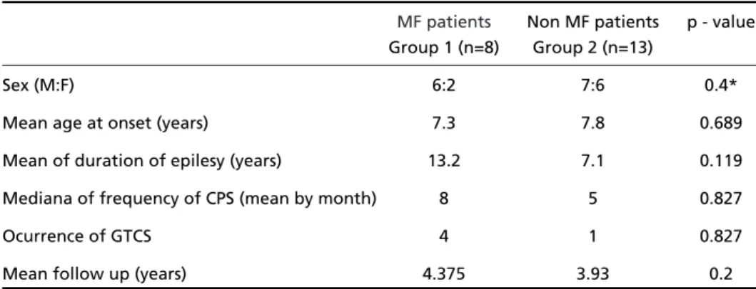
Wilcoxon’s rank-sum test showed no statiscal-ly significant differences between the mirror focus and the non-mirror focus groups in relation to age of onset, duration of seizure disorder and frequen-cy of seizures. Fisher’s test showed statistically sig-nificant difference for history of tonic-clonic seizures (p=0.047) in Group 1 (Table 2).
DISCUSSION
control-ling the seizures completely. Like other authors6,10,11, we focused our attention on patients with benign tumors expecting that, without kindling or second-ary epileptogenesis, epileptiform discharges would occur only in the cortex invaded by or in the imme-diate neighborhood of the benign tumor and cer-tainly not at a distance from the tumor.
The results of the series published in literature show that 21 to 38% of patients with epilepsy caused by tumors were reported to develop a
mir-ror focus9-11. In this series 38% had mirror focus con-firming these numbers. The occurrence of bilater-al independent foci may bilater-also be influenced by the state in which the EEG is obtained14. If we consid-ered only patients who developed the mirror focus during awakefulness, excluding the effect of pro-pagation of epileptiform discharges during non-REM sleep, this number would decrease to 23.8%. In our cases, as well as in those published in literatu-re, the interictal epileptiform discharges in the
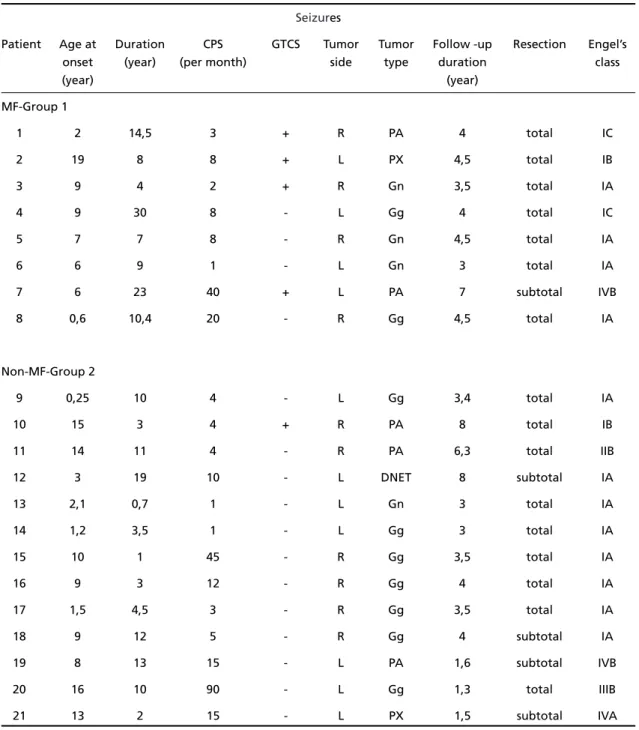
se-Table 1. Summary of 21 patients with temporal lobe tumors.
Seizures
Patient Age at Duration CPS GTCS Tumor Tumor Follow -up Resection Engel’s
onset (year) (per month) side type duration class
(year) (year)
MF-Group 1
1 2 14,5 3 + R PA 4 total IC
2 19 8 8 + L PX 4,5 total IB
3 9 4 2 + R Gn 3,5 total IA
4 9 30 8 - L Gg 4 total IC
5 7 7 8 - R Gn 4,5 total IA
6 6 9 1 - L Gn 3 total IA
7 6 23 40 + L PA 7 subtotal IVB
8 0,6 10,4 20 - R Gg 4,5 total IA
Non-MF-Group 2
9 0,25 10 4 - L Gg 3,4 total IA
10 15 3 4 + R PA 8 total IB
11 14 11 4 - R PA 6,3 total IIB
12 3 19 10 - L DNET 8 subtotal IA
13 2,1 0,7 1 - L Gn 3 total IA
14 1,2 3,5 1 - L Gg 3 total IA
15 10 1 45 - R Gg 3,5 total IA
16 9 3 12 - R Gg 4 total IA
17 1,5 4,5 3 - R Gg 3,5 total IA
18 9 12 5 - R Gg 4 subtotal IA
19 8 13 15 - L PA 1,6 subtotal IVB
20 16 10 90 - L Gg 1,3 total IIIB
21 13 2 15 - L PX 1,5 subtotal IVA
condary epileptogenic zone were not time-locked to the spikes in the vicinity of the tumor (primary epileptogenic zone). This would suggest that the secondary epileptogenic focus had achieved rela-tive independence, which means the phase of independent secondary epileptogenesis1. However, even if there is no direct proof of dependence in these cases we cannot exclude the possibility that spikes in the primary epileptogenic region were actually triggering the discharges in the second-ary epileptogenic region15. In these cases we should consider the possibility that the primary epilepto-genic spikes might be undetectable with the record-ing techniques used in these cases. The EEG recor-ded only through scalp electrodes may not detect deep-seated foci. We recorded independent scalp-recorded EEG seizure patterns arising from the contralateral side of the tumor in 37% of the pa-tients. This result is comparable to those of Gilmore et al.11. Some factors may be important in the me-chanisms of secondary epileptogenesis. The age at seizure onset was not important in our series, a fin-ding also similar to others studies11,16. Morrell et al.2, reported a series of 123 patients with tempo-ral lobe tumors and observed that patients under 30 years of age at seizure onset had more chances of developing mirror foci, especially the patients with larger duration of their disorders.
In animal models, duration of the epilepsy is also an important factor in the mechanism of second-ary epileptogenesis1,12. We did not find a signifi-cant relationship between the duration of seizure disorder and the mirror foci. Once more, our find-ing is similar to that of Gilmore et al.11who could not demonstrate this relation in a sample of 22 patients. Gupta et al.17 reported that temporal lobe seizure patients with bilateral, independent interictal abnormalities had a longer duration of
illness. In their non- tumoral patients the possibil-ity that multiple primary lesions might have con-founded conclusions about secondary epilepto-genesis.
The frequency of seizures as a factor implied in the development of mirror foci and their persist-ence after surgery is attractive and stressed by Morrell7. There was no statistically significant dif-ference between our two groups of patients. Fal-coner et al.10and Gilmore et al.11also observed this tendency. In their series, however, there was no sta-tistic significance and prognostic correlation. The occurrence of tonic-clonic seizures was a factor that showed statistic significance in our patients in contrast with others4,16. Also, there are other fac-tors that might be important in the mechanism of secondary epileptogenesis and which may interact with one another. Seeking simplistic correlations among these factors may underestimate the com-plexity of epileptogenesis.
Our patients were considered at a reversible stage of secondary epileptogenesis, characterized by independent secondary ictal and interictal foci; excision of the primary focus resulted in disappea-rance of the secondary foci and epileptic seizures originated from them. One patient with pylocitic astrocytoma associated with cortical dysplasia per-sisted with seizures. After 7 years of follow-up he still presented seizures and an ictal EEG recording showed seizures originating in the region of the primary foci. We believe that these seizures could be related to two factors: the eventual presence of cortical dysplasia and/or primary foci reminiscen-ces. Gilmore et al.11, in their series similar to our results, also observed only this reversible stage of intermediate secondary epileptogenesis.
The occurrence of dependent and intermediate
Table 2. Summary of statistical analysis.
MF patients Non MF patients p - value Group 1 (n=8) Group 2 (n=13)
Sex (M:F) 6:2 7:6 0.4*
Mean age at onset (years) 7.3 7.8 0.689
Mean of duration of epilesy (years) 13.2 7.1 0.119
Mediana of frequency of CPS (mean by month) 8 5 0.827
Ocurrence of GTCS 4 1 0.827
Mean follow up (years) 4.375 3.93 0.2
secondary epileptogenesis is a factor that has to be considered in the electroencephalographic diag-nosis of epilepsy. The electroencephalogram can give “misleading” information regarding the loca-tion and extent of the epileptic zone. In patients with a focal tumor demonstrated by MRI, despite bilateral EEG abnormalities that could represent dependent or intermediate secondary epilepto-genesis, the focal resection of brain tumor may lead to complete disappearance of epileptic seizures.
Morrell6reported eight cases, which he inter-preted as clear examples of independent secondary epileptogenesis. The main evidence for persisten-ce of seizures arising from the contralateral side of the tumor was actually clinical semiology. After resection of the tumor, seizure semiology suggest-ed that the remaining seizures were coming from the contralateral side. We agree with Lüders15who stated that in most cases even careful analysis of video recordings do not permit reliable differen-tiation of seizures coming from the opposite tem-poral lobe in any given patient. Morrell6 present-ed evidence of a recordpresent-ed seizure arising from the contralateral side of the tumor in only one case.
Careful analysis of the recording revealed that the tracing is unusual for an EEG seizure pattern with almost identical EEG patterns at left and right side of temporal chain electrodes15. If we accept that occasional cases of independent secondary epileptogenesis occur, the difficulty in finding well-documented cases clearly indicates that such cas-es must be extremely rare. Once more, we can not find any case of secondary epileptogenesis in the irreversible phase. If the intermediate secondary epileptogenesis was the highest stage of secondary epileptogenesis possible in humans, the necessity for early aggressive seizure control would depend
primarily on the impact of seizures on the quality of life. The occurrence of mirror focus is not a con-traindication to surgery even when interictal epilep-tiform activity predominates contralaterally to the tumor and neither when seizures appear to arise from the mirror focus on scalp EEG. Good surgical outcome is expected despite scalp/sphenoidal EEG findings that may conflict with tumor location.
REFERENCES
1. Morrell F. Secondary epileptogenic lesions. Epilepsia 1959/60;1:538-560. 2. Morrell F, Rasmussen T, Gloor P, De Toledo-Morrell L. Secondary epileptogenic foci in patients with verified temporal lobe tumors. Eletroencephalogr Clin Neurophysiol 1983;54:26.
3. Blume WT, Girvin JP, Kaufmann JCE. Childhood brain tumors present-ing as chronic uncontrolled focal seizure disorders. Ann Neurol 1982;12:538-541.
4. Hughes JR, Zak SM. EEG and clinical changes in patients with chron-ic seizures associated with slowly growing brain tumors. Arch Neurol 1987;44:540-543.
5. Morrell F, Rasmussen T, Toledo-Morrell L, Quesney LF, Gloor P. Frontal lobe epilepsy of neoplastic etiology: incidence of secondary epilepto-genesis. Epilepsia 1984;25:654-655.
6. Morrell F. Varieties of human secondary epileptogenesis. J Clin Neurophysiol 1989;6:227-275.
7. Morrell F. Secondary epileptogenesis in man. Arch Neurol 1985;42:318-335. 8. Morrell F. The role of secondary epileptogenesis on human epilepsy.
Arch Neurol 1991;48:1221-1224.
9. Morrell F, Toledo-Morrell L. From mirror focus to secondary epilepto-genesis in man: an historical review. Adv Neurol 1999;81:11-23. 10. Falconer MA, Kennedy WA. Epilepsy due to small focal temporal
lesions with bilateral independent spike-discharging foci. J Neurol Neurosurg Psychiatry 1961;24:205-212.
11. Gilmore R, Morris II H, Van Ness C, Gilmore-Pollak W, Estes M. Mirror focus: function of seizure frequency and influence on outcome after sur-gery. Epilepsia 1994;35:258-263.
12. Wilder BJ, King RL, Schmidt RP. Comparative study of secondary epileptogenesis. Epilepsia 1968;9:275-289.
13. Goddard DV. Development of epileptic seizures through brain stimu-lation at low intensity. Nature 1967;214:1020-1021.
14. Sammaritano M, Gigli GL, Gotman J. Interictal spiking during wake-fulness and sleep and the localization of foci in temporal lobe epilep-sy. Neurology 1991;41:290-297.
15. Lüders HO. Clinical evidence for secondary epileptogenesis. Int Rev Neurobiol. 2001;45;469-480.
16. Lim SH, So NK, Lüders H, Morris HH, Turnbull J. Etiologic factors for unitemporal vs bitemporal epileptiform discharges. Arch Neurol 1991;48:1225-1228.