CHRONIC PROGRESSIVE EXTERNAL OPHTHALMOPLEGIA
II. A Q U A L I T A T I V E A N D Q U A N T I T A T I V E E L E C T R O N M I C R O S C O P Y S T U D Y O F S K E L E T A L M U S C L E S
ELZA DIAS-TOSTA *
S U M M A R Y — T h i s s t u d y q u a n t i f i e s t h e m a i o r e l e c t r o n m i c r o s c o p i c c h a n g e s in l i m b m u s c l e
b i o p s i e s f r o m 31 o u t o f 34 p a t i e n t s w i t h t h e s y n d r o m e o f c h r o n i c p r o g r e s s i v e e x t e r n a l
o p h t h a l m o p l e g i a . P a t i e n t s w e r e d i v i d e d i n t o t h r e e c l i n i c a l g r o u p s — A ) 10 s p o r a d i c c a s e s
w i t h m u s c l e w e a k n e s s o n l y ; B ) 9 f a m i l i a l c a s e s w i t h m u s c l e w e a k n e s s o n l y ; C ) 15 c a s e s
w i t h m u s c l e w e a k n e s s a n d o n e o r m o r e o f t h e f o l l o w i n g f e a t u r e s : p i g m e n t a r y r e t i n o p a t h y ,
c e r e b e l l a r a t a x i a , p y r a m i d a l s i g n s a n d p e r i p h e r a l n e u r o p a t h y . E l e c t r o n m i c r o s c o p i c m i t o
-c h o n d r i a l a b n o r m a l i t i e s w e r e f o u n d in all g r o u p s (8 p a t i e n t s f r o m g r o u p A , 3 f r o m g r o u p B ,
14 f r o m g r o u p C ) . Q u a n t i t a t i v e m e a s u r e m e n t s o f c e r t a i n m u s c l e f i b r e c o n s t i t u e n t s , u s i n g
a p o i n t - c o u n t i n g t e c h n i q u e , r e v e a l e d d e c r e a s e d m y o f i b r i l v o l u m e - f r a c t i o n s a n d i n c r e a s e d
v o l u m e - f r a c t i o n s o f m i t o c h o n d r i a , g l y c o g e n a n d l i p i d i n s o m e b i o p s i e s f r o m e a c h g r o u p .
M i t o c h o n d r i a l v o l u m e - f r a c t i o n s c o r r e l a t e d p o s i t i v e l y w i t h l i p i d c o n t e n t , t h e p r o p o r t i o n of
t y p e 1 f i b r e s , a n d t h e p e r c e n t a g e o f f i b r e s w i t h i n c r e a s e d o x i d a t i v e e n z y m e a c t i v i t y . T h e
t h r e e g r o u p s d e f i n e d c l i n i c a l l y s h o w e d n o s i g n i f i c a n t d i f f e r e n c e s in t e r m s o f t h e r e l a t i v e
p r o p o r t i o n s o f t h e s e m e a s u r e d c o n s t i t u e n t s .
Oftalmoplegia externa crônica progressiva: II. Estudo qualitativo e quantitativo por micros-copia eletrônica de músculos esqueléticos.
R E S U M O — T r a t a - s e d e e s t u d o q u a n t i t a t i v o d a s p r i n c i p a i s a l t e r a ç õ e s e n c o n t r a d a s e m
m i c r o s c o p i a e l e t r ô n i c a d e b i ó p s i a d e m ú s c u l o s s o m á t i c o s d e 31 e n t r e 34 p a c i e n t e s c o m
s í n d r o m e d e o f t a l m o p l e g i a e x t e r n a c r ô n i c a e p r o g r e s s i v a ( O E C P ) . O s p a c i e n t e s f o r a m d i v i d i d o s
e m t r ê s g r u p o s c l í n i c o s — A ) 10 c a s o s e s p o r á d i c o s , c o m O E C P e f r a q u e z a m u s c u l a r ;
B ) 9 c a s o s c o m h i s t ó r i a f a m i l i a r p o s i t i v a , O E C P e f r a q u e z a m u s c u l a r ; C ) 15 c a s o s c o m
O E C P , f r a q u e z a m u s c u l a r e a l g u n s d o s s e g u i n t e s s i n t o m a s : r e t i n o p a t i a p i g m e n t a r , a t a x i a
c e r e b e l a r , s i n a i s p i r a m i d a i s e n e u r o p a t i a p e r i f é r i c a . F o r a m e n c o n t r a d a s a l t e r a ç õ e s e m t o d o s
o s g r u p o s (8 d o g r u p o A , t r ê s d o g r u p o B e 14 d o g r u p o C ) .
A s a v a l i a ç õ e s q u a n t i t a t i v a s d e c e r t o s c o n s t i t u i n t e s d a s f i b r a s m u s c u l a r e s , u s a n d o a t é c n i c a
d e c o n t a g e m d e p o n t o s r e v e l o u : d i m i n u i ç ã o d a f r a ç ã o - v o l u m é t r i c a d e m i o f i b r i l a s , a u m e n t o
d a s f r a ç õ e s - v o l u m é t r i c a s d e m i t o c ô n d r i a , g l i c o g ê n i o e l i p í d e o s . A s f r a ç õ e s - v o l u m é t r i c a s d e
m i t o c ô n d r i a c o r r e l a c i o n a m p o s i t i v a m e n t e c o m o c o n t e ú d o l i p í d i c o , c o m a p r o p o r ç ã o d e f i b r a s
d o t i p o 1 e c o m a p e r c e n t a g e m d e f i b r a s c o m a u m e n t o d a a t i v i d a d e e n z i m á t i c a o x i d a t i v a
( d e f i n i d a s e m e s t u d o a n t e r i o r ) . O s t r ê s g r u p o s d e f i n i d o s c l i n i c a m e n t e n ã o m o s t r a r a m
d i f e r e n ç a s s i g n i f i c a t i v a s e m t e r m o s de. p r o p o r ç õ e s r e l a t i v a s d o s c o n s t i t u i n t e s a n a l i s a d o s .
N e u r o c y t o l o g y L a b o r a t o r y , N a t i o n a l H o s p i t a l f o r N e r v o u s D i s e a s e s , U n i v e r s i t y o f L o n d o n , Q u e e n S q u a r e , L o n d o n , E n g l a n d : * M . D . , P h . D .
A variety of clinical presentations has been described in association with chronic
progressive external ophthalmoplegia (CPEO). According to light microscopy criteria
the presence of ragged-red fibres indicates mitochondrial abnormalities. On this basis
two groups of patients (A and B ) with CPEO were chosen as representing a
mito-chondrial myopathy, together with another group ( C ) who did not have a mitomito-chondrial
abnormalities
1 2. In the latter group the pathological changes found were the "rimmed
vacuoles". Electronmicroscopy has demonstrated that the "rimmed vacuoles" were
filled with "myelin figures" and other sequestrated products of muscle fibre
break-down 36. The combined use of histochemical and electronmicroscopy techniques has
confirmed the existence of mitochondrial abnormalities both in sporadic and familial
cases of CPEO, and also in patients with signs confined to the muscles or with
associated signs of other system involvement
2,3,6,21,32,35,40.These previous studies have shown that the CPEO patients present with at
least two groups of myopathological changes, but because of their multiple clinical
presentations there are still doubts whether each group represents one single entity
or different nosological entities. In an attempt to solve this problem, this study was
designed to quantify the subcellular constituents of the muscle fibre from biopsies
of patients with CPEO and relate them with the clinical and histochemical data,
M A T E R I A L A N D M E T H O D S
Specimens from 34 patients were studied and 31 of these were suitable for the quantitative study. In each case the muscle sample was taken at resting length using a paired forceps and immediately placed in 3% glutaraldehyde buffered with O . I M sodium cacodylate ( p H 7.3 7.4), postfixed in 1% osmium tetroxide similarly buffered, and embedded in epoxi resin. Sections were cut at lum , stained with toluidine blue for 30 sec
and examined under the light microscope to select an area with bluish granular appearance. Thin sections (0.06 0.09^m) were cut and stained with a saturated solution of uranyl acetate in absolute methanol followed b y 0.4% aqueous lead citrate, and examined with a Jeol 120 C X electronmicroscope at 80 K W . T o study the variation of the cellular com-ponents between the muscle fibres, 20 microscopical fields were sampled in each biopsy; to study the variation within one fibre the cross sectioned muscle fibres were then sub sampled in their peripheral and central zones.
Volumetric analysis was performed by the point counting technique. T h e test system consisted of a grid on transparent acetate paper having a squared network of 144 points, 1.5cm apart. T h i s test grid was placed over the electronmicrographs enlarged to a final magnification of x 28C00. Each time one point coincided with mitochondrion, glycogen, lipid or myofibril, one score was made for that muscle fibre constituent. T h e volume occupied by each element was represented as a percentage of the total volume of the fibre. Because of the preferential zones of distribution of mitochondria in the muscle fibres and I band ( J t f ) , care
was taken always to measure a band of the same depth beneath the muscle plasma mem-brane. The volumes attributable to mitochondria, myofibrils, glycogen within the sarcoplasm, and lipids were measured individually and the mean relative volumes of these components were calculated together with their standard deviations, variances and standard errors. These data were compared using: 1) one w a y analysis of variance, to evaluate the distri-bution of these data in relation to the muscle biopsied, s e x of patients and clinical groups; 2) correlation coefficient, to evaluate the relationship between these data and the patient's age when the biopsies were performed, the percentage of fibres with increased oxidative enzyme activity and the proportions of type 1 fibre (the light microscopic data were shown in the previous paper). Correlation coefficients were also applied to compare the relation-ships between individual components of the muscle fibres.
R E S U L T
Mitochondria
Cases Paracrystalline Concentric Abnormal Lipofuscin Myelin figures inclusions cristae size
A l
+ + +
+
+
—
—
A3
+
+
+
4—
A4 t
—
4 4A7 4 -Ο- 4 4
A8
+
4 4 4A9 4 4
+
—
A10
—
4 4-+
A l l ——i —t— —t— 4- 4- 4
—
E4 4- + 4- r : .
+
4B7 4 4
4-BIO + + 4- 4- 4 4
CI + 4- 4-
+
4-+
4C2
+ + +
4 4- .—.
—
C3
+
+ +
+
4-+
C4
+ + +
+
4-+
—
C6
+ + +
4 +
4—
C7
+
+ +
+
+
+
—
C8
+
4 —
+
—
CIO
+
4—
+
4C l l
+
+
—.+
—
C12
—
+
4- 4 4 .C13 + 4 - 4 - 4
+
4C14 4 .
+
4-+
4-C15
+
+
—
+
—
C16
+ + +
+
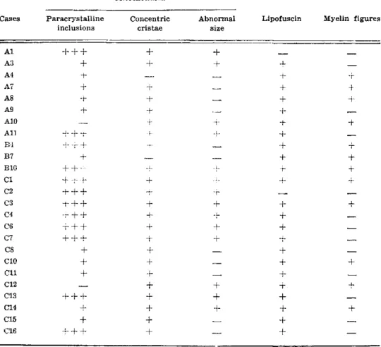
4-Table 1 — Samples showing mitochondrial changes: + + 4 - , abundant paracrystalline inclu-sions; + , few paracrystallme incluinclu-sions; A2, B9, C9, no electronmicroscc-py.
changes in mitochondrial ultrastructure. T h e paracrystalline inclusions consisted of the «intramural type» and the «parkinglot type» (Fig. 1 ) . The inclusions occurred either as a single structure, or in groups linked together b y the remaining cristae and matrix, in a longitudinal arrangement (Fig. 2) or in parallel (Fig. 3 ) . The cristae of the mitochondria in twentythree biopsies showed obvious morphological abnormalities. These included angu lated branching of the cristae, the presence of one single concentric crista and multiple layers of concentric cristae (Fig. 2 ) . In the commonest form, the mitochondrion was composed of multiple concentric layers of alternating cristae and matrix. Mitochondria could be found with one single ringshaped crista, surrounding a rather dense matrix core, or as multiple concentric cristae with either matrix material or matrix and some preserved transverse cristae at the centre (Fig. 2 ) . T h e Combination of concentric cristae and paracrystalline inclusions were seen either in a number of small mitochondria or in one enormously enlarged mitochondrion. On a few occasions elongated paracrystalline inclusion were found closely aligned with the outer mitochondrial membranes. Apart from paracrystalline inclusions, some mitochondria possessed homogeneous electrondense inclusions, electronlucent inclusion probably representing lipids, and others contained central aggre-gations of glycogen granules.
other mitochondrial abnormalities (15 out of 25). In general, the abnormal accumulations of mitochondria were subsarcolemmal and perinuclear, but agglomerations of these organelles were sometimes seen at the centre of the muscle fibre.
were longitudinally running zones of local disruption of myofibrillar structure in which the Z discs were difficult to discern; there was a loss of mitochondria and presence of dilated tubules and glycogen amongst the remaining filaments. Necrosis and phagocytosis — necrotic myofibres were present in 6 biopsies (three from group A, one from group B and two from group C) but these were never the main features of the disease process, but mast cells were a common feature of the intersticial connective tissue (16 cases). In specimens which showed «rimmed» vacuoles by light microscopy these were filled with variable amounts of cellular debris and «myelin figures*. Satellite cells were seen in 20 biopsies as a large nucleous surrounded by a narrow rim of cytoplasm. Satellite cells with slight changes from normal morphology were also detected; some contained lipofuscin, others showed nonspecific mitochondrial abnormalities, or accumulation of debris between the cell and adjacent muscle fibre. Other inclusions and structural changes — lipofuscin was the most common inclusion, found in all but three of the specimens (one from group A and two from group C ) . In 14 biopsies the sarcotubular system appeared dilated. Tubular aggregates were abundant in one case from group C and occurred sporadically in another 4 (two from group A , one from group B and one from group C ) . «Honeycomblike» eleborations of the « T » system and rods were present in one muscle each. «Filamentous bodies* was seen in three specimens and «cylindrical laminated bodies* in another case.
Thickening of the basal lamina of blood capillaries was a very common observation, either
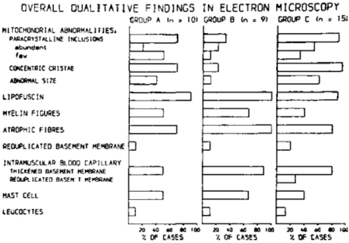
alone or associated with reduplication. A phospholipid inclusion in an otherwise normal looking endothelial cell mitochondrion was found in one case from group C. F e w intra-muscular nerve terminals, or intrafascicular nerve bundles were found, two of the latter chowed a loss of nonmyelinated fibres (two cases from group B ) . Fig. 6 represents an overall view of the qualitative findings.
O V E R A L L Q U A L I T A T I V E F I N D I N G S I N E L E C T R O N M I C R O S C O P Y
GROUP A (n = 10) GROUP 8 (n = 9) CROUP C <n = 15) MITOCHONDRIAL ABNORMALITIES.
P A R A C R Y S T A L L I NE I N C L U S I O N S a b u n d a n t
CONCENTRIC C R I S T A S
ABNORMAL S I Z E
LIPOFUSCIN MYELIN FIGURES ATROPHIC FIBRES
RE0UPLICATE0 BASEMENT MEMBRANE H
I N T R A M U S C U L A R BL0O0 C A P I L L A R Y THICKENED BASEMENT MEMBRANE R E D U P L I C A T E D BASEM T MEMBRANE
MAST CELL IEUC0CYTES
Z 3
•
1Z 3
•
i iZ 3
•
i1
Z 3
•
1
Z 3
•
1
Z 3
•
lZ 3
•
1
1 11
i i1
1
1J
Z I
1 i I
! 1
1 I 1
•
•
M »0 »0 K 100 20 40 60 00 100 » «0 «0 ( 0 1
V. OF CASES '/. OF CASES X OF CASES
Fig. 6 — Overall qualitative findings in electronmicroscopy.
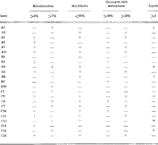
B. Quantitative studies — Comparison between values from normal controls, measured by other observers (5,8,16,18) permitted the establishment of normal limits for the various components: 1) F o r mitochondrial volumefraction: V v m ^ 5 normal; marginally increased 5 V v m < 7; definitely increase V v m ^ 7. 2) F o r myofibrillar volumefractions: V v m y > 80 normal; decreased V v m y ^ 80. 3) F o r glycogen volumefractions: V v g ^ 10 normal; marginally increased 10 < V v g < 12; definitely increased V v g ^ 12. 4) F o r lipid volumefraction: V v l ^ 1 normal; increased V v l > 1.
In this study the mitochondrial volumefractions showed a broad range of values (3 to 13%). Of the 31 biopsies measured, 20 (64%) presented one or more abnormal values: 10 (32%) with an increase of mitochondrial volumefraction, 16 (51%) with a low myofibril content, 10 (32%) with a high glycogen volumefraction, and 6 (19%) with high values for lipids (Table 2 ) . T h e most frequent abnormality found w a s a decrease in myofibril content, the next most freauent being an increase in mitochondria and glycogen. The least affected component was the lipid.
Glycogenrich
Mitochondria Myofibrils sarcoplasm Lipids
Cases > 5 % > 7 % < 8 0 % > 1 0 % > 1 2 %
A l
—
A3
—
A5
+
A6
+
A7
+
A l l
+
B2
—
B3
+
B4
—
B5
+
B6
—
B8
—
BIO
—
CI
—
C3
+
C6
—
C7
—
CIO
4-C l l
+
C13
—
C14
+
C15
—
C16
+
+
+
+
4-+
+
+
+
4-4
4
Data on ultrastructural abnormalities from quantitative studies.
biopsied, age of onset of the disease or age at the biopsy, or with any of the clinical groups. However of 5 extensor muscles from the forearm sampled 4 showed an increased mitochondrial content. A direct relationship was found between muscles with type 1 pre-dominance by light microscopy and an increase in mitochondrial volumefraction. The overall quantitative electronmicroscopy findings are shown in Fig. 7.
U V t - R A L L Q U A N T I TAT I V h F I N D I N G S I N E L E C T R O N M I C R O S C O P Y GROUP A (n . 9) G R O U P B I n = 8) G R O U P C In = H )
M I T O C H O N D R I A L V O L U M E - F R A C I I O N I NCREASEO i v . h
MARGINALLY INCREASED (S<V,?i NORMAL IV.51
M Y O F I B R I L S VOLUME F R A U I O N DECREASED (v.eO) NORMAL IV>B0l
G L Y C O G E N VOLUME I-RAC I I UN INCREASED ( V. l P l NORMAL (V*!*1)
L I P I D S VOLUME I R A U K I N INCREASED I V ,n NORMAL I V .n
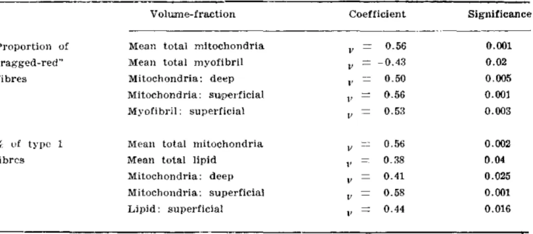
One way analysis of variance was used to compare the differences in mean volume fractions in different muscles biopsied: left or right side, males or females, and the threq clinical groups. None of these factors showed a difference in mean volumefractions at the 95% level of significance. Using the Pearson correlation coefficient, positive correlations were found when values for the proportion of fibres having increased oxidative enzyme activity at light microscopy ( J 2 ) were tested against the mean of the total mitochondrial volumefraction (p==0.001), the total volumefraction of the deep sited mitochondria ( p = 0 . 0 0 5 ) , and the volumefraction of the superficial mitochondria ( p = 0 . 0 0 1 ) . A negative correlation was found between the proportion of those «raggedred» fibres and the mean volumefraction of the total myofibrils ( p = 0 . 0 2 ) , and a higher negative correlation with the volumefraction of the superficial myofibrils ( p = 0 . 0 0 3 ) . T h e proportion of type 1 fibre at light microscopy ( J * ? )
correlated positively with the mean of the total mitochondrial volumefraction (p 0.002), the total lipid volumefraction (p = 0.04), the deeply placed mitochondrial volumefraction
(p = 0.05), the superficial mitochondrial volumefraction (p — 0.001) and the superficial lipid volumefraction (p = 0.016) (Table 3 ) . The mean of the total volumefraction of mitochondria correlated positively with the mean of the total lipid volumefraction (p = 0.001), but negatively with the mean of the total myofibril volumefraction (p = 0.17). The mean of the total myofibril volumefraction showed a negative correlation with the mean of the total glycogen volumefraction (p < = 0.001) (Table 4 ) .
Volumefraction Coefficient Significance
Proportion of Mean total mitochondria
V
=
0.56 0.001"raggedred" Mean total myofibril
V 0 . 4 3 0.02
fibres Mitochondria: deep
V
=
0.50 0.005Mitochondria : superficial
V
=
0.56 0.001Myofibril : superficial
V — 0.53 0.003
% of type 1 Mean total mitochondria
V 0.56 0.002
fibres Mean total lipid
V 0.38 0.04
Mitochondria: deep
V
=
0.41 0.025Mitochondria: superficial
V 0.58 0.001
Lipid: superficial
V
=
0.44 0.016Table 3 — Correlation coefficients: light and electronmicroscopy.
Volumefraction Coefficient Significance
Mean total Mean total lipids V
=
0.56 0.001mitochondria
Mean total Mean total myofibrils v — 0 . 4 2 0.017
mitochondria
Mean total Mean total glycogen v = 0 . 0 8 0.001
myofibril
Table k — Correlation coefficient: proportion of total volume-fractions of the muscle fibre constituents.
C O M M E N T S
mality of mitochondria 22. Another criterion used in assessing mitochondrial
abnor-malities is the alteration in the volume proportions of mitochondria relative to other
subcellular components, as determined by quantitative methods of analysis
(volume-fraction). The adoption of such methods is made necessary because of the normal
variation in the number of mitochondria found in the different sites within individual
myofibres and in different muscle fibre types
37,39,42.Only values greater than one
standard deviation above the mean mitochondrial volume-fraction found in normal
muscles, used as controls, were regarded as abnormal. No single criterion could be
used alone, however, to define the presence of mitochondrial abnormalities.
Accumu-lations of mitochondria as shown by light microscopy as "ragged-red" fibres with
the modified Gomori trichrome method or by the increased succinic dehydrogenase
activity, have been widely described in muscle biopsies. The abnormal structure of
the mitochondria has been shown by electronmicroscopy. These abnormalities have
been reported sporadically in cases where there is no reason to suspect a metabolic
mitochondrial defect, but they are likely to be due to secondary degenerative changes
in the mitochondria. However, in certain conditions there is now good evidence to
indicate that changes in mitochondrial structure do reflect an abnormality in one of
the pathways of the oxidative metabolism. Some of these cases had already a defined
biochemical defect such as in mitochondrial substrate transport 9, mitochondrial substrate
utilization
2325,defect in electron transfer chain
31,33,34,45,47,and a defect of energy
conservation and transduction
1.10,14,15,28. A variety of clinical presentations has beendescribed in association with a biochemical defined mitochondrial defect, including
cases of early or late onset myopathy and fatigability, and cases of myopathy and
signs of multisystem involvement such as cataracts, growth retardations, cerebellar
ataxia, neurosensory loss, pigmentary retinal changes, optic atrophy, mental retardation
or regression, and episodic severe metabolic acidosis. In our studies, the mitochondrial
morphological abnormalities have been associated with all of different clinical
presen-tations of CPEO. These confirmed earlier studies that showed mitochondrial
abnor-malities in sporadic and early onset 3.6, sporadic and late onset 38, familiar and early
onset with involvement of other organs or systems
17,27,43and familial and late onset
with muscle weakness only 20. The present data support the concept that the presence
of "ragged red" fibres and their ultrastructure counterparts, when they are the maior
or exclusive feature in a muscle biopsy from patients with the syndrome of CPEO,
indicate mitochondrial myopathy.
Qualitative electronmicroscopy studies confirmed the presence of mitochondrial
abnormalities in biopsies containing fibres with increased oxidative enzyme activity
characterized on histochemical preparations, and permitted the identification of two
further patients having mitochondrial abnormalities in the absence of those fibres.
One of these cases was a genetically determined myopathy with vacuolated fibres
in light microscope, evident mitochondrial abnormalities was found in his biopsy on
electronmicroscope, and further confirmed by the quantitative analysis. Similar case
has been reported 20 on qualitative studies. Quantitative electronmicroscopy also
revealed increase in the volume-fraction of normal looking mitochondria in another
case. The difficulties experienced when attempting to classify patients with CPEO
is exemplified by one of our cases, which by clinical, light microscopy, and qualitative
electronmicroscopy criteria, appeared to fall within group B, and yet showed a
consi-derable increase in mitochondrial volume-fraction (on quantitative analysis). It may
be postulated that the increase in mitochondrial volume-fraction detected is related
to the predominance of type 1 fibres in the muscle sampled, but other biopsies having
a similar high pronort'on of tvpe 1 fibre have yielded lower values for mitochondrial
volume-fraction. Mitochondrial abnormalities and vacuoles filled with myelin figures
may co-exist in the same biopsy indicating that neither is a specific feature of any
particular clinical group. While it is accepted that "ragged-red" fibres can present
in the muscles of some patients as a non-specific degenerative change 26, this does
not explain the increased mitochondrial volume-fraction found in quantitative studies
in one of our cases from group B. It is therefore not possible to fix precise Iinrts
for the clinico-pathological groups defined earlier, and overlapping cases (one out
of group A and four from group B) will occur where more than one criterion is used
for classification. Nevertheless groups A and C were homogenous groups on the basis
of the qualitative light and electronmicroscopy criteria chosen.
in Sudan black-stained preparations, suggesting the existence of a disturbance of lipid
metabolism in the group of patients with an abnormal mitochondrial morphology.
It has been pointed out 7 that the abnormal storage of neutral lipids in the muscle
fibres of these patients may be an expression of an impaired capacity of the abnormal
mitochondria to metabolize neutral fats. The overall increase in "glycogen content,
while not correlating directly with the increased mitochondrial volume-fractions found,
suggests a disturbance of muscle fibre metabolism involving either impaired glycogen
breakdown or utilization. Several mild non-specific features of muscle fibre
degene-ration were found in some cases belonging to all three clinical groups: atrophic fibres
(some rounded and others angulated), folded and reduplicated basement membrane
and mild dilatation of the sarcotubular system. Only a few biopsies showed a more
advanced stage of frank degeneration with necrotic fibres containing macrophages.
Although atrophic angulated muscle fibres are usually described as a sign of
dener-vation, no direct evidence of denervation was found in the few intramuscular nerve
terminals examined. The significance of the loss of nonmyelinated fibres observed
in the intrafascicular nerve bundles of two cases from group B is not known and
no loss of sensation or vasomotor regulation was detected in these two cases.
There-fore our data do not support the findings of denervation found by other observers
4 1.
The miscellaneous collections of inclusions found within the myofibres of a number
of biopsies were non-specific. The consistent thickening and reduplication of the
capillary basal lamina has been reported as a non-specific finding in inflammatory
myopathies !9, and in metabolic and degenerative diseases
4 6. The findings of
redu-plication of basal lamina, mast cells and leucocytes may indicate a minor inflammatory
component in some stage of this disease, confirming the light microscopy findings in
one case reported
4.
The morphological abnormalities seen on light and electronmicroscopy are
definitely indicative of at least two different groups of disease mechanisms on patients
presenting as chronic progressive external ophthalmoplegia. Although in those patients
with positive family story without ragged-red fibres (group B in our study) have
been reported nuclear inclusions, said to be characteristic of the disease
29.30,44the
morphological basis are not enough to differenciate between the groups of mitochondrial
myopathies n.12,30. Each additional step of our morphological analysis showed that
different disease entities could be found within CPEO group and it is suggested that
further techniques are required to define fully the pathogenic mechanisms in these
patients.
Aknowledgements — G r a t e f u l a k n o w l e d g e m e n t i s m a d e t o D r . D . N . L a n d o n a n d D r .
J . A . M o r g a n - H u g h e s f o r c o l l a b o r a t i o n in t h i s s t u d y w i t h t h e i r l a b o r a t o r y f a c i l i t i e s a n d h e l p f u l d i s c u s s i o n . I a m g r a t e f u l t o M i s s E . P a u l f o r h e r h e l p w i t h t h e s t a t i s t i c s a n a l y s i s , t o M r . B . Y o u n g a n d M i s s L . C o l l i n s f o r e x c e l e n t t e c h n i c a l a s s i s t a n c e . I a l s o t h a n k t h e p h y s i c i a n s o f t h e N a t i o n a l H o s p i t a l f o r N e r v o u s D i s e a s e s w h o a l l o w e d m e t o s t u d y p a t i e n t s u n d e r t h e i r c a r e . T h e a u t h o r w a s s u p p o r t e d d u r i n g p r e p a r a t i o n o f t h i s w o r k b y t h e B r a z i l i a n G o v e r n m e n t .
R E F E R E N C E S
1. A f i f i A K , I b r a h i m M Z M , B e r g m a n R A , H a y d a r N A , M i r e J, B a h u t N , K a y l a n i F — M o r p h o l o g i c f e a t u r e s o f h y p e r m e t a b o l i c m i t o c h o n d r i a l d i s e a s e : a l i g h t m i c r o s c o p i c , h i s t o c h e m i c a l a n d e l e c t r o n m i c r o s c o p i c s t u d y . J N e u r o l S c i 15 : 271, 1972.
2. B a s t i a e n s e n L A K , J o o s t e n E M G , d e R o o i j J A M , H o m m e s O R , S t a d h o u d e r s A M , J a s p a r H H J , V e e r k a m p J H , B o o k e l m a n H , v a n H i n s b e r g h V W M — O p h t h a l m o p l e g i a - p l u s , a r e a l n o s o l o g i c a l e n t i t y . A c t a N e u r o l S c a n d 58 : 9, 1978.
3. B e r e n b e r g R A , P e l l o c k J M , D i M a u r o S, S c h o t t a n d D L , B o n i l l a E , E a s t w o o d A , H a y s A , V i c a l e C T , B e h r e n M , C h u t o r i a n A , R o w l a n d L P — L u m p i n g o r s p l i t t i n g ? « O p h t h a l -m o p l e g i a - p l u s » o r K e a r n s - S a y r e . s y n d r o -m e ? A n n N e u r o l 1 : 37, 1977.
4. B o s h E P , G o w a n s J D C , M u n s a t J — I n f l a m m a t o r y m y o p a t h y i n o c u l o p h a r y n g e a l d y s t r o p h y . M u s c l e N e r v e 2 : 73, 1979.
6. C a s t a i g n e P , L h e r m i t t e F , E s c o u r o l l e R , C h a i n F , F a r d e a u M , H a u w J J , C u r e t J,
F l a v i g n y C — E t u d e a n a t o m o - c l i n i q u e d ' u n e o b s e r v a t i o n d' « o p h t h a l m o p l e g i a - p l u s » a v e c a n a l y s e d e s l é s i o n s m u s c u l a i r e s , n e r v e u s e s c e n t r a l e s , o c u l a i r e s , m y o c a r d i q u e s e t t h y r o ο
-d i e n n e s . R e v N e u r o l 133 : 369, 1977.
7. C o l e m a n R F , N i e n h u i s A W , B r o w n W J , M u n s a t T L , P e a r s o n C M — N e w m y o p a t h y
w i t h m i t o c h o n d r i a l e n z y m e h y p e r a c t i v i t y . J A M A 199 :118, 1967.
8. C u l l e n M J , W e i g h t m a n D . — T h e u l t r a s t r u c t u r e o f n o r m a l h u m a n m u s c l e in r e l a t i o n s
o f f i b r e t y p e . J N e u r o l Sci 25 : 43, 1975.
9. D i D o n a t o S, C o r n e l i o F , B a l e s t r i n i M R , B e r t a g n o l i o B , P e l u c h e t t i D — M i t o c h o n d r i a —
l i p i d - g l y c o g e n m y o p a t h y , h i p e r l a c t a c i d e m i a a n d c a r n i t i n e d e f i c i e n c y . N e u r o l o g y 28 : 1110,
1978.
10. D i M a u r o S, B o n i l l a E , L e e C P , S c h o t l a n d D L , S c a r p a A , C o n n H , C h a n c e B . — L u f t ' s
d i s e a s e : f u r t h e r b i o c h e m i c a l a n d u l t r a s t r u c t u r a l s t u d i e s o f s k e l e t a l m u s c l e i n t h e s e c o n d
c a s e . J N e u r o l S c i 27 : 217, 1976.
11. D i M a u r o S, B o n i l l a E , Z e v i a n i M , N a k a g a w a M , D e V i v o D C — M i t o c h o n d r i a l m y o p a t h i e s . A n n N e u r o l 17 : 521, 1985.
12. D i a s - T o s t a E . — C h r o n i c p r o g r e s s i v e e x t e r n a l o p h t h a l m o p l e g i a : I . A q u a n t i t a t i v e h i s t o - ¬
c h e m i c a l s t u d y o f s k e l e t a l m u s c l e s . A r q N e u r o - P s i q u i a t ( S ã o P a u l o ) 46 : 133, 1988.
13. E i s e n b e r g B , K u d a A , P e t e r J B — S t e r e o l o g i c a l a n a l y s i s o f m a m m a l i a n s k e l e t a l m u s c l e :
1. S o l e u s m u s c l e o f t h e a d u l t g u i n e a p i g . J C e l l B i o l 60 : 732, 1974.
14. E r n s t e r L , I k k o s D , L u f t R . — E n z y m i c a c t i v i t i e s o f h u m a n s k e l e t a l m u s c l e m i t o
-c h o n d r i a : a t o o l in -c l i n i -c a l m e t a b o l i -c r e s e a r -c h . N a t u r e 184 : 1851, 1959.
15. H a y d a r N A , C o n n H L , A f i f i A , W a k i d N , B a i l a s S, F a w a y K — S e v e r e h y p e r -m e t a b o l i s -m w i t h p r i -m a r y a b n o r -m a l i t y o f s k e l e t a l -m u s c l e -m i t o c h o n d r i a . A n n I n t M e d
74 : 548, 1971.
16. H o p p e l e r H , L ٧ t h i P , C l a a s s e n H , W e i b e l E R , H o w a l d H . — T h e u l t r a s t r u c t u r e o f t h e
n o r m a l h u m a n s k e l e t a l m u s c l e . P f l ٧ g e r s A r c h 344 : 217, 1973.
17. J a n k o w i c z E , B e r g e r H , K u r a s z S, W i n o g r o d z k a W , E l g a s z L — F a m i l i a l p r o g r e s s i v e
e x t e r n a l o p h t h a l m o p l e g i a w i t h a b n o r m a l m u s c l e m i t o c h o n d r i a . E u r o p N e u r o l 15 : 318, 1977.
18. J e r u s a l e m F , E n g e l A G , P e t e r s o n H A — H u m a n m u s c l e f i b r e f i n e s t r u c t u r e : m o r p h o - ¬
m e t r i c d a t a o n c o n t r o l s . N e u r o l o g y 25 : 127, 1975.
19. J e r u s a l e m F , R a k u s a M , E n g e l A G , M a c D o n a l d R D — M o r p h o m e t r i c a n a l y s i s o f s k e l e t a l
m u s c l e c a p i l l a r y u l t r a s t r u c t u r e in i n f l a m m a t o r y m y o p a t h i e s . J N e u r o l S c i 23:391, 1974.
20. J u l i e n J, V i t a l C, V a l l a t J M , V a l l a t M , D e B l a n c M — O c u l o p h a r y n g e a l m u s c u l a r d y s t r o p h y :
a c a s e w i t h a b n o r m a l m i t o c h o n d r i a a n d « f i n g e r p r i n t » i n c l u s i o n s . J N e u r o l S c i 21 : 165, 1974.
21. K a m i e n i e c k a Z — M y o p a t h i e s w i t h a b n o r m a l m i t o c h o n d r i a : a c l i n i c a l h i s t o l o g i c a l a n d
e l e c t r o p h y s i o l o g i c a l s t u d y . A c t a N e u r o l S c a n d 55 : 57, 1976.
22. K a m i n i e c k a Z , S c h m a l b r u c h H — N e u r o m u s c u l a r d i s o r d e r s w i t h a b n o r m a l m u s c l e m i t o
-c h o n d r i a . I n t R e v C y t o l 65 : 321, 1980.
23. K a r k R A P , R o d r i g u e z - B u d e l l i M — T h e s p e c t r u m o f a t a x i c s y n d r o m e s d u e t o l i p o a m i d e
d e h y d r o g e n a s e d e f i c i e n c y . N e u r o l o g y 27 : 359, 1977.
24. K a r k R A P , R o d r i g u e z - B u d e l l i M — P y r u v a t e d e h y d r o g e n a s e d e f i c i e n c y in s p i n o c e r e b e l l a r
d e g e n e r a t i o n . N e u r o l o g y 29 : 126, 1979.
25. K a r k R A P , R o d r i g u e z - B u d e l l i M — C l i n i c a l c o r r e l a t i o n s o f p a r t i a l d e f i c i e n c y o f l i p o a m i d e
d e h y d r o g e n a s e . N e u r o l o g y 29 : 1006, 1979.
27. L e v e i l l e A S , N e w e l l F W — A u t o s o n a l d o m i n a n t K e a r n s - S a y r e s y n d r o m e . O p h t h a l m o l
87 : 99, 1980.
28. L u f t R , I k k o s D , P a l m i e r i G, E r n s t e r L , A f z e l i n s B — A c a s e o f s e v e r e h y p e r m e t a b o l i s m
o f n o n t h y r o i d o r i g i n w i t h a d e f e c t in t h e m a i n t e n a n c e o f m i t o c h o n d r i a l r e s p i r a t o r y c o n t r o l : a c o r r e l a t e d c l i n i c a l , b i o c h e m i c a l a n d m o r p h o l o g i c a l s t u d y . J C l i n I n v 41 : 1776, 1962.
29. M a r t i n J J R , C e n t e r i c k C M , M e r c e l i s R J — N u c l e a r i n c l u s i o n s in o c u l o p h a r y n g e a l m u s c u l a r d y s t r o p h y . M u s c l e N e r v e 5 : 735, 1982.
30. M i t s u m o t o H , A p r i l l e J R , W r a y S, N e m n i R , B r a d l e y W G — C h r o n i c p r o g r e s s i v e e x t e r n a l
o p h t h a l m o p l e g i a ( C P E O ) : c l i n i c a l , m o r p h o l o g i c a n d b i o c h e m i c a l s t u d i e s . N e u r o l o g y 3 3 : 452, 1983.
31. M o n e n s L , G a b r e e l s F , W i l l e m s J L — A m e t a b o l i c m y o p a t h y a s s o c i a t e d w i t h c h r o n i c l a c t i c a c i d e m i a , g r o w t h f a i l u r e a n d n e r v e d e a f n e s s . J P e d i a t r 86 : 983, 1975.
32. M o r g a n - H u g h e s J A — M i t o c h o n d r i a l m y o p a t h i e s . I n M a s t a g l i a F L , W a l t o n J N ( e d s ) :
S k e l e t a l M u s c l e P a t h o l o g y . C h u r c h i l l L i v i n g s t o n e , L o n d o n , 1982, p g 309.
33. M o r g a n - H u g h e s J A , D a r v e n i z a P , K a h n S N , L a n d o n D N , S h e r r a t R M , L a n d J M , C l a r k
J B — A m i t o c h o n d r i a l m y o p a t h y c h a r a c t e r i z e d b y a d e f i c i e n c y in r e d u c i b l e c y t o c h r o m e b . B r a i n 100 : 617, 1977.
34. M o r g a n - H u g h e s J A , D a r v e n i z a P , L a n d o n D N , L a n d J M , C l a r k J B — A m i t o c h o n d r i a l
m y o p a t h y w i t h a d e f i c i e n c y o f r e s p i r a t o r y c h a i n N a D H - C o Q r e d u c t a s e a c t i v i t y . J N e u r o l S c i 43 : 27, 1979.
35. M o r g a n H u g h e s J A , M a i r W G P — A t y p i c a l m u s c l e m i t o c h o n d r i a i n o c u l o s k e l e t a l m y o p a -t h y . B r a i n 96 :215, 1973.
36. N e v i l l e H E , B r o o k e M H — M u s c l e b i o p s y in t h e d i a g n o s i s o f o c u l o p h a r y n g e a l m y o p a t h y .
J N e u r o p a t h o l E x p N e u r o l 33 : 193, 1974.
37. O g a t a T , M u r a t a F — C y t o l o g i c a l f e a t u r e s o f t h r e e f i b r e t y p e s in h u m a n s t r i a t e d m u s c l e .
T o h o k u J E x p M e d 99 : 225, 1969.
38. P a l m u c c i L , B e r t o l o t t o A , C a v i c c h i o l i D , M o n g a G, S c h i e f f e r D — S p o r a d i c o c u l o p h a r y n g e a l m y o p a t h y w i t h a b n o r m a l m i t o c h o n d r i a . A c t a N e u r o l B e l g 78 : 373, 1978.
39. P a y n e C M , S t e r n s L Z , C u r l e s s R G , H a n n a p e l L K — U l t r a s t r u c t u r a l f i b r e t y p i n g in
n o r m a l a n d d i s e a s e d h u m a n m u s c l e . J N e u r o l S c i 25 : 99, 1975.
40. P e l l e g r i n i G, V a l l i G, S e r g i P , M o g g i o M , S c a r l a t o G — O p h t h a l m o p l e g i a - p l u s : a
m u l t i s y s t e m d i s o r d e r o f u n k n o w n e t i o p a t h o g e n e s i s . I t a l J N e u r o l S c i 2 : 85, 1980.
41. P r o b s t A , T a c k m a n n W , S t o e c k l i H R , J e r u s a l e m F , U l r i c h J — E v i d e n c e f o r a c h r o n i c
a x o n a l a t r o p h y in o c u l o p h a r y n g e a l « m u s c u l a r d y s t r o p h y » . A c t a N e u r o p a t h o l 57 : 209, 1982.
42. S h a f i q S A , G o r y c k i M A , G o l d s t o n e L , M i l h o r a t A T — F i n e s t r u c t u r e o f f i b r e t y p e s in
n o r m a l h u m a n m u s c l e . A n a t Rec 156 : 283, 1966.
43. S c h n i t z l e r E R , R o b e r t s o n W C — F a m i l i a l K e a r n s - S a y r e s y n d r o m e . N e u r o l o g y 29 : 1172, 1979.
44. T o m é F M S , F a r d e a u M — N u c l e a r i n c l u s i o n s in o c u l o p h a r y n g e a l d y s t r o p h y . A c t a N e u r o - ¬
p a t h o l o g i c a 49 : 85, 1980.
45. v a n B i e r v l i e t J P G M , B r u i n v i s L , K e t t i n g D , D e B r e e P K , v a n d e r H e i d e n C, W a d m a n
S K , W i l l e m s J L , B o o k e l m a n H , v a n H a e l s t V , M o n n e n s A H — H e r e d i t a r y m i t o c h o n d r i a l m y o p a t h y w i t h l a c t i c a c i d e m i a , a D e T o n i - F a n c o n i - D e b r é s y n d r o m e a n d a d e f e c t i v e
r e s p i r a t o r y c h a i n in v o l u n t a r y s t r i a t e d m u s c l e s . P e d i a t r R e s 11 : 1088, 1977.
46. V r a c k o R — S k e l e t a l m u s c l e c a p i l l a r i e s in d i a b e t i c s : a q u a n t i t a t i v e , a n a l y s i s . C i r c u l a t i o n 41 : 271, 1970.
47. W i l l e m s J D , M o n n e n s L A H , T r i j b e l s J M F , V e e r k a m p J H , M e y e r A E F H , v a n D a m K ,