Correspondence: Profa. Alexandra Mussolino de Queiroz, Departamento de Clínica Infantil, Odontologia Preventiva e Social, Faculdade de Odontologia de Ribeirão Preto, Universidade de São Paulo, Avenida do Café S/N, 14040-904 Ribeirão Preto, SP, Brasil. Tel: +55-16-3602-4116. Fax: +55-16-3633-0999. e-mail: [email protected]
Biological Restorations as a Treatment Option
for Primary Molars with Extensive Coronal
Destruction - Report of Two Cases
Karina SANCHES
Fabrício Kitazono de CARVALHO Paulo NELSON-FILHO
Sada ASSED
Francisco Wanderley Garcia de Paula e SILVA Alexandra Mussolino de QUEIROZ
Department of Pediatric Clinics, Preventive and Community Dentistry, School of Dentistry of Ribeirão Preto, University of São Paulo, Ribeirão Preto, SP, Brazil
This article reports the cases of two young children aged 4 and 5 years, in whom biological restorations using tooth fragments were placed in primary molars with severely damaged crowns due to extensive carious lesions. After radiographic and clinical evaluation, tooth fragments obtained from extracted teeth in stock were autoclaved, adjusted to the prepared cavity and bonded to the remaining tooth structure with either adhesive system (Case 1) or dual-cure resin-based cement (Case 2) over a calcium hydroxide layer and a glass ionomer cement base. Occlusal adjustment was performed and topical sodium fluoride was applied to tooth surface. Periodical clinical and radiographic controls were carried out and the restored teeth were followed up for 4 and 3 years, respectively, until exfoliation. In these two reports, the technical aspects are described and the benefits and disadvantages of biological restorations as an alternative treatment for rehabilitation of severely destroyed primary molars are discussed.
Key Words: biological restoration, primary teeth, fragment bonding, tooth fragment.
INTRODUCTION
Dental caries is the most prevalent disease in humans and manifests with an extremely high index in several countries, especially during early childhood. Primary molars with extensive carious lesions are rou-tinely observed in clinical practice and should be prop-erly restored to reestablish their anatomy and hence their masticatory, phonetic, esthetic and space-maintainer functions in the dental arches. In an attempt to widen, as biologically and conservatively as possible, the treat-ment options to rehabilitate severely destroyed tooth crowns, several authors have suggested the use of tooth structure as a restorative material (1-7).
The expression “biological restoration” was coined
by Santos and Bianchi (8), in 1991. This technique consists of bonding sterile dental fragments to teeth with large coronal destruction. Cavity preparation should be non-retentive and the fragment is retained with adhesive materials. Fragments obtained either from the patient or from a tooth bank may be used as a safe and reliable alternative to restore dental anatomy and func-tion with excellent biomechanical properties (2,9).
effectiveness, preservation of sound tooth structure, prevention of physiological wear, and no need of com-plex material resources (3,5,6,9,10).
This article describes two cases in which biologi-cal restorations using tooth fragments were placed in primary molars with severely damaged crowns due to extensive carious lesions.
CASE REPORT
Case 1
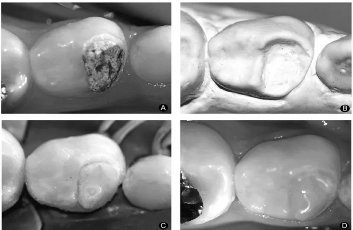
A 5-year-old patient was admitted to the Pediat-ric Dentistry Clinic of the School of Dentistry of Ribeirão Preto, University of São Paulo, Brazil, with an extensive carious lesion in the primary mandibular left first molar underneath a glass ionomer provisional restoration (Fig. 1A). After clinical and radiographic examinations, local anesthesia was given and a rubber dam was placed for isolation of the operative field. Cavity preparation was limited to removal of the glass ionomer restoration and carious tissue and flattening of cavity walls and margins. Retentive areas were eliminated. As the tooth had vital pulp, the cavity floor was protected with a calcium hydroxide cement layer (Dycal; Dentsply Ind. e Com. Ltda., Petrópolis, RJ, Brazil) and a glass ionomer cement base was built (Vitrebond, 3M/ESPE, St. Paul, MN, USA).
The rubber dam was removed and an impres-sion of the mandibular arch was taken using irrevers-ible hydrocolloid material (Jeltrate Plus; Dentsply Ind. e Com. Ltda.). A stone cast was obtained (Fig. 1B) and the mesiodistal, cervico-occlusal and buccolingual dimensions of the tooth were measured using a com-pass, in order to select an extracted tooth from stock, whose coronal dimensions best fitted the prepared tooth. Color matching was also taken into account.
A tooth was selected, decoronated and the coronal fragment was adjusted with diamond points at high-speed under air/water spray coolant until it fitted the cavity. Articulating paper was interposed between the fragment and the cavity in the stone cast to demarcate the areas that needed further adjustments. The prepared fragment was autoclaved at 120°C for 20 min.
In a second clinical appointment, a rubber dam was placed and, after prophylaxis, the adaptation of the fragment to the tooth was checked (Fig. 1C). Both the cavity and the fragment were etched with a 37%
phosphoric acid gel (Acid Gel; Dentalville, Joinville, SC, Brazil) during 30 s, rinsed and dried. A polyester matrix strip was placed and maintained with a wedge and Scotchbond multipurpose adhesive system (3M/ ESPE) was applied to the cavity and fragment, accord-ing to the manufacturer’s instructions. The fragment was adapted to the tooth and each surface was light-cured for 60 s (Fig. 1D). Small imperfections were corrected with light-curing composite resin (Z-250, 3M/ ESPE) and the occlusion was checked with articulating paper. Fluoride gel (Sultan-Topex; DFL, Rio de Janeiro, RJ, Brazil) was topically applied to tooth surfaces.
Case 2
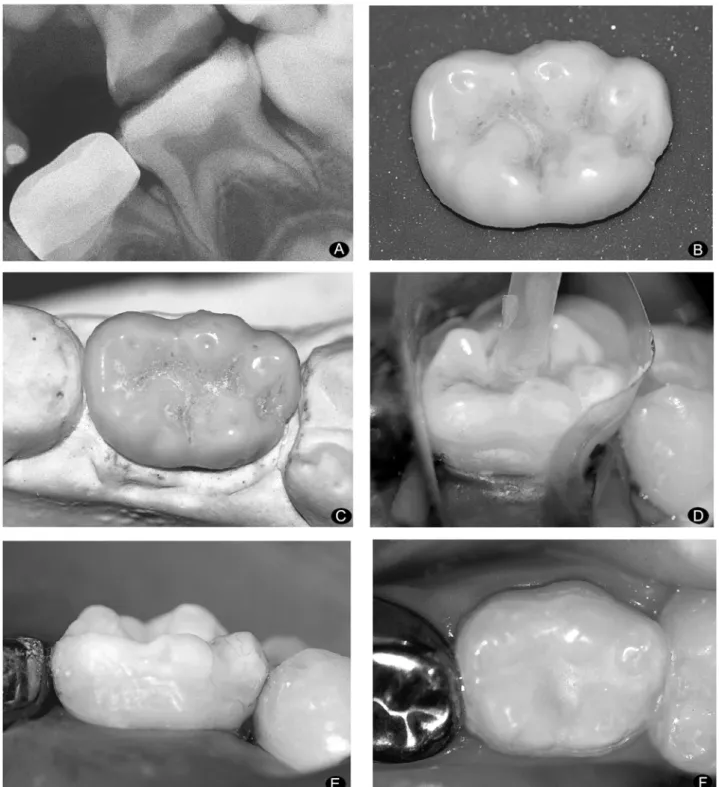
A child aged 4 years and 10 months was brought to our Pediatric Dentistry Clinic with a severely dam-aged primary mandibular left second molar due to an extensive carious lesion (Fig. 2A). Local anesthetic was administered and the tooth was isolated with a rubber dam. During caries excavation, it was noted that all cavity walls were involved, which did not allow placing an amalgam restoration. A biological restora-tion was proposed. The cavity floor was protected with a calcium hydroxide cement layer (Dycal, Dentsply Ind. Com. Ltda.) and a glass ionomer cement base (Vitrebond, 3M/ESPE) was built. In this case, how-ever, retention grooves were prepared because the amount of remaining tooth structure was not sufficient for adhesion. Impressions were taken from the maxillary and mandibular arches and an interocclusal wax record was obtained. The casts were mounted in a non-adjust-able articulator. The steps for selection of a tooth compatible with the remaining tooth structure, cutting and adaptation of the fragment to the stone cast (Figs. 2B and 2C) were the same as described for Case 1.
(Sultan-Topex, DFL) was applied (Fig. 2F).
In both cases described in this paper, the steps, benefits and disadvantages of the technique were fully explained to the parents and a signed, written informed consent was obtained. The parents were instructed to bring the children periodically for clinical and radio-graphic controls. The restored teeth were followed up for 4 and 3 years, respectively, until exfoliation. Post-treatment course was uneventful.
DISCUSSION
The use of bonded tooth fragments as biological restorations constitutes a viable restorative alternative for teeth with extensive coronal destruction. The tech-nique is simple, allows the preservation of sound tooth structure and provides excellent esthetics compared to composite resins and stainless steel crowns, especially regarding translucency. In addition, it allows mainte-nance of pulp vitality (11) and has low cost (12). An
advantage of using tooth fragments as restorative materials is that the enamel has physiologic wear and offers superficial smoothness and cervical adaptation compatible with those of surrounding teeth (6,9,13,14). Biological restorations not only mimic the missing part of the oral structures, but are also biofunctional (15). Clinical chairtime for fragment bonding proce-dures is relatively short, which is very interesting when treating pediatric patients (2,3,5,6,15). However, as any indirect restorations, biological restorations require a laboratorial phase that may become a critical step if not properly handled. Hence, in spite of being simple, the technique requires professional expertise to adequately prepare and adapt the natural crowns to the cavity.
Disadvantages of the biological restoration tech-nique include the difficulty in obtaining teeth with the required coronal dimensions and characteristics, prob-lems inherent to indirect restorations and matching fragment color with tooth remnant color. Also, having fragments from other people’s teeth in their mouth is
not a pleasant idea for some patients and many of them refuse to receive this treatment (2). However, all these factors are not contraindications of the technique.
It is important that the parents are informed that the tooth fragments used for biological restoration are previously submitted to a rigorous sterilization process
that completely eliminates any risk of contamination or disease transmission to the child receiving the fragment. Presently, secure methods of sterilization and storage are available to ensure the safety of teeth or tooth fragments coming from tooth banks (2,16,17).
Several materials have been used for bonding dental fragments to cavities, e.g., adhesive systems, composite resins, glass ionomer cements and dual-cure resin cements (2). In the cases hereby presented, the choice for each bonding material was based on frag-ment dimensions and bonding agent layer thickness. In Case 1, the fragment had small dimensions and hence the use of an adhesive system was the best choice because a thin bonding agent layer was required not to interfere with fragment adaptation. Scotchbond multi-purpose adhesive system produces a good homog-enous hybrid layer and similar characteristics, involv-ing resin penetration of peritubular and intertubular dentin matrix (18). In Case 2, a larger and thicker fragment was used and there was concern that optimal light-curing would not be achieved at the cavity gingival margin. Thus, a dual-cure resin-based cement was used to enhance polymerization at this region in addition to filling any possible gaps existing at tooth/fragment interface (7). An important point is that, regardless of the material used for fragment bonding, rubber dam placement is essential for a high-quality restoration. Periodical clinical-radiographic follow-up until primary tooth exfoliation is mandatory for long-term success. Based on the positive results in the literature (2,3,5-7,9) and on our own clinical experience, it may be concluded that the biological restoration technique using tooth fragments has a practical clinical applicabil-ity and is a viable, cost-effective restorative procedure for primary teeth with severely damaged crowns.
RESUMO
Este artigo descreve dois casos clínicos de reconstrução de molares decíduos com extensa destruição coronária por meio de restaurações biológicas, em crianças de 4 e 5 anos. Após avaliação clínica e radiográfica, os fragmentos dentais heterógenos foram submetidos à colagem ao remanescente dental preparado usando sistema adesivo (Caso 1) ou cimento resinoso de presa dual (Caso 2) sobre uma camada de hidróxido de cálcio e uma base de ionômero de vidro. Foi realizado ajuste oclusal e aplicação tópica de flúor sobre a superfície dentária. Controles clínico e radiográfico foram realizados periodicamente e os dentes restaurados foram acompanhados por 4 e 3 anos respectivamente, até a exfoliação. Por meio destes dois relatos, os autores discutem os aspectos técnicos, além das vantagens
e desvantagens das restaurações biológicas como tratamento alternativo para restauração de molares decíduos.
REFERENCES
1 . Andreasen FM, Noren JG, Andreasen JO, Engelhardtsen S, Lindh-Stromberg U. Long-term survival of fragment bonding in the treatment of fractured crowns: a multicenter clinical study. Quintessence Int 1995;26:669-681.
2 . Busato ALS, Loguercio AD, Barbosa NA, Sanseverino MCS, Macedo RP, Baldissera RA. Biological restorations using tooth fragments. Am J Dent 1998;11:46-48.
3 . Ramires-Romito ACD, Wanderley MT, Oliveira MDM, Imparato JCP, Côrrea MSNP. Biologic restoration of primary anterior teeth. Quintessence Int 2000;31:405-411. 4 . Olsburgh S, Jacoby T, Krejci I. Crown fractures in the
perma-nent dentition: pulpal and restorative considerations. Dent Traumatol 2002;18:103-115.
5 . Barcelos R, Neves AA, Primo L, Souza IPR. Biological resto-rations as an alternative treatment for primary posterior teeth. J Clin Pediatr Dent 2003;27:305-310.
6 . Mandroli PS. Biologic restoration of primary anterior teeth: a case report. J Indian Soc Pedod Prev Dent 2003;21:95-97. 7 . Terry DA. Adhesive reattachment of a tooth fragment: the biological restoration. Pract Proced Aesthet Dent 2003;15:403-409.
8 . Santos J, Bianchi J. Restoration of severely damaged teeth with resin bonding systems: case reports. Quintessence Int 1991;22:611-615.
9 . Pegoraro CN, Domingues LA, Trassi PMMM. Biological onlay: an alternative technique for restoration of severely damaged posterior tooth. A case report. Rev Dent Press Estét 2006;3:114.
10. Chosack ABDS, Eidelman EDO. Rehabilitation of a fractured incisor using the patient’s natural crown - case report. J Dent Child 1964;31:19-21.
11. Chu FCS, Yim TM, Wei SHY. Clinical considerations for reattachment of tooth fragments. Quintessence Int 2000;31:385-391.
12. Ehrmann EH. Restoration of a fractured incisor with exposed pulp using original tooth fragment: report of a case. J Am Dent Assoc 1989;118:183.
13. Sengun A, Ozer F, Unlu N, Ozturk B Shear bond strengths of tooth fragments reattached or restored. J. Oral Reabil 2003;30:82-86.
14. Imparato JCP, Tollara MN, Trindade CP, Bertolini PFR, Bussadori SK. Biological restorations an alternative for reha-bilitation of primary teeth, case report. Rev Paul Odont 2002;24:4-8.
15. Kapur A, Chawla HS, Goyal A, Gaube K. An esthetic point of view in very young children. J Clin Pediatr Dent 2005;30:99-103. 16. Yang ZP, Chang CS. A 3-year follow-up of a
homotransplanted tooth from a tooth bank. J Endod 1990;16:34-37.
17. Cru E, Carpenter WM. Extracted teeth - decontamination, disposal and use. J Cal Dent Assoc 1997;25:801-804. 18. Macari S, Gonçalves M, Nonaka T, Santos JM. Scanning
electron microscopy evaluation of the interface of three adhesive systems. Braz Dent J 2002;13:33-38.