Timing and sequence of primary tooth eruption
in children with cleft lip and palate
!"#!$!%&'(#)*%+,-.&./011, Márcia Ribeiro GOMIDE2, Cleide Felício de Carvalho CARRARA3
1- DDS, MSc, PhD student, Department of Pediatric Dentistry, Orthodontics and Public Health, Bauru School of Dentistry, University of São Paulo, Bauru, SP, Brazil.
2- DDS, MSc, PhD, Pediatric Dentistry Sector, Hospital for Rehabilitation of Craniofacial Anomalies, University of São Paulo, Bauru, SP, Brazil. 3- DDS, MSc, PhD student, Pediatric Dentistry Sector, Hospital for Rehabilitation of Craniofacial Anomalies, University of São Paulo, Bauru, SP, Brazil.
Corresponding address: Cleide Felício de Carvalho Carrara - Rua Silvio Marchione, 3-20 - 17012-900 - Setor de Odontopediatria - HRAC/USP - Bauru, SP - Brasil - Phone: (14) 32358141 / Fax (14) 3234-7828 - e-mail: [email protected]
2343#5367%/38"39:3(%;<=%;>>?%@%A*6#B4!"#*$7%C'DE%<F=%;>>F%@%.4438"367%G3:('!(E%<H=%;><>
O
bjective: To determine the timing and sequence of eruption of primary teeth in children with complete bilateral cleft lip and palate. Material and Methods: This cross-sectional study was conducted at the Hospital for Rehabilitation of Craniofacial Anomalies of the University of São Paulo, Bauru, SP, Brazil, with a sample of 395 children (128 girls and 267 boys) aged 0 to 48 months, with complete bilateral cleft lip and palate. Results: Children with complete bilateral clefts presented a higher mean age of eruption of all primary teeth for both arches and both genders, compared to children without clefts. This difference!"#"$!$%"$%&!''(#"%)*%+&!*$#,-.#!''#$//$01#/2&/3$#,-.#$0/#4!2%''!.(#+."$#4-'!.5#6/!*#!)/#-,#
eruption of most teeth was lower for girls compared to boys. The greatest delay was found for the maxillary lateral incisor, which was the eighth tooth of children with clefts of both genders. Analyzing by gender, the maxillary lateral incisor was the eighth tooth to erupt in girls and the last in boys. Conclusion: The results suggest an interference of the cleft on the timing and sequence of eruption of primary teeth.
Key words: Tooth eruption. Primary dentition. Cleft lip. Cleft palate.
ABSTRACT
INTRODUCTION
The period of eruption of primary teeth is %*78/*&/9# :(# "/;/.!'# ,!&$-."1# "8&0# !"# '/*)$0# -,#
gestational period25, disease7, gender3,14, race14,15,
nutrition and general growth10. In addition, cleft
lip and palate may also influence the timing
and sequence of eruption of both primary4,11,12,18
and permanent teeth2,13,21. This malformation
causes injury to the face in many different manners, depending on its occurrence, resulting in morphological and functional incapacity to variable extents. However, as the alveolar ridge is not always affected in all cleft types, it might be assumed that the different types of clefts may not exert the same %*78/*&/# -*# $--$0# /.83$%-*# %*# $0/"/# 3!$%/*$"1# !"#
observed by some authors5,22,23.
Only few studies have mentioned the timing of eruption of primary teeth in children with clefts.
Fishaman5 (1970) evaluated both primary and
permanent dentitions and observed an evident delay in the timing of tooth eruption in patients with different types of clefts, the greatest delay
occurring for bilateral clefts and for the maxillary lateral incisor on the cleft side.
Analyzing the influence of the cleft on the
eruption of maxillary incisors, Kramer, et al.12
<=>?>@#-:"/.;/9#!#"%)*%+&!*$#9/'!(#%*#$0/#/.83$%-*# of the lateral incisor on the cleft side. The delay increased when a palatal cleft was also present. A delay of approximately 8 months was observed in the eruption of primary maxillary lateral incisors when positioned at the distal segment in children with cleft lip and alveolus and 13 months in children with cleft lip and palate; when the lateral incisor is positioned at the premaxilla, the delay was
approximately 4 months longer. Kramer, et al.12
(1993) studied the eruption of primary canines and 4-'!."1#!*9#-:"/.;/9#$0!$#$0/#-*'(#"%)*%+&!*$#9/'!(# !,,/&$/9#$0/#+."$#4-'!."#-*#$0/#&'/,$#"%9/1#:-$0#%*# the maxilla and mandible, in complete unilateral
cleft lip and palate11.
Pöyry and Ranta19 (1985) evaluated the eruption
the cleft area erupted later when compared to their homologues on the noncleft side.
In order to contribute to the studies on tooth eruption, this study investigated the chronology and sequence of eruption of primary teeth in white Brazilian children with complete bilateral cleft lip and palate. The expected results for this study would be identifying differences between children with children complete bilateral cleft lip and palate, and children without cleft, on the issues studied.
MATERIAL AND METHODS
The study sample of this cross-sectional study was composed of 395 white Brazilian children (128 girls and 267 boys) aged 0 to 48 months with complete bilateral cleft lip and palate, attending the Hospital for Rehabilitation of Craniofacial Anomalies of the University of São Paulo (HRAC/USP), Bauru, SP, Brazil. The duration of the surgical protocol for correction of the lip is about 3 months and the palate is more than 12 months.
Syndromic children were not included in this
study because of possible influence on tooth eruption inherent to certain syndromes. The research project was approved by the HRAC/
USP’s institutional review board. Individuals were
considered white if they presented skin color ranging from white to dark and straight to lightly
curly or curly hair type1.
A previously trained single observer performed the examination under natural light. The teeth were considered as erupted whenever there was any portion of the crown showing above the gingival barrier. Teeth that were not present in the oral cavity were considered as unerupted unless the child’s caretaker provided information on tooth extraction. Natal and neonatal teeth were not considered in this study.
After data collection, children were assigned to 49 age and gender groups. The mean age of eruption for each tooth was calculated using the AB.:/.#4/$0-91#!"#4-9%+/9#:(#C!(/"#!*9#6!*$/'9
(1958). The results obtained for children with bilateral clefts were compared to the results of
Vono, et al.26 (1972). Children without clefts were
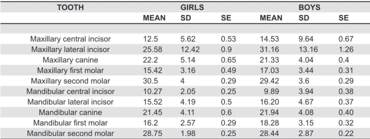
TOOTH GIRLS BOYS
MEAN SD SE MEAN SD SE
Maxillary central incisor 12.5 5.62 0.53 14.53 9.64 0.67 Maxillary lateral incisor 25.58 12.42 0.9 31.16 13.16 1.26 Maxillary canine 22.2 5.14 0.65 21.33 4.04 0.4 !"#$$!%&'(%)*'+,$!% 15.42 3.16 0.49 17.03 3.44 0.31 Maxillary second molar 30.5 4 0.29 29.42 3.6 0.29 Mandibular central incisor 10.27 2.05 0.25 9.89 3.94 0.38 Mandibular lateral incisor 15.52 4.19 0.5 16.20 4.67 0.37 Mandibular canine 21.45 4.11 0.6 21.94 4.08 0.40 !-.#/0$!%'(%)*'+,$!% 16.2 2.57 0.29 18.28 3.15 0.32 Mandibular second molar 28.75 1.98 0.25 28.44 2.87 0.22
Table 1- Mean, Standard Deviation (SD), and Standard Error (SE), in months, of eruption of maxillary and mandibular primary teeth in girls and boys
TOOTH GIRLS BOYS p Value
Maxillary central incisor 12.5 14.53 0.0181 Maxillary lateral incisor 25.58 31.16 0.0004
Maxillary canine 22.2 21.33 0.25ns
!"#$$!%&'(%)*'+,$!% 15.42 17.03 0.0061 Maxillary second molar 30.5 29.42 0.0088 Mandibular central incisor 10.27 9.89 0.40ns
Mandibular lateral incisor 15.52 16.20 0.27ns
Mandibular canine 21.45 21.94 0.50ns
!-.#/0$!%'(%)*'+,$!% 16.2 18.28 0.0000 Mandibular second molar 28.75 28.44 0.3529ns
Table 2-'1,+2!%#),-',3'+4!-'!54)',3'4%02*#,-',3'2%#+!%&'*44*6'3,%'5#%$)'74%)0)'/,&)8'9*!*#)*#:!$$&')#5-#(:!-*'.#334%4-:4' at p<0.05
taken as the control group.
Statistical analysis was performed using the D$89/*$E"# F$G# $/"$# !$# !# "%)*%+&!*&/# '/;/'# -,# H5HI# (p<0.05) to assess possible differences in the age of eruption of primary teeth between genders and between the cleft and control groups, for both the maxilla and mandible.
RESULTS
From the 395 patients examined in this study, 32.4% were girls and 67.6% were boys. The results obtained for the mean ages of eruption of primary teeth are presented in Table 1. Tables 2-4 present the results of comparisons of mean ages of eruption between genders and between children with and
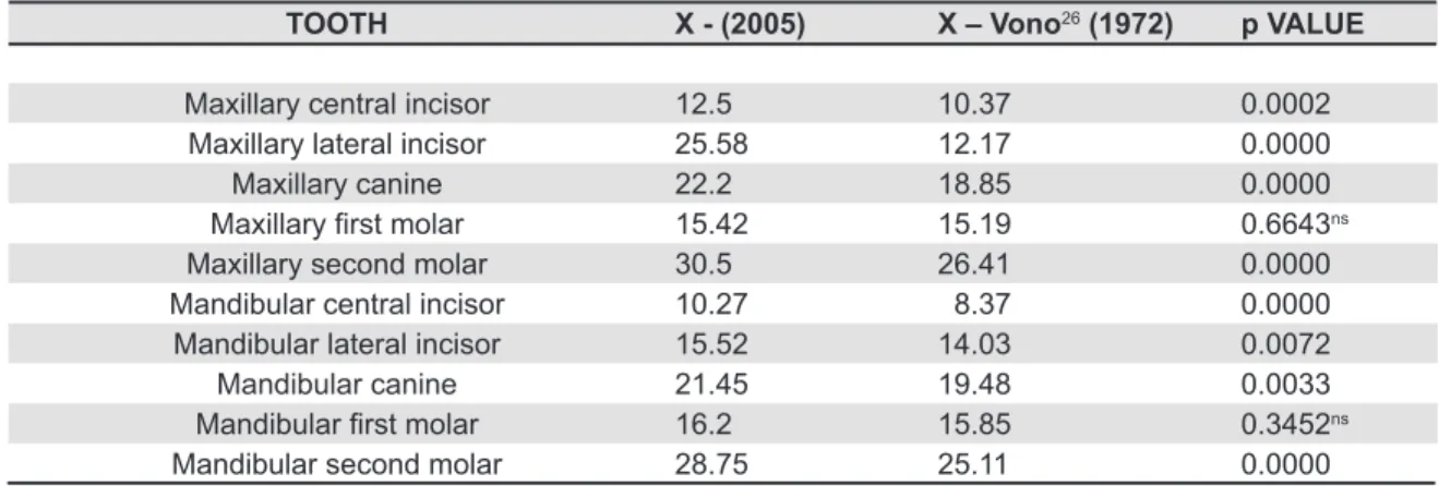
TOOTH X - (2005) X – Vono26 (1972) p VALUE
Maxillary central incisor 12.5 10.37 0.0002 Maxillary lateral incisor 25.58 12.17 0.0000
Maxillary canine 22.2 18.85 0.0000
!"#$$!%&'(%)*'+,$!% 15.42 15.19 0.6643ns
Maxillary second molar 30.5 26.41 0.0000
Mandibular central incisor 10.27 8.37 0.0000 Mandibular lateral incisor 15.52 14.03 0.0072
Mandibular canine 21.45 19.48 0.0033
!-.#/0$!%'(%)*'+,$!% 16.2 15.85 0.3452ns Mandibular second molar 28.75 25.11 0.0000
!"#" $ "!%& %'() *
Table 3- Comparison of mean ages of eruption of primary teeth of girls with complete bilateral cleft lip and palate (X-2005) versus girls without clefts (X-Vono26;'<=>?@8'9*!*#)*#:!$$&')#5-#(:!-*'.#334%4-:4'!*'2AB8BC
TOOTH X – (2005) X – Vono26 (1972) p VALUE
Maxillary central incisor 14.53 9.47 0.0000 Maxillary lateral incisor 31.16 11.21 0.0000
Maxillary canine 21.33 18.18 0.0000
!"#$$!%&'(%)*'+,$!% 17.03 15.62 0.0001 Maxillary second molar 29.42 26.72 0.0000 Mandibular central incisor 9.89 8.00 0.0000 Mandibular lateral incisor 16.20 13.00 0.0000
Mandibular canine 21.94 19.13 0.0000
!-.#/0$!%'(%)*'+,$!% 18.28 16.07 0.0000 Mandibular second molar 28.44 25.67 0.0000
Table 4- Comparison of mean ages of eruption of primary teeth of boys with complete bilateral cleft lip and palate (X-2005) versus boys without clefts (X-Vono26;'<=>?@8'9*!*#)*#:!$$&')#5-#(:!-*'.#334%4-:4'!*'2AB8BC
SEQUENCE GIRLS BOYS
1st Mandibular central incisor Mandibular central incisor 2nd Maxillary central incisor Maxillary central incisor D%.'' ' ' ' ' ' ' !"#$$!%&'(%)*'+,$!%' ' ' ' ' ' ' ' ' !-.#/0$!%'$!*4%!$'#-:#),% E*6'' ' ' ' ' ' ' !-.#/0$!%'$!*4%!$'#-:#),%' ' ' ' ' ' ' !"#$$!%&'(%)*'+,$!% C*6'' ' ' ' ' ' ' !-.#/0$!%'(%)*'+,$!%' ' ' ' ' ' ' ' !-.#/0$!%'(%)*'+,$!%
6th Mandibular canine Maxillary canine
7th Maxillary canine Mandibular canine
8th Maxillary lateral incisor Mandibular second molar 9th Mandibular second molar Maxillary second molar 10th Maxillary second molar Maxillary lateral incisor
without clefts. The sequence of eruption obtained for each gender is shown in Figure 1.
DISCUSSION
The timing and sequence of eruption of primary teeth may differ somewhat among populations. Studies have also shown a delay in eruption in patients with malformations and
malocclusion2,4,6,14,17,24.
A delay in eruption of primary teeth has been demonstrated in previous studies with children with
cleft lip and palate2,4,5,6,12.
There are several studies on tooth eruption and formation in patients with clefts, in which the authors did not separate the samples according
to cleft type5,8,20, which impairs comparisons of
the present results. Therefore, only patients with complete bilateral cleft lip and palate were evaluated in this study, so that the data obtained for the mean ages of tooth eruption were compared just between the genders and between the cleft and control groups. Considering that the presence of &'/,$#!*9#&0%'9E"#)/*9/.#4%)0$#%*78/*&/#$0/#$%4%*)# of tooth eruption, data were separated between genders.
Girls showed a lower mean age of eruption for most maxillary and mandibular teeth compared to :-("1# %$0#"%)*%+&!*$#9%,,/./*&/"#,-.#$0/#4!2%''!.(# &/*$.!'#%*&%"-."1#'!$/.!'#%*&%"-."#!*9#+."$#4-'!."#!*9# 4!*9%:8'!.#+."$#4-'!."5#J0/#4!2%''!.(#"/&-*9#4-'!.# /.83$/9# /!.'%/.# %*# :-("1# 0%&0# !"# "%)*%+&!*$'(# different (Table 2).
Gender differences in the timing of tooth eruption in children with bilateral clefts (i.e. earlier eruption in girls) were in agreement with previous "$89%/"#-,#&'/,$#3-38'!$%-*"1#&-*+.4%*)#$0/#*//9#-,#
quantitative rules for each gender2,4. However, some
teeth showed earlier eruption in boys, which can be K8"$%+/9#:(#$0/#'- /.#*84:/.#-,#)%.'"#/;!'8!$/9#%*# relation to the number of boys, due to the higher frequency of complete bilateral cleft lip and palate
among boys2,4.
Comparison of data between the study and control groups revealed that, in girls, eruption of !''# $//$0# !"# "%)*%+&!*$'(# 9/'!(/9# %*# $0/# ).-83# with cleft, except for the maxillary and mandibular +."$#4-'!."#<J!:'/#L@5#M*#$0/#-$0/.#0!*91#%*#:-("1# /.83$%-*#-,#!''#$//$0# !"#"%)*%+&!*$'(#9/'!(/9#%*#$0/# cleft group (Table 4).
J0/# 4-"$# "%)*%+&!*$# 9/'!(# %*# $0/# &'/,$# ).-83# compared to the control group was observed for the maxillary lateral incisor, which erupted 13.41 and 19.95 months later in girls and boys, respectively.
The delay in the mean age of eruption of the maxillary lateral incisor may have a multifactorial etiology and can also be related to the intrinsic and extrinsic factors that contribute with tooth
development of individuals to clefts. Local factors include scar tissue after surgical repair, delayed formation of crown and root, poor blood circulation
after surgery19, lack of space in the maxilla17 and
lack of bone support16.
The sequence of eruption of primary teeth in the study group varied between genders, as shown in Figure 1. The last tooth to erupt in girls was the maxillary second molar and the maxillary lateral incisor in boys. The sequence of tooth eruption in the mandible did not differ from the control group.
In the maxillary arch of girls, the canine erupted before the lateral incisor, whereas in the maxillary arch of the control group and boys, the canine and second molar erupted before the lateral incisor.
CONCLUSION
J0/#+*9%*)"#-,#$0/#3./"/*$#"$89(#"8))/"$#$0!$# the cleft affects the timing and sequence of eruption -,#3.%4!.(#$//$01# 0%&0#&-..-:-.!$/"#$0/#+*9%*)"# in the literature evaluating the chronological sequence of eruption on reliable samples of patients
with clefts2,4. Knowledge of the mean timing and
sequence of eruption of primary teeth in children with complete bilateral cleft lip and palate adds importance information to professionals working in the rehabilitation process of patients with malformations, in order to provide parents with information about inherent characteristics of these patients.
REFERENCES
1- Bastos de Avila J. Antropologia física. Rio de Janeiro: Agir; 1958. 2- Carvalho Carrara CF, Oliveira Lima JE, Carrara CE, Gonzalez Vono B. Chronology and sequence of eruption of the permanent teeth in patients with complete unilateral cleft lip and palate. Cleft Palate Craniofac J. 2004;41(6):642-5.
3- Dermirjian A, Levesque GY Sexual differences in dental development and prediction of emergence. J Dent Res. 1980;59(7):1110-22.
4- Duque C, Dalben GS, Aranha AM, Carrara CF, Gomide MR, Costa B. Chronology of deciduous teeth eruption in children with cleft lip and palate. Cleft Palate Craniofac J. 2003;41(3):73-7.
5- Fishman LS. Factors related to tooth number, eruption time, and tooth position in cleft palate individuals. ASDC J Dent Child. 1970;37(4):303-6.
6- Fuchslocher G, Blanco R. Analysis of permanent tooth eruption in cleft palate and normal individuals. Odontol Chil. 1988;36(1):27-32.
7- Galili G, Rosenzweig KA, Klein H. Eruption of primary teeth and general pathologic conditions. ASDC J Dent Child. 1969;36(1):51-4.
8- Haring FN. Dental development in cleft and noncleft subjects. Angle Orthod. 1976;46(1):47-50.
11- Kramer GJ, Hoeksma JB, Prahl-Andersen B. Emergence of the deciduous canines and molars in CLP children. Eur J Orthod. 1993;15(1):65-71.
12- Kramer GJ, Hoeksma JB, Prahl-Andersen B. Emergence of the deciduous incisors in CLP children. Eur J Orthod. 1989;11(3):265-70.
13- Loevy HT, Aduss H. Tooth maturation in cleft lip, cleft palate, or both. Cleft Palate J. 1988;25(4):342-7.
14- Lysell L, Magnusson B, Thilander B. Relations between the times of eruption of primary and permanent teeth. A longitudinal study. Acta Odont Scand. 1969;27(3):271-81.
15- Magnússon TE. Emergence of primary teeth and onset of dental stages in Icelandic children. Community Dent Oral Epidemiol. 1982;10(2):91-7.
16- Peterka M, Müllerová Z, Penkava J. Causes of the development of orthodontic anomalies in patients with a total unilateral cleft. Cesk Stomatol. 1980;80(2):100-9.
17- Peterka M, Tvrdek M, Müllerová Z. Tooth eruption in patients with cleft lip and palate. Acta Chir Plast. 1993;35(3-4):154-8. 18- Pöyry M. Prenatal factors and tooth eruption in children with oral clefts. ASDC J Dent Child. 1986;53(6):436-8.
19- Pöyry M, Ranta R. Emergence of deciduous teeth in children with oral clefts. Proc Finn Dent Soc. 1985;81(3):171-6. 20- Pöyry M, Ranta R. Formation of anterior maxillary teeth in 0-3-year-old children with cleft lip and palate and prenatal risk factors for delayed development. J Craniofac Genet Dev Biol. 1986;6(1):15-26.
21- Ranta R. A review of tooth formation in children with cleft lip/palate. Amer J Orthod Dentofacial Orthop. 1986;90(1):11-8. 22- Ranta R. Associations of some variables to tooth formation in children with isolate cleft palate. Scand J Dent Res. 1984;92(6):496-502.
23- Ranta R. Comparison of tooth formation in noncleft and cleft-affected children with or without hypodontia. ASDC J Dent Child 1982;49(3):197-9.
24- Ranta R. Eruption of premolars and canines and factors affecting it in unilateral cleft lip and palate cases: an orthopantomographic study. Swom Hammaslaak Toim. 1971;67(6):350-5.
25- Seow WK. Effects of preterm birth on oral growth and development. Aust Dent J. 1997;42(2):85-91.