Root canal irrigants are used to minimize the negative effects of smear layer on endodontic sealer retention. The aim of this study was to evaluate the efficacy of agitation of 17% ethylenediaminetetraacetic acid (EDTA) with ultrasonic, 1064-nm Nd:YAG and 980-nm diode laser on the retention of an epoxy-based sealer to the root canal walls. Forty single-rooted bovine teeth were instrumented with ProTaper rotary system and divided into four groups according to the final irrigation protocol (n = 10): (1) 17% EDTA (control); (2) 17% EDTA with 50-s ultrasonic agitation; (3) 17% EDTA with 50-s diode laser (2-W) agitation; and (4) 17% EDTA with 50-s Nd:YAG (1.5-W) laser agitation. After endodontic filling with gutta-percha F5 master cone and Sealer 26, the roots were sectioned at the cervical, middle, and apical root thirds to obtain 1.5-mm slices. Push-out tests were performed using a universal testing machine at a 1 mm/min crosshead speed. Data were analyzed using two-way ANOVA and Tukey’s tests (α=0.05). Apical root thirds had significant higher retention values than cervical and middle thirds (p < 0.05). EDTA with 1064-nm Nd:YAG or 980-nm diode laser presented the highest retention values and was significantly different from EDTA with ultrasonic agitation and EDTA only (p < 0.05). Adhesive failures were predominant to EDTA only group. Mixed failures were predominant to all agitation groups. 1064-nm Nd:YAG and 980-nm diode laser EDTA agitation enhanced the retention of the epoxy-based sealer to the root canal walls compared with that due to EDTA only or EDTA with ultrasonic agitation.
1064-nm Nd:YAG and 980-nm Diode
Laser EDTA Agitation on the Retention
of an Epoxy-Based Sealer to Root Dentin
Helena Suleiman de Macedo, Danielle Cristine Furtado Messias, Fuad Jacob Rached-Júnior, Ligia Teixeira de Oliveira, Yara Teresinha Correa Silva-Sousa, Walter Raucci-Neto
UNAERP - University of Ribeirão Preto, Ribeirão Preto, SP, Brazil
Correspondence: Prof. Dr. Walter Raucci Neto, Avenida Constábile Romano 2200, Ribeirânea, 14096-000, Ribeirão Preto, SP, Brazil. Tel: +55-16-3603-6763. e-mail: [email protected]
Key Words: laser irradiation; endodontics; lasers diode; lasers Nd-YAG.
Introduction
The biomechanical preparation of root canals led to a residual layer (smear layer) of organic and inorganic materials formed by cutting and detrition of root dentin (1). Advances in root canal chemical treatment have focused on minimizing the smear layer effects on sealers penetration to enhance the endodontic sealer retention to root canal (2).The endodontic sealer should have good retention because, under static conditions, it contributes to minimize or eliminate spaces that could serve as pathways for infiltration of fluids between the sealer and the radicular dentin walls (2).
Sodium hypochlorite (NaOCl) has been widely used as root chemical treatment due to its bactericidal effects and great capacity for organic tissue dissolution (3). However, due to its limited effects on inorganic materials from the smear layer and its cytotoxicity at higher concentrations (4), alternative solutions have been developed to complement root canal irrigation (5,6). Ethylenediamine tetraacetic acid (EDTA) was developed as a predictable means of canal irrigation for smear layer removal and a complement to NaOCl interaction with root canal systems, without significantly modifying dentin ultrastructure (7,8).
Previously, the enhanced action of irrigating agents has been demonstrate using different agitation strategies with the possibility of improve the contact of these substances to the root canal walls and promote changes in their chemical and physical properties (9,10).
Laser irradiation has already been evaluated on root dentin smear layer and debri removal (9,11), root canal system disinfection (12), and the root fracture resistance (13). Recently, the 1064-nm Nd:YAG and 980-nm diode laser EDTA agitation were evaluated considering the microhardness and roughness alteration. It was observed significant microhardness reduction and substantial modification of dentin topography (14). However, there is still some concern on the protocol and type of laser most suitable for promoting changes in the root dentin, without adversely compromising filling material retention (15,16). Considering its use as an alternative protocol in endodontic therapy, it would be interesting to evaluate how root dentin ultrastructure modifications affect the filling material retention.
Nd:Y
A
G and 980-nm diode laser EDT
A agitation
retention of an epoxy-based sealer to the root canal walls. The null hypothesis was that there are no differences in the push-out retention of filling materials at the different irrigations protocols.
Material and Methods
Study Design
The variable being examined was the sealer retention to the root canal walls. Additionally, the root thirds retention was also compared. There were 4 experimental groups (n
= 10): (1) 17% EDTA; (2) 17% EDTA with 50 s ultrasonic agitation; (3) 17% EDTA with 50 s 980-nm diode laser (2W) agitation; and (4) 17% EDTA with 50 s 1064-nm Nd:YAG (1.5 W) laser agitation.
Laser Systems
A 980-nm wavelength gallium-aluminum-arsenide diode laser (SIROlaser 2.2, SIRONA Dental, Bensheim, Germany) system equipped with a 20-W power source was used. The laser delivery system used in the study was a 300-µm sized fiber-optic cable, at 2 W in pulse mode (10 ms–100 Hz). The actual energy density of the parallel fiber-optic tip was 272.60 J/cm2 (14).
A 1064-nm wavelength yttrium-aluminum-garnet neodymium laser (SmartFile, DEKA, Italy) system was also used. The laser delivery system consisted of a fiber-optic cable with a 300-µm quartz fiber, at 1.5 W and in pulse mode (100 µs-15 Hz). The actual energy density of the parallel fiber-optic tip was 124.3 J/cm2 (14,17).
Specimen Preparation
Freshly extracted, single-rooted, bovine mandibular anterior teeth were stored in 0.1% thymol for disinfection and then washed in running water for 24 h. The teeth were examined under a stereomicroscope to select those with similar size, similar root morphology, and absence of cracks and structural defects. Periodontal tissues and calculus were removed mechanically from the root surfaces with a periodontal scaler. Forty specimens were selected (1.54 ± 0.42 mm of buccal-lingual root canal diameter and 0.47 ± 0.24 mm of mesiodistal root canal diameter) and sectioned transversally near the cementoenamel junction with a water-cooled diamond disc (KG Soresen, Barueri, Brazil) at slow speed, so as to remove the crown and standardize the root lengths at 18-mm. A #10K-file (Dentsply-Maillefer, Ballaigues, Switzerland) was passively introduced into each root canal to confirm the working length (17-mm). Teeth with laterally displaced foramina and/or canal length less than 17-mm were replaced. The canals were instrumented with the ProTaper-Universal (Dentsply-Maillefer). The cervical-third was prepared with the instrument SX and the middle and apical-thirds were
prepared with the instruments S1, S2, F1, F2, F3, F4 and F5. The canals were irrigated with 2-mL of 2.5%NaOCl at each file change using NaviTip needles coupled to plastic syringes (Ultradent-Products Inc.,South Jordan, Utah, USA). VDW Silver (VDW GmbH, Munich, Germany) was used to operate all files.
Irrigation Protocols
The root apex was closed with wax to prevent the irrigant from overflowing. The root was then dried with paper points (CellPack; Dentisply Ind. e Com, Petrópolis, RJ, Brazil) and final flushed as follows:
1) 17% EDTA only: The root canal was irrigated for 120 s with 10 mL 17% EDTA (Biodinâmica, Ibiporã, Brazil) as a final flush. Then the specimens were irrigated with 5 mL 2.5% NaOCl (Farmácia da Terra, Ribeirão Preto, Brazil) for 120 s. This was followed by a final rinse with 5 mL distilled water. The total volume of EDTA solution was 10 ml, the total exposure time to EDTA solution was 120 s (9,14), and the total volume of all irrigants was 20 mL.
2) 17% EDTA with ultrasonic agitation: The root canal was filled with 5 mL 17% EDTA, an E1 – Irrissonic tip (Helse Dental Technology – Santa Rosa do Viterbo, Brazil) was placed, and the solution was agitated for 50 s. The specimens were then irrigated with 5 mL 17% EDTA for 70 s, followed by 5 mL 2.5% NaOCl for 120 s. A final rinse was performed with 5 mL of deionized water. The total volume of EDTA solution was 10 mL, the total exposure time to EDTA solution was 120 s, and the total volume of all irrigants was 20 mL.
3) 17% EDTA with 980-nm diode laser: The root canal was filled with 1 mL 17% EDTA, and the laser fiber agitated the solution with 2 W in continuous waves for 10 s. This procedure was repeated five times, so that the total agitation was 50 s. The laser fiber had helical motion from the apical to cervical third. The specimens were then irrigated with 5 mL 17% EDTA for 70 s, followed by 5 mL 2.5% NaOCl for 120 s. A final rinse was performed with 5 mL of deionized water. The total volume of EDTA solution was 10 mL, the total exposure time to EDTA solution was 120 s, and the total volume of all irrigants was 20 mL.
H.S. de Macedo et al.
Root Canal Filling
The root canals were filled with F5 gutta-percha master cones (Dentsply-Maillefer) as single cone technique with Sealer 26 (Dentsply-Maillefer). The root sealer was manipulated according to the manufacture intructions and inserted into root canal with a lentulo spiral attached to a low-speed handpiece (Dabi-Atlante, Ribeirão Preto, Brazil) to avoid bubble formation. After insertion of the main gutta-percha cone, which was previously tested for apical fit, they were cut with a heated instrument and vertically condensed with a plugger (Maillefer, Ballaigues, Switzerland). Sealer excess was removed with cotton pellets. The canal entrances were sealed with a quick-setting temporary restorative filling material (Cimpat; Septodont Ltda., Barueri, Brazil) and the teeth were placed immediately at 37°C and 95% humidity for a period 3 times greater than the regular setting time of the sealer (135 min) before the retention test.
Push-out Test
The roots were fixed on acrylic plates using wax (Kota-Import, São Paulo, Brazil) and sectioned in a precision cutting machine (Minitom-Struers, Westlake, USA) at 375-rpm and water cooling. Nine 1.5-mm-thick slices were obtained from each root (3 per root-third), resulting in a total of 360 specimens.
The first slice of each third was selected and a stainless steel support was used to hold the specimens in an Instron 3345 universal testing machine (Instron-Corporation, Canton, USA) in such a way that the side with the smaller diameter of the root canal faced upwards and was aligned to the shaft that would exert pressure load on the sealer (apical-coronally) until debonding occurred. A 6-mm long shaft with tip diameter of 0.6-mm for the apical-third, 0.8-mm for the middle-third and 1-mm for the coronal-third were used. This method assured the alignment of the specimen in an accurate and reproducible manner and also maintained the shaft centralized to avoid its contact with the dentin during testing. The force needed to dislodge the filling material (F in kN) was transformed into tension (σ in
MPa) by dividing the force by the adhesive area of the filling material (SL in mm2), using the following equation: σ=F/A
After push-out test, the slices were examined with a stereomicroscope (ZEISS, Stemi 2000-C, Germany) at 25× to determine the failure pattern. Failure was considered adhesive if the sealer was totally separated from dentin (dentin surface without sealer), cohesive if the fracture occurred within the sealer (dentin surface totally covered by the sealer), and mixed when a mixture of adhesive and cohesive modes (dentin surface partially covered by the sealer) occurred.
Statistical Analysis
The Kolmogorov–Smirnov statistical test for normality revealed normal distribution for push-out data. Two-way ANOVA (α=0.001) and Tukey’s (α=0.05) tests were carried out for statistical analyses. All statistical analyses were performed using the SigmaStat software, version 3.5 (Systat Software Inc., Chicago, Illinois, USA). The failure pattern was qualitatively evaluated.
Results
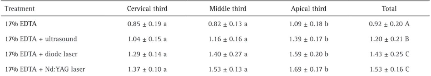
ANOVA showed significant statistical differences between irrigants protocols and the filling material retention (p=0.001). It was also observed significant differences between root canal thirds push-out values (p=0.001), the apical-third presented higher retention values compared to cervical and middle-thirds. EDTA+Diode (1.43±0.25) and EDTA+Nd:YAG (1.53±0.16) presented higher retention values than EDTA (0.92±0.20) (p=0.001). EDTA+Ultrasonic (1.20±0.21) presented lower retention values than laser groups and higher values than EDTA only group (0.92±0.20). The mean and standard deviations of the retention values (in MPa) for the displacement of the filling materials during the push-out test are presented in Table 1.
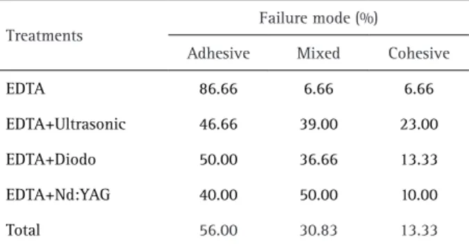
Analysis of the mode of failure showed a predominance of adhesive failures (86,66%) for EDTA only group, particularly at the sealer/dentin interface. There were many mixed failures in all EDTA agitation groups (ultrasonic,
Table 1. Mean and standard deviations of the retention values (in MPa) for the displacement of the filling materials during the push-out test in the cervical, middle and apical root thirds
Treatment Cervical third Middle third Apical third Total
17% EDTA 0.85 ± 0.19 a 0.82 ± 0.13 a 1.09 ± 0.18 b 0.92 ± 0.20 A
17% EDTA + ultrasound 1.04 ± 0.15 a 1.16 ± 0.16 a 1.39 ± 0.17 b 1.20 ± 0.21 B
17% EDTA + diode laser 1.29 ± 0.14 a 1.40 ± 0.27 a 1.59 ± 0.20 b 1.43 ± 0.25 C
17% EDTA + Nd:YAG laser 1.37 ± 0.10 a 1.53 ± 0.13 a 1.69 ± 0.17 b 1.53 ± 0.16 C
Nd:Y
A
G and 980-nm diode laser EDT
A agitation
diode and Nd:YAG). Cohesive failures were observed in 23% of the ultrasonic agitation specimens. The distribution of failure modes is presented in Table 2.
Discussion
In light of the test results, the null hypothesis that there is no difference in the push-out retention of filling materials at the different irrigations protocols has to be rejected. The root canal sealers retention depends on a multitude of interacting factors including the surface energy of the dentinal walls, the sealer’s surface tension, its ability to wet the surfaces, and the cleanliness of the adherent surface (18). As the agitation of irrigating solutions is used in endodontics to enhance physical and chemical changes in the ultrastructure of the root dentin, higher retention of filling materials was expected. Previous studies have already evaluated the effects of laser-activated irrigation (LAI) on root dentin microhardness (14), smear layer removal (9), dentinal debris removal (11), disinfection (12), and dentin roughness (14). However, there are divergent results on the agitation solution to the filling material retention to root canal walls.
The push-out test has been shown to be effective and reliable in the assessment of sealers retention to dentin, as it evaluates the material dislocation resistance (15). Furthermore, the push-out test causes failure parallel to the dentin/material interface, which is similar to that observed in clinical circumstances (16). In this study, the push-out test was used to compare the retention of different EDTA agitation techniques using an epoxy-based sealer.
The effectiveness of the NaOCl-EDTA combination in removing the smear layer from the root canal walls is well documented in endodontics (7,8,9).However, the EDTA agitation promotes improved penetration and enhanced changes in dentin chemical and physical properties (10,14,19). According to the results of this study, ultrasonic and/or laser agitation promoted higher push-out retentions compared to the EDTA only. These findings suggest the
EDTA effect on dentinal walls can be enhanced by different methods and has a positive effect on the filling material retention to the root canal. Interestingly, the analysis of failure mode confirmed the retention results, as mixed and cohesive failures were predominant with all EDTA agitation groups. The prevalence of adhesive failures of EDTA only group can be explained by a weaker retention of filling material to the root dentin.
The agitation time can be considered an essential parameter for laser agitation effects on dentin ultrastructure modifications. It was previously reported that 808-nm diode laser agitation of EDTA only for 40-s caused more reduction in root dentin microhardness, compared with EDTA associated with ultrasonic agitation (19). Recently, it was observed that 50-s of laser agitation with 980-nm diode and 1,064-nm Nd:YAG lasers induce significant reduction in microhardness and increased dentin roughness than EDTA alone or associated with manual agitation, which also could be related to the EDTA heating during the agitation. In the present study the time for laser and ultrasonic agitation was set at 50 s and it was observed a positive effect on sealer retention to root dentin (14). These results, compared with the previous studies, indicate that these parameters, as irradiation wavelength, power output, and time, can be considered to clinical use as did not compromise the root dentin ultrastructure and can favor the filling material retention.
It must also be mentioned that the two wavelengths used in the present study presented similar sealer push-out strength. Probable explanations include light/irrigant interaction and similar degrees of water absorption caused by Nd:YAG (1064-nm) and diode (980-nm) lasers. In a previous study (14) it was also observed similar dentin modifications with the same wavelength and laser parameters of the present study.
The laser application within the root canal usually requires caution considering the temperature increase with the laser/tissue interaction. It was previously reported that Nd:YAG laser at 100 Hz/15 W caused an average temperature elevation of less than 9 °C on the root surfaces and this temperature change would cause minimal damage in bone and periodontal tissues (20). As the present study used 15 Hz/1.5 W for Nd:YAG lasers, the temperature increase on the root surface would be minimal. Similar results could be expected for diode lasers, as both lasers used similar wavelengths and tissue interaction (14).
In the present study, it was also observed that for all groups the push-out strength of apical root third was higher than middle and cervical thirds. According to Tao and Pashley (21), there are some structural differences in the root dentin, such as higher tubular density/diameter, and collagen fiber density in the cervical-third. Therefore,
Table 2. Distribution of the mode of failure (%) after the displacement of the filling material from the specimens in each group during the push-out test.
Treatments
Failure mode (%)
Adhesive Mixed Cohesive
EDTA 86.66 6.66 6.66
EDTA+Ultrasonic 46.66 39.00 23.00
EDTA+Diodo 50.00 36.66 13.33
EDTA+Nd:YAG 40.00 50.00 10.00
H.S. de Macedo et al.
sealer retention should be different at cervical to apical third. However, Tartari et al. (22) and Macedo et al. (14) reported that despite differences in the structure of the roots thirds, the resulting alterations were found to be similar. Our results differ from the previous evaluations and could be explained by the differences within the internal root anatomy, as the apical third present a narrow filled space and could represent a difficult area to the sealer dislodgment. Also, as the apical root third presents higher mineral content, the EDTA agitation could significantly affect the root dentin at this area compared to middle and cervical thirds, which are less mineralized. Nevertheless, as was recently discussed, root canal geometry has direct effect on push-out retention of an epoxy resin based-sealer to root dentin using single-cone technique (23), and the higher percentage gutta-percha filling of apical root third possible contributed to the increased material retention.
Overall, in the present study, better results were seen when EDTA was laser agitated, as these groups presented increased push-out strength than ultrasonic agitation and EDTA only. The increased EDTA agitation effect compared to ultrasonic agitation could be related to the laser dentin interaction. According to Esteves-Oliveira et al. (17), these wavelengths increase root dentin permeability, therefore, the demineralizing effect of the EDTA used could also be increased, with deeper mineral removal. Corroborating with these results, previous studies already observed the positive effect of different lasers systems on retention of resin-based root canal sealers (24,25). Recently, Lopes et al. (26) observed that high power lasers combined to NaOCl and EDTA change the root dentin inorganic/organic ratio and that could be a contributive factor to sealers retention to root dentin. Nevertheless, further studies are still required to assess the effects of lasers/irrigant/dentin interactions on the fracture resistance of the root canal, using different solutions regimes.
Within the limitations of this study, it is possible to conclude that the 1064-nm Nd:YAG and 980-nm diode laser EDTA irradiation enhanced the retention of the epoxy-based sealer to all root thirds, compared with EDTA only or EDTA with ultrasonic agitation, also, the apical root third was significantly affected by the irrigations methods.
Resumo
Irrigantes para canais radiculares são usados para minimizar os efeitos negativos da camada de smear na retenção do cimento obturador. O objetivo deste estudo foi avaliar a eficácia da agitação do ácido etilenodiaminotetracético a 17% (EDTA) com ultrassom, Nd:YAG 1064-nm e laser diodo 980-nm na retenção de um cimento obturador à base de resina epóxica nas paredes do canal radicular. Quarenta dentes bovinos unirradiculares foram instrumentados com o sistema rotatório ProTaper e divididos em quatro grupos de acordo com o protocolo de irrigação final (n = 10): (1) EDTA 17% (controle); (2) EDTA 17% com agitação ultrassônica por 50-s; (3) EDTA 17% com agitação com laser diodo (2-W) por 50-s;
e (4) EDTA 17% com agitação com laser Nd:YAG (1,5-W) por 50-s. Após obturação endodôntica com cone principal F5 e cimento Sealer 26, as raízes foram seccionadas nos terços radiculares cervical, médio e apical para obtenção de slices de 1,5-mm. Testes de push-out foram realizados utilizando uma máquina universal de ensaios com velocidade de carga de 1 mm/min. Os dados foram analisados utilizando os testes two-way ANOVA e Tukey’s (α=0,05). Os terços radiculares apicais tiveram força de adesão significantemente maior que os terços cervical e médio (p<0,05). EDTA com Nd:YAG 1064-nm ou laser diodo 980-nm apresentaram os maiores valores de força de adesão e foram significantemente diferentes do EDTA com agitação ultrassônica e EDTA apenas (p < 0,05). Falhas adesivas foram predominantes apenas no grupo EDTA. Falhas mistas foram predominantes em todos os grupos de agitação. A agitação do EDTA com Nd:YAG 1064-nm e laser diodo 980-1064-nm aumentou a força de adesão do cimento à base de resina epóxica às paredes do canal radicular comparado com a obtida com EDTA apenas ou EDTA com agitação ultrassônica.
Acknowledgements
The authors are grateful to FAPESP (Fundação de Apoio à Pesquisa do Estado de São Paulo - Grant #2013/23535-7) for financial support.
References
1. De-Deus G, Reis C, Paciornik S. Critical appraisal of published smear layer-removal studies: methodological issues. Oral Surg Oral Med Oral Pathol Oral Radiol Endod 2011;112:531-543.
2. Tagger M, Tagger E, Tjan AHL, Bakland LK. Measurement of retention of endodontic sealers to dentin. J Endod 2002;285:351-354. 3. De-Deus G, de Berredo Pinho MA, Reis C, Fidel S, Souza E, Zehnder M.
Sodium hypochlorite with reduced surface tension does not improve in situ pulp tissue dissolution. J Endod 2013; 39:1039-1043. 4. Marending M, Luder HU, Brunner TJ, Knecht S, Stark WJ, Zehnder M.
Effect of sodium hypochlorite on human root dentine - mechanical, chemical and structural evaluation. Int Endod J 2007;40:786-793. 5. Dutta A, Saunders WP. Comparative evaluation of calcium hypochlorite
on soft tissue dissolution. J Endod 2012:38:1395-1398.
6. Oliveira JS, Raucci Neto W, Faria NS, Fernandes FS, Miranda CE, Abi Rached-Junior FJ. Quantitative assessment of root canal roughness with calcium-based hypochlorite irrigants by 3D CLSM Braz Dent J 2014:25:409-415.
7. Mancini M, Armellin E, Casaglia A, Cerroni L, Cianconi L. A comparative study of smear layer removal and erosion in apical intraradicular dentine with three irrigating solutions: A scanning electron microscopy evaluation. J Endod 2009:35:900–903.
8. Cruz-Filho AM, Sousa-Neto MD, Savioli RN, Silva RG, Vansan LP, Pecora JD. Effect of chelating solutions on the microhardness of root canal lumen dentin. J Endod 2011:37:358–362.
9. Arslan H, Ayrancı LB, Karatas E, Topçuoglu HS, Yavuz MS, Kesim B. Effect of agitation of EDTA with 808-nanometer diode laser on removal of smear layer. J Endod 2013:39:1589-1592.
10. Guerisoli DM, Marchesan MA, Walmsley AD, Lumley PJ, Pecora JD. Evaluation of smear layer removal by EDTAC and sodium hypochlorite with ultrasonic agitation. Int Endod J 2002:35:418–421.
11. Deleu E, Meire MA, De Moor RJ. Efficacy of laser-based irrigant activation methods in removing debris from simulated root canal irregularities. Lasers Med Sci 2015:30:831-835.
12. Christo JE, Zilm PS, Sullivan T, Cathro PR. Efficacy of low concentrations of sodium hypochlorite and low-powered Er,Cr:YSGG laser activated irrigation against an Enterococcus faecalis biofilm. Int Endod J 2016:49:279-286.
13. Faria MIA, Sousa-Neto MD, Souza-Gabriel AE, Alfredo E, Romeo U, Silva-Sousa YTC. Effects of 980-nm diode laser on the ultrastructure and fracture resistance of dentine. Lasers Med Sci 2013:28:275-280. 14. Macedo HS, Colucci V, Messias DC, Rached-Júnior FJ, Fernandes FS,
Silva-Sousa YT, Raucci-Neto W. Effect of Nd:YAG (1064-nm) and diode laser (980-nm) EDTA agitation on root dentin ultrastructure properties. Photomed Laser Surg 2015:33:349-356.
Nd:Y
A
G and 980-nm diode laser EDT
A agitation
of the push-out test in Endodontic research. Int. Endod. J 2015:48:498-500.
16. Carneiro SM, Sousa-Neto MD, Rached FA Jr, Miranda CE, Silva SR, Silva-Sousa YT. Push-out strength of root fillings with or without thermomechanical compaction. Int Endod J 2012:45:821-828. 17. Esteves-Oliveira M, de Guglielmi CA, Ramalho KM, Arana-Chavez VE,
de Eduardo CP. Comparison of dentin root canal permeability and morphology after irradiation with Nd:YAG, Er:YAG, and diode lasers. Lasers Med Sci 2010:25:755-760.
18. Akisue E, Araki AT, Michelotto AL, Moura-Netto C, Gavini G. Effect of chemical and Er:YAG laser treatment on retention of root canal resin-based sealers. Lasers Med Sci 2013:28:253-258.
19. Arslan H, Yeter KY, Karatas E, Yilmaz CB, Ayranci LB, Ozsu D. Effect of agitation of EDTA with 808-nm diode laser on dentin microhardness. Lasers Med Sci 2015:30:599-604.
20. Strakas D, Franzen R, Kallis A, Vanweersch, Gutknecht N. A comparative study of temperature elevation on human teeth root surfaces during Nd:YAG laser irradiation in root canals. Lasers Med Sci 2013:28:1441-1444.
21. Tao L, Pashley D. Shear retentions to dentin: effect of surface treatments, depth and position. Dent Mater 1988:4:371-378.
22. Tartari T, Almeida Rodrigues Silva e Souza P, Vila Nova de Almeida B, Carrera Silva Júnior JO, Facíola Pessoa O, Silva e Souza Junior MH. A new weak chelator in endodontics: effects of different irrigation regimens with etidronate on root dentin microhardness. Int J Dent 2013:2013:743018.
23. Pereira RD, Brito-Júnior M, Leoni GB, de Sousa-Neto MD. Evaluation of retention in single-cone fillings of canals with different cross-sections. Int Endod J 2015 [Epub ahead of print. doi: 10.1111/iej.12607]. 24. Ehsani S, Bolhari B, Etemadi A, Ghorbanzadeh A, Sabet Y, Nosrat A.
The effect of Er,Cr:YSGG laser irradiation on the push-out retention of RealSeal self-etch sealer. Photomed Laser Surg 2013:31:578-585. 25. Ayrancı LB, Köseoglu M. The evalution of the effects of different
irrigating solutions and laser systems on retention of resin-based root canal sealers. Photomed Laser Surg 2014:32:152-159.
26. Lopes FC, Roperto R, Akkus A, Akkus O, Souza-Gabriel AE, Sousa-Neto MD. Effects of different lasers on organic/inorganic ratio of radicular dentin. Lasers Med Sci 2016;31:415-420.