Rafael Massunari MAENOSONO(a) Odair BIM JÚNIOR(a)
Marco Antonio Húngaro DUARTE(a) Regina Guenka PALMA-DIBB(b) Linda WANG(a)
Sérgio Kiyoshi ISHIKIRIAMA(a)
(a)Universidade de São Paulo – USP, Bauru School of Dentistry, Department of Operative Dentistry, Endodontics and Dental Materials, Bauru, SP, Brazil.
(b)Universidade de São Paulo – USP, Ribeirão Preto School of Dentistry, Department of Restorative Dentistry, Ribeirão Preto, SP, Brazil.
Diode laser irradiation increases
microtensile bond strength of dentin
Abstract: Laser irradiation after the immediate application of dentin bonding systems (DBSs) and prior to their polymerization has been pro-posed to increase bond strength. The objective of this study was to eval-uate the effect of diode laser irradiation (λ = 970 nm) on simpliied DBSs through microtensile bond strength tests. Forty healthy human mo-lars were randomly distributed among four groups (n = 10) according to DBSs used [Adper™ SingleBond 2 (SB) and Adper™ EasyOne (EO)], and the respective groups were irradiated with a diode laser (SB-L and EO-L). After bonding procedures and composite resin build-ups, teeth were stored in deionized water for 7 days and then sectioned to obtain stick-shaped specimens (1.0 mm2). The microtensile test was performed
at 0.5 mm/min, yielding bond strength values in MPa, which were eval -uated by two-way ANOVA followed by Tukey’s test (p < 0.05) for indi -vidual comparisons. For both adhesive systems, diode laser irradiation promoted signiicant increases in bond strength values (SB: 33.49 ± 6.77; SB-L: 43.69 ± 8.15; EO: 19.67 ± 5.86; EO-L: 29.87 ± 6.98). These results sug -gest that diode laser irradiation is a promising technique for achieving better performance of adhesive systems on dentin.
Keywords: Dentin; Lasers; Tensile Strength.
Introduction
One of the main challenges in adhesive dentistry is obtaining a reliable link between composite restorations and dentin,1 especially with the simplified dentin bonding systems (DBSs) that are consid-ered less technique sensitive and less time consuming,2,3,4,5 but are usually based on complex hydrophilic monomers that determine higher permeability6,7 and consequently more rapid degradation in the oral environment.8
For better adhesion to dentin, some authors have suggested the use of lasers for dentin conditioning, as laser irradiation can provide an appar-ently microretentive surface, free of a smear layer and open dentinal tubules.9,10 However, despite this notable morphology, laser-treated den-tin has been reported to provide bond strength values only equal to or lower than those of dentin treated in the traditional manner.11,12
Recently, a different approach was suggested to promote better adhe-sion to dentin via lasers. Gonçalves et al.13 proposed the laser irradiation of already iniltrated dentin adhesive systems, prior to their polymeriza -tion. This technique promoted increased bond strength values of
simpli-Declaration of Interests: The authors certify that they have no commercial or associative interest that represents a conflict of interest in connection with the manuscript.
Corresponding Author: Sérgio Kiyoshi Ishikiriama E-mail: [email protected]
DOI: 10.1590/1807-3107BOR-2015.vol29.0004
Submitted: Mar 28, 2014
ied DBSs irradiated with the Nd:YAG laser in micro -tensile14 and shear bond strength tests.15 According to those authors, laser irradiation could increase bond strength by determining a higher penetration of DBSs into dentin14 or promoting the development of a substrate where dentin and adhesive are fused by the action of the laser.15
As an alternative, the diode laser also provides a near-infrared irradiation (wavelength = 970 nm) with parameters similar to those provided by Nd:YAG, but with more attractive usage and avail -ability, such as lower size, weight, and cost. Because the inluence of diode laser irradiation associated with simpliied DBSs has not been well established, the aim of this study was to evaluate, through the microtensile bond strength test, the effect of diode laser irradiation on two simpliied DBSs applied to human dentin.
The null hypothesis is that laser irradiation may not determine different bond strength values to den-tin with simpliied DBSs.
Methodology
Experimental DesignThis was a factorial 2 × 2 in vitro study wherein the factors were two adhesive systems [(SB) Adper™ SingleBond 2, 3M ESPE, St. Paul, USA; and (EO) Adper™ EasyOne, 3M ESPE, St. Paul, USA] with two levels of laser irradiation [CONTROL, no irradiation; (L) LASER, irradiation with a diode laser, SiroLaser, Sirona, Bensheim, Germany].
The experimental sample consisted of 40 teeth, randomized by crown size and assigned to one of the above four groups (n = 10). The quantitative response variable was bond strength to dentin in megaPas -cals (MPa), obtained from microtensile specimens tested for each tooth.
Sample Preparation
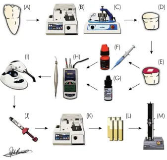
Forty sound human third molars (Figure 1A) indi -cated for extraction were obtained from the dental emergency clinic of Bauru School of Dentistry, Uni-versidade de São Paulo — USP, after approval by the
Ethics Committee of Research on Human Beings at the same institution (CAAE 05737812.7.0000.5417). Immediately after extraction, the teeth were cleaned with periodontal curettes and stored in a 0.1% thymol solution (ISO TR 11405), renewed weekly. They were kept immersed until the specimens were prepared.
Each selected tooth had its crown sectioned trans-versely on the occlusal third by means of a section-ing machine (Figure 1B) (Isomet™ Low Speed Saw®,
Buehler, Lake Bluff, USA) with a water-cooled dia -mond disc (Extec Corporation, Enfield, USA) to expose supericial dentin for adhesive procedures, after which it was subjected to wear on a polish-ing machine (Figure 1C) (Arotec, Cotia, Brazil) with 320-grit silicon carbide paper (Carbimet Paper Discs, Buehler) for the eventual removal of enamel remain-ing on the surface. The teeth were then polished for 30 s with a 600-grit SiC paper to simulate smear layer formation.
Over the lat dentin (Figure 1D), a 6 mm × 6 mm condensation-cured silicon mold (Zetalabor, Zher-mack, São Paulo, Brazil) was positioned centrally in each specimen, and nail varnish was applied all around to delimit the testing area (Figure 1E), after which each group received the respective treatment according to the manufacturer’s instructions (Table 1, Figures 1F and 1G) followed by gentle air-drying for 10 s for solvent evaporation.
In the laser groups, after application of the den-tin bonding system, a diode laser (Figure 1H) was activated in contact mode, and the full testing area was scanned for 30 s with the tip perpendicular to
Table 1. Application technique for each dentin bonding system according to its manufacturer’s instructions.
Adhesive system Application technique
Adper™ Single Bond 2 3M ESPE, St. Paul, MN, USA
Etch dentin surface with phosphoric acid (37%) for 15 s Rinse for 15 s
Gently dry with absorbent paper Apply adhesive with a microbrush
Gently air-dry for 10 s Adper™ EasyOne
3M ESPE, St. Paul, MN, USA
the specimen’s surface. Prior to laser application, the operator was trained to standardize the scan-ning speed, ensuring that the entire testing area was equally irradiated. Other parameters used for laser irradiation are listed in Table 2.
After laser irradiation, the specimens were light-cured for 20 s with light-emitting diodes (LED) (Blue Star 2, Microdont, São Paulo, Brazil) (Figure 1I) with 1 W/cm2 power density positioned perpendicular to
the dentin surface as closely as possible. Composite
resin (Filtek™ Z250, 3M ESPE) (Figure 1J) was then applied with the incremental technique with three consecutive 1.5-mm thick increments, each also light-cured for 20 s. Specimens were immersed in deion -ized water at 37°C for 7 days.
After storage, specimens were sectioned (Figure 1K) perpendicular to the bonding surface to create mul-tiple beam-shaped sticks with a cross-sectional sur-face area of approximately 1.0 mm2 (Figure 1L).
Microtensile test
Prior to being tested, the cross-sectional area of each specimen’s adhesive interface was measured with a digital caliper (Digimatic Caliper, Mitutoyo Sul Americana, Rio de Janeiro, Brazil), and the val -ues were entered into the BlueHill Lite software (BlueHill® Materials Testing Software, Norwood,
USA). Specimens were then attached individually with a cyanoacrylate-based adhesive (Loctite Super Bonder Gel Control, Henkel Ltda, São Paulo, Brazil) to a microtensile dispositive similar to Bencor Multi T (Danville Engineering, San Ramon, USA) adapted to a universal testing machine (Figure 1M) (Instron 3342, Illinois Tool Works, Norwood, USA). The spec -imen’s adhesive interface was positioned perpen-dicular to the tensile forces generated by the test-ing machine. Testtest-ing was performed at 0.5 mm/min speed (ISO TR 11405) with a 500 N load cell. Bond strength values (MPa) were calculated on the basis of the maximum force supported by each specimen (in N) divided by the cross-sectional area ratio of each specimen (in mm2).
Failure mode analysis
After being tested, both segments of the frac-tured specimens were analyzed with a digital por-table microscope (Dino Lite Microscope 100×, AnMo Electronics Corp, New Taipei City, Taiwan) and clas-siied by failure modes: Adhesive (A) for failures at the adhesive interface; Cohesive in Dentin (CD) for failures on the dentin substrate; Cohesive in Resin (CR) for failures in the resin composite; and Mixed (M) when none of the types predominated. The per -centage of each type of failure was obtained.
Table 2. Diode laser parameters used for irradiation of the testing areas.
Parameter Value
Energy per pulse 80 mJ
Frequency 10 Hz
Power 0.8 W
Testing area 36 mm2
Irradiation time 30 s
Total energy 24 J
Figure 1. (A) Human third molar; (B) Sectioning machine; (C) Polishing machine; (D) Cut and polished specimen; (E) Speci-men with delimited testing area; (F) Two-step etch-and-rinse adhesive system; (G) One-step self-etch adhesive system; (H) Diode laser; (I) Light-emitting diode; (J) Microhybrid composite resin; (K) Sectioning machine; (L) Beam-shaped stick speci-mens; (M) Universal testing machine.
(A) (B) (C) (D)
(E) (F)
(H) (I)
(J) (K) (L) (M)
Statistical analysis
Data were subjected to two-way ANOVA (p ≤ 0.05)
followed by Tukey’s test (p < 0.05) for individual com -parisons. Statistical analysis was performed with the software Statistica 10.0 (StatSoft, Inc., Tulsa, USA).
Results
Both factors (DBSs and laser) were signiicant by two-way ANOVA. For the adhesive factor, higher bond strength values were obtained when the etch-and-rinse adhesive system (SB) was used. For the laser factor, both groups (SB-L and EO-L) yielded higher bond strength values (Table 3). Adhesive failures were predominant in all groups. The distribution of each type of failure mode is described in Table 4.
Discussion
Results obtained in this in vitro evaluation showed an increase in bond strength in both adhesive sys-tems when the diode laser was irradiated, rejecting the null hypothesis. Similar results have already been reported with the Nd:YAG laser irradiation of sim -pliied DBSs,14,15 suggesting that near-infrared laser irradiation is a promising technique for achieving a more reliable link to dentin; however, the underly -ing mechanism is still unknown.
Some authors have speculated that laser irradia-tion promotes “the development of a new substrate, in which dentin substrate and adhesive would be fused
by laser action,”15 thus obtaining higher bond strength values. As hydroxyapatite melting occurs only at a very high temperature (approximately 800°C),16 the intra-pulp-chamber temperature is another issue that must be investigated.
Another attempt to explain the immediate increase in bond strength is the heat provided directly by laser irradiation, which could favor adhesive penetration14 and solvent evaporation. It was recently found that a warm air stream can also provide immediate increases in bond strength values of simpliied DBSs,17 which could also explain the favorable results obtained with the laser irradiation technique.
The pronounced but local heat generation caused by laser irradiation may also determine a higher degree of conversion of the adhesives already inil -trating the dentin, especially if the diode laser wave-length is well absorbed by the adhesives. Fourier transform infrared spectrometry and near-infra-red spectrometry may be notable instruments for these evaluations.
Despite the favorable results, the immediate increase in bond strength does not guarantee that the resin composite restorations with margins in dentin will last longer, especially in posterior tooth restora-tions, because the main cause of failures is the deg-radation of the hybrid layer over time.1 Conversely, the immediate increase in bond strength reveals that some favorable interaction is proceeding, which must stimulate more investigations in this ield.
Laser irradiation may represent one more step in dentin hybridization; however, the results provide evidence that simplified DBSs can also establish high bond strength values to dentin. Other studies should be performed to investigate the underlying mechanism by which laser irradiation can promote increased bond strength values and also the longev-ity of the laser-treated hybrid layer after water stor-age and before clinical usstor-age.
Conclusion
Based on the results of this study, the associa-tion of diode laser irradiaassocia-tion with simpliied adhe -sive systems already applied to dentin, but prior to polymerization, is a promising alternative for achiev-ing higher bond strength values.
Table 4. Failure mode distribution (%) for each group.
A CR CD M
SB 94% 3% 3% 0%
EO 76% 3% 0% 21%
SB-L 65% 25% 5% 5%
EO-L 96% 0% 0% 4%
(A) Adhesive; (CR) Cohesive in Resin; (CD) Cohesive in Dentin; (M) Mixed.
Table 3. Microtensile bond strength of dentin bonding sys-tems with or without irradiation using a diode laser [Mean (MPa) ± standard deviations].
Adhesive System Control Diode Laser
SB 33.49 ± 6.77Aa 43.69 ± 8.15Ba
EO 19.67 ± 5.86Ab 29.87 ± 6.98Bb
N = 10
Acknowledgments
This manuscript is part of a dissertation submit-ted by the irst author to the Bauru School of Den -tistry, Universidade de São Paulo– USP, in partial ful
-illment of the requirements for the Master’s degree in Restorative Dentistry. This study was supported by Conselho Nacional de Desenvolvimento Cientíico e
Tecnológico– CNPq (Process #134356/2011-0).
1. Spencer P, Ye Q, Park J, Topp EM, Misra A, Marangos O, et al. Adhesive/Dentin interface: the weak link in the composite restoration. Ann Biomed Eng. 2010 Jun;38(6):1989-2003.
2. Tay FR, Gwinnett JA, Wei SH. Micromorphological spectrum from overdrying to overwetting acid-conditioned dentin in
water-free acetone-based, single-bottle primer/adhesives. Dent Mater. 1996 Jul;12(4):236-44.
3. Pashley DH, Carvalho RM, Sano H, Nakajima M, Yoshiyama M, Shono Y, et al.. The microtensile bond test: a review. J Adhes Dent. 1999 Winter;1(4):299-309.
4. Melo MA, Goes DC, Moraes MD, Santiago SL, Rodrigues LK.
Effect of chlorhexidine on the bond strength of a self-etch adhesive system to sound and demineralized dentin. Braz
Oral Res. 2013 May-Jun;27(3):218-24.
5. Sánchez-Ayala A, Farias-Neto A, Vilanova LS, Gomes JC, Gomes OM. Marginal microleakage of class V resin-based
composite restorations bonded with six one-step self-etch
systems. Braz Oral Res. 2013 May-Jun;27(3):225-30.
6. Tay FR, Pashley DH, Suh BI, Carvalho RM, Itthagarun A. Single-step adhesives are permeable membranes. J Dent. 2002 Sep-Nov;30(7-8):371-82.
7. Tay FR, Pashley DH, Yoshiyama M. Two modes of nanole
-akage expression in single-step adhesives. J Dent Res. 2002 Jul;81(7):472-6.
8. De Munck J, Van Landuyt K, Peumans M, Poitevin A, Lam
-brechts P, Braem M, et al. A critical review of the durability of adhesion to tooth tissue: methods and results. J Dent Res. 2005 Feb;84(2):118-32.
9. Visuri SR, Gilbert JL, Wright DD, Wigdor HA, Walsh JT Jr. Shear strength of composite bonded to Er:YAG laser-pre
-pared dentin. J Dent Res. 1996 Jan;75(1):599-605.
10. Dostálová T, Jelínková H, Kucerová H, Krejsa O, Hamal K, Kubelka J, et al. Noncontact Er:YAG laser ablation: clinical evaluation. J Clin Laser Med Surg. 1998 Oct;16(5):273-82. 11. De Munck J, Van Meerbeek B, Yudhira R, Lambrechts P,
Vanherle G. Micro-tensile bond strength of two adhesives to Erbium:YAG-lased vs. bur-cut enamel and dentin. Eur J Oral Sci. 2002 Aug;110(4):322-9.
12. Oliveira MT, Reis AF, Arrais CAG, Cavalcanti AN, Aranha AC, de Paula Eduardo C, Giannini M. Analysis of the in -terfacial micromorphology and bond strength of adhesive
systems to Er:YAG laser-irradiated dentin. Lasers Med Sci. 2013 Jul;28(4):1069-76.
13. Gonçalves SEP, Araujo MAM, Damiao AJ. Dentin bond strength: influence of laser irradiation, acid etching, and hy
-permineralization. J Clin Laser Med Surg. 1999 Apr;17(2):77-85. 14. Franke M, Taylor AW, Lago A, Fredel MC. Influence of
Nd:YAG laser irradiation on an adhesive restorative proce
-dure. Oper Dent. 2006 Sep-Oct;31(5):604-9.
15. Marimoto AK, Cunha LA, Yui KC, Huhtala MF, Barcel
-los DC, Prakki A, et al. Influence of Nd:YAG Laser on
the Bond Strength of Self-etching and Conventional Adhesive Systems to Dental Hard Tissues. Oper Dent.
2013 Jul-Aug;38(4):447-55.
16. Zuerlein MJ, Fried D, Featherstone JDB. Modeling the modi -fication depth of carbon dioxide laser-treated dental enamel.
Lasers Surg Med. 1999 25(4):335-47.
17. Reis A, Wambier L, Malaquias T, Wambier DS, Loguercio AD.
Effects of warm air drying on water sorption, solubility, and
adhesive strength of simplified etch-and-rinse adhesives. J Adhes Dent. 2013 Feb;15(1):41-6.
![Table 3. Microtensile bond strength of dentin bonding sys- sys-tems with or without irradiation using a diode laser [Mean (MPa) ± standard deviations].](https://thumb-eu.123doks.com/thumbv2/123dok_br/15411705.586457/4.892.62.405.957.1059/table-microtensile-strength-dentin-bonding-irradiation-standard-deviations.webp)
