Cervical necrotizing fasciitis (CNF) is an uncommon, potentially fatal soft tissue infection with rapid progression characterized by necrosis in the subcutaneous tissue and fascia. A case of CNF of odontogenic origin in a diabetic patient, complicated by alcohol dependence and tobacco abuse, is presented with a literature review. The emergency procedure comprised hydration, colloid administration, glycemic control and broad spectrum antibiotic therapy, followed by aggressive surgical debridement. Necrosis in the platysma muscle was verified by histopathologic analysis. Reconstructive surgery was performed after suppressing the infection, and the wound was closed with an autologous skin graft. The patient had a long hospital stay, in part because the substance abuse led to a difficult recovery. The principles of early diagnosis, aggressive surgical debridement, broad-spectrum antibiotic therapy and intensive supportive care in the treatment of CNF were confirmed in the present case. It was concluded that given the occurrence of CNF in the presence of diabetes mellitus and abuse of substances such as alcohol and tobacco, the health care professional should consider a stronger response to treatment and longer hospitalization.
Cervical Necrotizing Fasciitis of
Odontogenic Origin in a Diabetic Patient
Complicated by Substance Abuse
Rubens Camino Junior1, Maria G. Naclerio-Homem1, Lecy Marcondes Cabral2, João Gualberto C. Luz1
1Department of Oral and Maxillofacial
Surgery, São Paulo School of Dentistry, USP - University of São Paulo, São Paulo, SP, Brazil
2Integrated Clinic of Plastic
Surgery, São Paulo, SP, Brazil Correspondence: Prof. Dr. João Gualberto C. Luz, Rua Duarte de Azevedo 284, s.22, 02036-021 São Paulo, SP, Brasil. e-mail: [email protected]
Key Words: necrotizing fasciitis, bacterial infections, debridement, substance-related disorders.
Introduction
Necrotizing fasciitis is a soft tissue infection with potential to be fatal, characterized by excessive necrosis and gas formation in the subcutaneous tissue and fascia (1,2). It is uncommon in the head and neck and most cases are odontogenic or pharyngeal in origin (2). This infection spreads along the fascial planes with rapid progression and systemic toxicity (3). The ability to move rapidly is secondary to its polymicrobial origin and the synergistic effect of the bacterial enzymes (4). Myonecrosis and mottling of the skin may occur due to thrombosis of the feeding vessels (1). Involvement of the underlying bone has been reported (5). An underlying systemic illness may predispose or be an aggravating factor in patients suffering from necrotizing fasciitis (6).
The clinical manifestations of cervical necrotizing fasciitis (CNF) are nonspecific but are often typical for diagnosis (7). Patients are febrile, tachycardic and dehydrated; the overlying skin is generally erythematous and tense (1,4). The management principles include fast identification of the necrotic tissue and its radical surgical debridement as there is a potentially high morbidity, combined with high-dose antimicrobial therapy (6,8).
A case of CNF of odontogenic origin in a diabetic patient, complicated by alcohol dependence and tobacco abuse is presented.
Case Report
A 65-year-old man was referred to the emergency department by the rescue medical service with an extensive toxic cellulitis of odontogenic origin and a provisional diagnosis of Ludwig’s angina. He reported that he had complained to his dentist of a toothache in the lower right posterior region three days before and had been medicated with oral amoxicillin, but he observed progressive worsening. His medical history revealed uncompensated diabetes mellitus, alcohol dependency and heavy smoking. The patient signed an informed consent form authorizing publication of this clinical report.
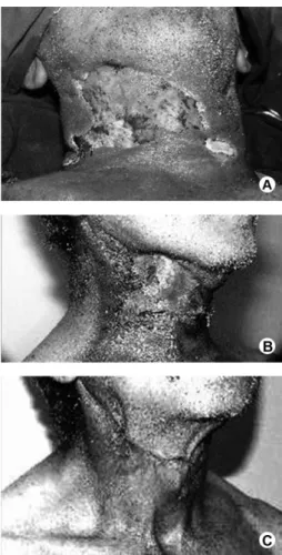
On examination he appeared generally ill and there was a diffuse, bilateral, consistent swelling with signs of inflammation in the submandibular and cervical region. Some areas of edematous erythema and vesicles were observed (Fig. 1A). Additional observations included tongue elevation, mandibular trismus, hyperthermia (39.8 °C), respiratory distress (RF 15/min) with an oxygen saturation of 92%, a heart rate of 90 bpm, blood pressure 130/80 mm Hg and, in some moments, a decreased level of consciousness. The patient was admitted to the hospital for emergency treatment.
Initial laboratory data revealed leukocytosis (22,400/ µL) with a left shift, increased serum C-reactive protein (CRP) (0.8 mg/dL) and hyperglycemia (350 mg/dL). The ISSN 0103-6440
Braz Dent J 25(1) 2014
70
R. Camino Jr et al.
remaining investigations were within normal limits. Computed tomography (CT) showed the extent of the process, demonstrating the presence of secretions in the submandibular, sublingual and submental spaces in addition to gas-formation in the deeper cervical spaces including the retropharyngeal and parapharyngeal spaces, with infiltration of the platysma and sternocleidomastoid muscles (Fig. 1B, C).
The emergency treatment consisted of intravenous hydration and colloid (albumin) administration, glycemic control (Dextro) and broad-spectrum antibiotic therapy. Crystalline penicillin G, metronidazole and amikacin sulfate were administered. Dexamethasone was used to control the swelling and tramadol hydrochloride was used for pain control. To normalize the hyperglycemia insulin
was administered and the patient was fed a balanced diet through a nasogastric tube.
The patient was taken to the operating room and, under general anesthesia and orotracheal intubation; he underwent extensive incision and drainage of the swelling with CT assistance, liberation of pus, and excision of the part of platysma muscle which appeared necrotic. Secretions were collected for culture and sensitivity testing. The mandibular right first and second molars’ roots were extracted. Subsequently, significant and wide debridement of all necrotic tissues of the submandibular and anterior cervical areas was performed, and all spaces were irrigated with an antiseptic solution (Fig. 2A). The wound was packed with dressings soaked in a topical preparation of chloramphenicol every 8 hours. The patient was transferred to the intensive care unit (ICU).
Culture identified Staphylococcus spp with penicillin resistance and anaerobic Peptococcus sp. A histopathologic analysis confirmed necrosis of the platysma muscle. The infectious disease service was consulted. Ampicillin, sulbactam and ciprofloxacin were prescribed, and metronidazole was continued. The patient remained in the
Figure 2. A: Broad surgical debridement in the affected regions. B: Postoperative aspect of the recipient area of the skin graft. C: Complete recovery of the affected area after one year.
Braz Dent J 25(1) 2014
71
Cervical necrotizing fasciitis of odontogenic origin
ICU for 45 days. Systemic improvement and formation of granulation tissue in the wound were observed.
Reconstructive surgery was performed 15 days after ceasing the infection. The wound was closed with a skin graft taken from the right thigh (Fig. 2B). The patient recovered and was discharged from the hospital 90 days after admission. Complete healing of the cervical and submandibular regions was obtained. After one year, the patient presented no esthetic or functional restrictions (Fig. 2C).
Discussion
A case of CNF secondary to an odontogenic infection that had compromised the prognosis due to uncompensated diabetes mellitus, associated with alcohol dependence and tobacco abuse is reported. The emergency procedure comprised hydration, colloid administration, glycemic control and broad-spectrum antibiotic therapy, followed by aggressive surgical debridement, emphasizing the principles of conduct in this entity (4,8,9).Necrotizing fasciitis often results in the sequestration of fluid which may lead to septic shock (6). Thus, the patient was admitted to the ICU for monitoring, wound checks, antibiotic therapy, fluid status monitoring and hemodynamic assessment (3,6).
Odontogenic infections usually originate from pulpal necrosis with bacterial invasion of the periapical tissue, producing the formation of purulent collections. Such infections are usually mild and easily treated with antibiotics but their complications are around 12.5% of cases (10). These processes still represent an important cause of morbidity in the population and their complications demand careful monitoring for prevention and application of appropriate management (11,12). Given the nonspecific nature of primary odontogenic infections, different combinations of virulence factors will cause different diseases in subjects. Knowledge of the main factors involved in the pathogenesis of apical periodontitis may help establish proper therapeutic measures to inactivate this bacterial attack (13).
Insulin-dependent diabetes is the most common predisposing systemic condition for CNF, observed in 18% to 72.3% of cases (1,6,7). Chronic hyperglycemia impairs leukocyte function leading to suppression of the immune system and susceptibility to exacerbation of an odontogenic infection (3,4). Additionally, alcoholism, tobacco abuse and CNF are also associated. However, this may be due to either a direct effect of these substances or the malnutrition and poor health status that is often observed in these patients (3). Alcohol abuse has also been associated with CNF and is observed in 22% to 45% of cases (1,4,7). Alcoholism alone or associated with malnutrition increases the risk of CNF (4).
A history of pain for less than one week with the
rapid exacerbation of symptoms is typical in cases of CNF (4,8). Mandibular molars are frequently the source of odontogenic infections prior to CNF development (1). The initial laboratory data revealed leukocytosis and increased C-reactive protein. Similar findings have been reported in a CNF series (1). Patients with CNF are febrile, with erythema and edema, hyperesthesia, mottled skin and sepsis (4,8). Recognizing a necrotizing soft tissue infection may be difficult. A simple subcutaneous abscess has a central necrotic portion, whereas necrotizing soft tissue infections are poorly localized, with signs of inflammation and necrosis extending deep to normal-appearing skin. Systemic findings are therefore of great help in its identification (6,7). CT demonstrated the presence of secretions in the submandibular, sublingual and submental spaces as well as in deeper cervical spaces, with infiltration of the cervical region, and gas-formation. There is no consensus on the requirement of CT, although it provides a major contribution in the early diagnosis of CNF (1,4).
The antimicrobial agents initially used were targeted toward the most frequently encountered microorganisms in an infection of odontogenic origin. The antibiotic regimen should be modified according to culture and sensitivity results (1,14). Additionally, a consultation with the infectious disease service is required (6). Prompt surgical intervention is essential to reduce morbidity and mortality rates associated with CNF (4,8). Surgical debridement is considered a cornerstone of its treatment (7,8), and its extent is determined by the gross findings (6). In this case, necrosis of the platysma muscle was histopathologically verified, which is considered the most accurate test for identification of this disease (8). Reconstruction is typically initiated when the patient has been stable for a number of days (6). Complete healing of the affected regions was obtained, and the patient presented without esthetic or functional restrictions at the end of treatment. The patient had a long hospital stay, in part because the associated alcohol abuse and smoking lead to a difficult recovery. A review of reported cases identified mean hospital stays between 24 and 42 days, with most days spent in the ICU (3). Patient death can be avoided by professionals who are at the first point of patient contact (6), although the window of opportunity for surgical treatment extends to the advanced stages of the disease (8).
Resumo
Braz Dent J 25(1) 2014
72
R. Camino Jr et al.
seguido de debridamento cirúrgico agressivo. Necrose no músculo platisma foi verificada por análise histopatológica. Cirurgia reconstrutiva foi feita após resolução da infecção e a ferida foi fechada com enxerto dérmico autógeno. O paciente teve um longo período de internação hospitalar, em parte devido ao abuso de substâncias, o que levou a uma recuperação difícil. Os princípios de diagnóstico imediato, debridamento cirúrgico agressivo, antibioticoterapia de amplo espectro e cuidados em terapia intensiva no tratamento da FNC foram confirmados no presente caso. Foi concluído que diante da ocorrência de FNC na presença de diabetes mellitus e de abuso de substâncias como álcool e tabaco, o profissional assistente deve considerar uma resposta mais difícil ao tratamento e maior tempo de internação.
References
1. Umeda M, Minamikawa T, Komatsubara H, Shibuya Y, Yokoo S, Komori T. Necrotizing fasciitis caused by dental infection: a retrospective analysis of 9 cases and a review of the literature. Oral Surg Oral Med Oral Pathol Oral Radiol Endod 2003;95:283-290.
2. Chidzonga MM. Necrotizing fasciitis of the cervical region in an AIDS patient: report of a case. J Oral Maxillofac Surg 2005;63:855-859. 3. Quereshy FA, Baskin J, Barbu AM, Zechel MA. Report of a case of
cervicothoracic necrotizing fasciitis along with a current review of reported cases. J Oral Maxillofac Surg 2009;67:419-423.
4. Whitesides L, Cotto-Cumba C, Myers RA. Cervical necrotizing fasciitis of odontogenic origin: a case report and review of 12 cases. J Oral Maxillofac Surg 2000;58:144-151.
5. Salins PC, Saxena S, John JK. Reconstruction of mandible and surrounding soft tissues in patient with necrotizing fasciitis. Int J Oral Maxillofac Surg 1996;25:98-100.
6. McMahon J, Lowe T, Koppel DA. Necrotizing soft tissue infections of the head and neck: case reports and literature review. Oral Surg Oral Med Oral Pathol Oral Radiol Endod 2003;95:30-37.
7. Lin C, Yeh FL, Lin JT, Ma H, Hwang CH, Shen BH, et al.. Necrotizing fasciitis of the head and neck: an analysis of 47 cases. Plast Reconstr Surg 2001;107:1684-1693.
8. Hanna BC, Delap TG, Scott K, Sinclair S. Surgical debridement of craniocervical necrotizing fasciitis: the window of opportunity. J Laringol Otol 2006;120:702-704.
9. Yadav S, Verma A, Sachdeva A. Facial necrotizing fasciitis from an odontogenic infection. Oral Surg Oral Med Oral Pathol Oral Radiol 2012;113:e1-4.
10. Gonçalves L, Lauriti L, Yamamoto MK, Luz JGC. Characteristics and management of patients requiring hospitalization for treatment of odontogenic infections. J Craniofac Surg 2013;24:e458-e462. 11. Carter LM, Layton S. Cervicofacial infection of dental origin presenting
to maxillofacial surgery units in the United Kingdom: a national audit. Br Dent J 2009;206:73-78.
12. Flynn TR. The swollen face: severe odontogenic infections. Emerg Med Clin North Am 2000;18:481-519.
13. Siqueira Jr JF, Rôças IN. Bacterial pathogenesis and mediators in apical periodontitis. Braz Dent J 2007;18:267-280.
14. Zhang WJ, Cai XY, Yang C, Zhou LN, Cai M, Lu XF, et al.. Cervical necrotizing fasciitis due to methicillin-resistant Staphylococcus aureus: a case report. Int J Oral Maxillofac Surg 2010;39:830-834.