SYNCHRONOUS OCCURRENCE OF ODONTOGENIC KERATOCYST
AND GIANT CELL GRANULOMA- A CASE REPORT
1 2
Soniya Adyanthaya Maji Jose
1
Corresponding Author: Soniya Adyanthaya, Senior Lecturer ,Dept of Oral Pathology&Microbiology, Yenepoya
Dental College , Derlakatte,Mangalore-575018,Karnataka,India.+919741231333, Email:
Abstract
An odontogenic keratocyst (OKC) and a giant cell granuloma (GCG) in the jaws are two well known entities, which have been reviewed extensively. However, the synchronous occurrence of these two lesions is exceptionally rare. We report a case of OKC exhibiting foci of CGC-like lesion.
Keywords : Odontogenic keratocyst, giant cell granuloma
2
Senior Lecturer , Professor ,Dept of Oral Pathology & Microbiology, Yenepoya Dental College , India
drsoniya1978@gmail.com
151
Oral & Maxillofacial Pathology Journal [ OMPJ ] Vol. 2 No. 2 Jul-Dec -2011 ISSN 0976 - 1225
sclerotic margin, in the left mandibular posterior
Introduction
region. The lesion was extending from the distal aspect of second molar till the ascending ramus. OKC is a clinicopathologically distinct
A provisional diagnosis of an OKC or form of odontogenic cyst, known for its
ameloblastoma was made. The lesion was pathognomonic microscopic features,
1,2
surgically removed and submitted to the Dept. aggressiveness and high recurrence rate. Many
of Oral Pathology, Yenepoya Dental College, studies have focused on intrinsic growth
3-5 Yenepoya University for histopathological
potential of epithelial lining of OKC and it
examination. has been reclassified as keratocystic
6 odontogenic tumor by the WHO.
The excised specimen was well Giant cell granuloma is thought to be a
non-circumscribed, encapsulated, brownish white in neoplastic, reparative or a reactive process. It
colour, and measured 3.5 X 2.5 X 1.2 cm. On consists of spindled fibroblasts that are
sectioning, the specimen appeared as a well admixed with collagen, areas of haemorrhage
circumscribed cystic lesion with a cystic cavity and numerous multinucleated osteoclast-type
7,8 surrounded by a well defined capsule. The cystic
giant cells.
capsule was found to be unusually thick in one portion of the specimen. Representative areas Although these two entities are
of specimen were sampled and subjected to relatively common, synchronous occurrence is
routine tissue processing. Four µm thick highly unusual. A thorough search of literature
9 paraffin sections were cut and stained with
had revealed only one such case report. We
hematoxylin and eosin. report another case of OKC exhibiting foci of
CGC-like lesion in a 29 year old man.
The histopathological examination showed a typical OKC lined by uniform
Case report
thickness of parakeratinized stratified squamous epithelium, with hyperchromatic and A 29 year old man visited a private
palisaded basal cells (Fig. 1). The luminal surface dentist, complaining of swelling on the right
of the epithelial lining revealed a corrugated posterior mandible since 4 months which is
appearance. The epithelium- connective tissue gradually increasing in size. He had no previous
interface was flat, without evidence of rete history of trauma related to the lesion. The
ridges. The connective tissue wall exhibited patient's past medical and family histories were
chronic inflammatory cells in some foci. non-contributory. The panoramic radiography
152
Synchronous Occurrence Of Odontogenic Keratocyst And Giant Cell Granuloma- A Case Report
Oral & Maxillofacial Pathology Journal [ OMPJ ] Vol. 2 No. 2 Jul-Dec -2011 ISSN 0976 - 1225
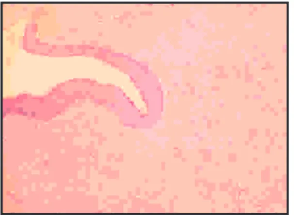
Fig 1: Photomicrograph showing characteristic OKC lining epithelium ( H&E, 10X)
In one portion of the section, the connective tissue component was highly cellular and vascular with many dilated RBC filled blood vessels. Multiple giant cells exhibiting eosinophilic cytoplasm and containing 10-20 nuclei were found to be distributed in this stroma, mimicking GCG( Fig.2). There was no evidence of any foreign body. The trabeculae of woven bone were also present within the more fibrous connective tissue wall.
Fig 2: Photomicrograph showing cystic cavity lined by
epithelium and foci of giant cell granuloma.( H& E, 4X)
Discussion
In the present report, the cystic lesion showed the characteristic histopathological features of an OKC. The presence of GCG like areas in many parts of the connective tissue wall is the most interesting feature in this lesion.
Giant cell reaction has been reported in odontogenic tumors like ameloblastoma and odontogenic fibroma. The authors have interpreted these combinations as an odontogenic tumor with reactive giant cell response, although the initiating stimulus was
8,10,11
not identified. An association between Giant
cell granuloma and other conditions like central osifying fibroma and benign fibro-osseous
12,13 lesions of the jaw has also been reported.
Similarly, OKC also can occur simultaneously with a traumatic bone cyst and
14,15,16
as well ameloblastoma. But OKC
exhibiting foci of GCG like lesion is very rare and only one case has been reported by Yoon
9 J.H et.al in a 10 year old boy.
The present lesion was unusual in its gross appearance. In contrast to typical OKC lining, the lesion had an unusually thick lining particularly in the posterior part. The same area e x h i b i t e d t h e C G C G l i k e f e a t u r e s microscopically. The author of previous similar case report had interpreted the giant cell granuloma like areas as reactive process of osteoclasts. As the CGCG like area was observed mainly in one part our case might be mere co-existence of two lesions which would have coalesced. However the possibility of overt proliferative reaction of bone resorbing osteoclasts should also be considered.
153
Synchronous Occurrence Of Odontogenic Keratocyst And Giant Cell Granuloma- A Case Report
Oral & Maxillofacial Pathology Journal [ OMPJ ] Vol. 2 No. 2 Jul-Dec -2011 ISSN 0976 - 1225
11. Kawakami T, Antoh M, Minemura T. Giant cell Acknowledgement
r e a c t i o n t o a m e l o b l a s t o m a : a n immunohistochemical and ultrastructural study of
Authors are grateful to Dr. Jagadish
a case. J Oral Maxillofac Surg
1989;47:737-Chandra, Professor ,Dept of Oral &
741.
Maxillofacial Surgery, Yenepoya Dental College
for providing the biopsy specimen of this case. 12. Ilana Kaplan I, Manor I, Yahalom I, Hirshberg
A. Central giant cell granuloma associated with
References central ossifying fibroma of the jaws: a
clinicopathologic study. Oral Surgery, Oral Medicine, Oral Pathology, Oral Radiology and 1. De Paula AMB, Carvalhais JN, Domingues MG,
Endodontology Volume 103, Issue 4 , Pages e35-Barreto DC, Mesquita RA. Cell proliferation
e41, April 2007 markers in the odontogenic keratocyst: effect of
inflammation. J Oral Pathol Med. 2000;29:477-2. . Crusoé-Rebello I, Torres MGG, Burgos V,
1
Oliveira C, dos Santos JN, Azevedo RA and 2. Stoelinga PJ. Long term follow-up on keratocysts
Campos PSF. Hybrid lesion: central giant cell treated according to a defined protocol. Int J Oral
granuloma and benign fibro-osseous lesion. Maxillofac Surg. 1996;25:124-9.
Dentomaxillofacial Radiology 2009;38: 421-3. Ogden GR, Chisholm DM, Kiddie RA, et al. p53
425. protein in odontogenic cysts: increased expression in
14. Maste JL, Beto LM, Fantasia JE, Fielding AF. some odontogenic keratocysts. J Clin Pathol.
Pathologic fracture of the mandible associated with 1992;45:1007-10.
simultaneous occurrence of an odontogenic 4. Shear M. The aggressive nature of the odontogenic
keratocyst and traumatic bone cyst. J Oral keratocyst: is it a benign cystic neoplasm? Part II.
Maxillofac Surg 1987;45:69-71. Proliferation and genetic studies. Oral Oncol.
2002;38: 323-31. 15. Siar CH,Ng KH. Combined ameloblastoma and odontogenic keratocyst or keratiniz ing 5. Shear M. The aggressive nature of the odontogenic
ameloblastoma. BR J Oral Maxillofac Surg keratocyst: is it a benign cystic neoplasm? Part III.
1993;31:183-186. Immunohistochemistry of cytokeratin and other
epithelial cell markers. Oral Oncol. 2002;38:407- 16. Fregnani ER, da Cruz Perez DE, Soares FA,
15. Alves FA. Synchronous ameloblastoma and
orthokeratinized odontogenic cyst of the mandible. 6. Barnes L, Eveson JW, Reichart P, Sidransky D,
J Oral Pathol Med 2006;35:573-575. editors. World Health Organization of tumours.
Pathology and genetics of head and neck tumours. Lyon: IARC Press; 2005.
7. Rosenberg A.E, Nielsen G.P.Giant cell containing lesions of bone and their differential diagnosis. Current Diagnostic Pathology 2001;7: 235-246. 8. Mosqueda Taylor A, Bermudez Flores V, Diaz
Franco MA. Combined central odontogenic fibroma and giant cell granuloma-like lesion of the mandible: report of a case and review of the literature. J Oral Maxillofac Surg 1999;57:1258-1262.
9. Yoon J.H, Kim S.G, Lee S.H, Kim J. Simultaneous occurrence of an odontogenic keratocyst and giant cell granuloma-like lesion in the mandible.
10. Allen CM, Hammond HL, Stimson PG. Central odontogenic fibroma, WHO type. A report of three cases with an unusual associated with giant cell reaction. Oral Surg Oral Med Oral Pathol Oral Radiol Endod 1992;73:62-66.
13