Universidade do Minho
Escola de Ciências
João Nuno de Sousa Machado
julho de 2016
The role of the small GTPase Saci_1563 in
archaellum regulation
João N
uno de Sousa Mac
hado The role of t he small GTP ase Saci_1 563 in archaellum regulation UMinho|20 16
Universidade do Minho
Escola de Ciências
João Nuno de Sousa Machado
julho de 2016
The role of the small GTPase Saci_1563 in
archaellum regulation
Trabalho realizado sob orientação da
Prof. Dr. Sonja-Verena Albers
e do
Professor Doutor João Carlos Marcos
Dissertação de Mestrado
Mestrado em Bioquímica Aplicada
ii
Declaração
Nome: João Nuno de Sousa Machado
Endereço eletrónico: [email protected] Telefone: +351915185448
Número do Cartão de Cidadão: 14031750 3 ZZ9
Título da dissertação de mestrado: The role of the small GTPase Saci_1563 in
archael-lum regulation
O papel da pequena GTPase Saci_1563 na regulação do arcaelo
Orientadora: Prof. Dr. Sonja-Verena Albers
Co-orientador: Professor Doutor João Carlos Marcos.
Ano de conclusão: 2016
Mestrado em Bioquímica Aplicada, Ramo de Bioquímica Molecular e Analítica
DE ACORDO COM A LEGISLAÇÃO EM VIGOR, NÃO É PERMITIDA A REPRODUÇÃO DE QUALQUER PARTE DESTA DISSERTAÇÃO
Universidade do Minho, ___/___/______
iii
Agradecimentos/Aknowledgements
In this thesis I am summarizing approximately 9 months of my life that were filled with enormous gains at the academic, scientific and personal levels, only possible because of all the help I have received.
First of all, I want to thank Prof. Dr. Sonja- Verena Albers for having allowed me to work in her laboratory. This period in your laboratory has allowed me to understand better which my scientific interests are, and the work discussions and seminars have contributed to the improvement of my critical thinking skills and to my development as a Biochemistry student. I am very grateful for that. Secondly, I want to thank Dr. Florencia Haurat. Your patience to teach me even when I was asking you the same thing for the third or fourth time is unmatched. A lot of the technical skills that I now possess are because of your dedication and competence, and as such I will always be in debt with you.
To all the people in the laboratory, thank you for the great environment we work in, for the helpful discussions and for being so kind to help me whenever I was in need. It has been a pleasure to get to know you all.
Matthias, I want to thank you for the companionship, for introducing me to Maultaschen and for the weeks I could stay at your place. My stay in Germany would have much duller if I hadn’t met you, if you pardon the pun.
Agradeço também a todos os meus amigos que deixei em Portugal, em particular – e por ordem alfabética –, à Bárbara, à Margarida, ao Miguel e à Ni, por todas as conversas à distância.
Finalmente, agradeço à minha família por todos os esforços, incentivo e carinho que me têm prestado, ao longo não só desta tese, mas desde que nasci.
“Biology is a study, not in being, but in becoming. For us, the fascination of the subject derives from the astounding fact that life exists at all and has developed such remarkable complexity.” - Woese and Goldenfeld 2009
v
O papel da pequena GTPase Saci_1563 na regulação do arcaelo
Reconhecido formalmente há cerca de 40 anos o domínio Arqueia é, dos três domínios da vida, o menos estudado. Os filos Euryarchaeota e Crenarchaeota compreendem os organismos melhor caracterizados deste domínio e, no filo Crenarchaeota, o termoacidófilo Sulfolobus acidocaldarius é um dos organismos modelo que tem permitido mais desenvolvimentos nesta área da microbiologia. Este organismo móvel e aeróbico tem por habitat ambientes terrestres vulcânicos e por condições ótimas temperaturas de cerca de 75°C e pH 3. O arcaelo – um apêndice funcionalmente semelhante ao flagelo bacteriano, mas estrutural e geneticamente distinto – é responsável pela locomoção arqueana. Atualmente sabe-se que certos fatores ambientais estimulam a expressão do arcaelo. Em S. acidocaldarius, em particular, condições de subnutrição levam à expressão do arcaelo. Algumas proteínas envolvidas na rede regulatória do arcaelo já se encontram identificadas. Duas dessas proteínas – ArnA e ArnB – reprimem a sua expressão. As propriedades de ambas as proteínas podem ser alteradas por fosforilação, o que indica que a regulação do arcaelo ocorre a vários níveis hierárquicos.
A proteína Saci_1563, anotada como uma GTPase, encontra-se conservada em várias espécies pertencentes ao filo Crenarchaeota e foi detetada após a sua co-purificação com ArnA. Dado o papel das GTPases na regulação de diversos processos celulares, é plausível que esta proteína de alguma forma module ArnA e, consequentemente, a expressão do próprio arcaelo.
De modo a caracterizar bioquimicamente esta proteína otimizaram-se protocolos para a sua purificação a partir de Escherichia coli e de S. acidocaldarius. As amostras obtidas foram utilizadas para avaliar a capacidade de hidrólise de GTP desta proteína.
A influência da ausência de nutrientes no meio na expressão de saci_1563 foi estudada numa estirpe de S. acidocaldarius. Uma estirpe mutante que não possui o gene para Saci_1563 foi também utilizada de modo a determinar de que modo a ausência desta proteína influencia a expressão de outras proteínas associadas ao arcaelo ou à regulação da sua expressão. O efeito da ausência de Saci_1563 na locomoção de S. acidocaldarius foi também determinado.
Os resultados aqui apresentados não são de modo algum definitivos. Apesar de não ter sido possível atribuír um papel a Saci_1563 no que toca à regulação do arcaelo, não é também possível excluir completamente um papel regulatório por parte desta proteína. Por este motivo, no final sugerem-se vias de investigação alternativas para compreender esta biomolécula.
vii
The role of the small GTPase saci_1563 in archaellum regulation
Formally recognized approximately 40 years ago, Archaea is still the least studied of the three domains of life. Euryarchaeota and Crenarchaeota are the two major phyla of Archaea, and the most well understood, particularly because of the existence of genetic techniques that allow for their study. An important crenarchaeal model organism is Sulfolobus acidocaldarius, a motile aerobic thermoacidophile first isolated from terrestrial volcanic environments that thrives at around 75°C and pH 3. The archaellum – an organelle functionally similar to the bacterial flagellum, but structurally and genetically different – is responsible for the locomotion of motile archaeal species. In recent years it has been found that the expression of this organelle depends on environmental factors, and starvation in particular is associated with archaella induction in S. acidocaldarius. The signal transduction of nutrient depletion to archaella expression is not fully understood, but recently some key players of the archaellum regulatory network have been uncovered. Specifically, ArnA and ArnB were found to be negative regulators of archaella expression. Moreover, both proteins were found to be phosphorylated, which points at the existence of a multi-level regulatory system. Saci_1563 is a protein conserved in several Crenarchaeota species that was found to be co-purified with ArnA. This protein is a putative GTP-binding protein, a class of proteins implicated in the regulation of several biological phenomena and in cell signalling. It is thus plausible that Saci_1563, by acting as a molecular switch, is implicated in the fine tuning of archaella expression in response to specific signals.
In this study we assessed the biological role of Saci_1563 and its biochemical characterization. Protocols for the heterologous purification of this protein in Escherichia coli and for the purification of over-expressed protein in S. acidocaldarius were optimised. The purified protein was then subject to phosphate release assays to test for its GTP-binding ability. The impact of saci_1563 deletion on S. acidocaldarius motility was tested, and the influence of starvation on saci_1563 expression was determined. Moreover, we analysed the effect of saci_1563 deletion on the expression of proteins belonging to the archaellum regulatory network and to the archaellum operon.
The results reported in this thesis are far from conclusive. A role for Saci_1563 in archaella regulation cannot be excluded, but other venues of research regarding this protein are suggested.
ix
Chapter 1 - Introduction
1. – Archaea ... 2
1.1. – Archaeal diversity ... 3
1.2. – Ecology ... 4
1.3. – Archaeal physiology and genetics ... 5
1.4. – Sulfolobus as a model organism ... 7
2. – Motility ... 9
2.1. – Setting the Archaellum and the Flagellum apart ... 9
2.2. – Structure of the archaellum ... 11
2.3. – Archaellum regulation ... 15
2.4. – The GTPase superfamily of proteins ... 19
3. – Objective ... 23
Chapter 2 - Materials and Methods 1. - Chemicals ... 26
2. – Strains and Growth ... 26
2.1. – Strains ... 26
2.2. – Liquid media ... 27
2.3. – Solid media ... 27
2.4. – Glycerol cryostocks of Sulfolobus acidocaldarius ... 28
2.5. – Transformation of Sulfolobus acidocaldarius cells ... 28
2.5.1. – Preparation of competent cells ... 28
2.5.2. – Transformation by electroporation... 29
2.6. – Transformation of Escherichia coli ... 29
2.6.1. – Preparation of competent cells ... 29
2.6.2. – Transformation by heat shock ... 30
2.7. – Plasmid isolation ... 30
x
4. – Gel electrophoresis ... 31
4.1. – Protein electrophoresis ... 31
4.1.1. – Gel staining ... 32
4.2. – DNA and RNA electrophoresis ... 32
5. – Western blotting ... 32
5.1. – Blotting ... 33
6. – Polymerase chain reaction (PCR) ... 34
6.1. – Preparation of samples for colony PCR ... 34
6.2. – PCR reaction and program ... 34
7. – Motility assays ... 35
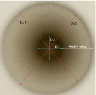
7.1. – Radii measurement ... 36
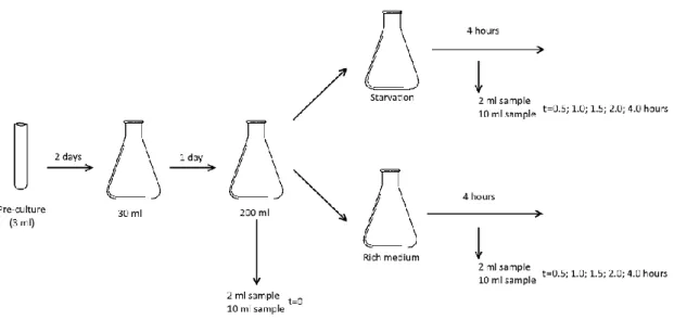
8. – Induction of starvation ... 37
8.1. – Cell growth ... 37
8.2. – Processing of samples for immunoblotting ... 38
8.3. – Processing of samples for RNA extraction ... 38
9. – RNA isolation and cDNA synthesis ... 38
9.1. – RNA purification ... 39
9.2. – cDNA synthesis ... 39
10. – qRT-PCR ... 40
11. – Heterologous protein purification ... 40
11.1. – Induction of expression and preparation of cell lysate ... 41
11.2. – Purification by gravity chromatography ... 42
11.3. – Purification with Profinia ... 42
12. – Preparation of soluble denatured proteins from E. coli ... 43
13. – Expression of saci_1563 in Sulfolobus acidocaldarius ... 43
13.1. – Induction of expression and preparation of cell lysate ... 43
xi
Chapter 3 - Results and Discussion
1. – Heterologous protein expression... 46
2. – Overexpression of saci_1563 in S. acidocaldarius ... 53
3. – Phosphate release assays ... 55
4. – Swimming behaviour of a saci_1563 knockout mutant ... 64
5. – saci_1563 deletion impact on the expression of selected genes ... 59
Chapter 4 - Conclusion 1. – Conclusion and future perspectives ... 68
References ... 73
xii
List of abreviations
5’-UTR 5’-untranslated region
ADP Adenoside diphosphate
ATP Adenoside triphosphate
BRE B recognition element
cDNA Complementary DNA
DNA Deoxyribonucleic acid
EDTA Ethylenediaminetetracetate
ePK Eukaryotic-like protein kinase
FHA Forkhead-associated
GAF GTPase-activating protein
GDP Guanoside diphosphate
GEF Guanine exchange factor
GppCp Guanosine-5’-[(β,γ)-methyleno]triphosphate
GTP Guanosine triphophsate
HTH Helix-turn-helix
IMAC Immobilized metal-affinity chromatography
IPTG Isopropyl β-D-1-thiogalactopyranoside
mRNA Messenger RNA
ORF Open reading frame
PCR Polymerase chain reaction
PTM Post-translational modifications
PVDF Polyvinylidene fluoride
RNA Ribonucleic acid
RNAP RNA polymerase
rRNA Ribosomal RNA
SDS-PAGE Sodium dodecyl sulfate polyacrylamide gel electrophoresis vWA von Willebrand
X-Gal 5-bromo-4-chloro-3-indolyl-β-D-galactopyranoside
List of tables
Page
Table 1 Archaeal and bacterial strains used in this work 26
Table 2 Plasmids used in this work to transform Sulfolobus acidocaldarius. 29
Table 3 Plasmids used in Escherichia coli 30
Table 4 Antibodies used in this work and the conditions at which the membrane was incubated
in the respective antibody solution. Antibody solutions were prepared in PBS.
33
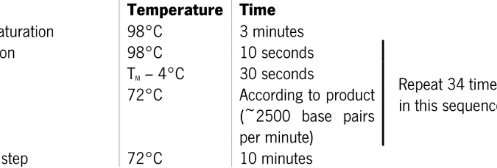
Table 5 Reaction mixture for polymerase chainreaction. 34
Table 6 PCR program 35
Table 7 Primers used in colony PCR or in standard PCR reactions 35
Table 8 Reaction mixture for qRT-PCR 40
Table 9 Composition of buffers and their variations used for protein purification by gravity IMAC
xiii
List of Figures
Page
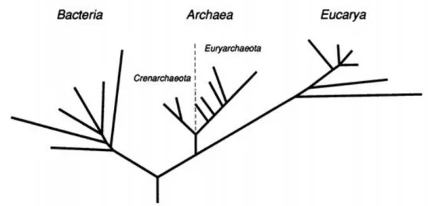
Fig. 1 First tree proposed with the phylogenetic relationship between the three primary domains.
The domain Archaea is here shown to be composed by two main branches, the Euryarchaeota and the Crenarchaeota. Figure adapted from Carl R. Woese, Kandler, & Wheeli.
3
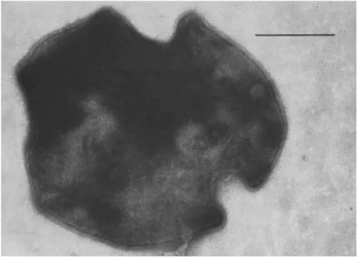
Fig. 2 Sulfolobus acidocaldarius cells are lobed, spherical cells. Bar represents 0.5 µm. Figure
from Brock et al., 1972. 8
Fig. 3 Schematic representation of different microbial surface structures. Proteins that share the
same colour in the archaellum and type IV pili scheme represent homologous proteins. Image from Ken F. Jarrell and Albers 2012.
11
Fig. 4 Organization of the fla operon in several species of the Crenarchaeota and Euryarchaeota.
Image adapted from Albers and Jarrell 2015. 13
Fig. 5 Current schematic representation of the archaellum. FlaB (B) forms the filament, which
with an average of 15 nm diameter is between the diameter of the flagellum and the type IV pili. FlaX (X) forms a platform in the membrane where the other proteins of the motor complex assem-ble. FlaF (F) is the stator of this nanomotor, anchoring the archaellum to the S-layer, composed by two proteins - SlaA and SlaB. FlaG (G) plays an essential but currently unknown role in the archaellum. PibD is a homologue of type IV prepilin peptidases and plays an essential role by cleaving the signal peptide from prearchaellins. Image from Banerjee et al. 2015.
14
Fig. 6 Summary of the currently known regulators of archaella expression. ArnR/R1 are
mem-brane-bound activators, and ArnR expression itself is stimulated in starvation conditions, pointing at the existence of another unknown regulator. ArnA and ArnB are negative regulators of the ar-chaellum that can be phosphorylated by ePKs and dephosphorylated by phosphatases like PP2A, which has been shown to influence the archaellum expression. AbfR1 is a biofilm regulator that activates the FlaB promoter. Image from Albers and Jarrell, 2015.
18
Fig. 7 Structure of Ras, a common prototype of the GTP-binding domain, bound to A the
non-hydrolysable GTP analogue GppCp and to B GDP. A1-A5 correspond to the 5 α-helices, B1-B6 corresponds to the 6 β-strands and G1-G5 to the 5 regions described in the text. The confor-mation of Switch I and Switch II is noticeably different between the GppCp- and the GDP-bound forms of the protein. Figure from Paduch, Jeleń and Otlewski 2001.
21
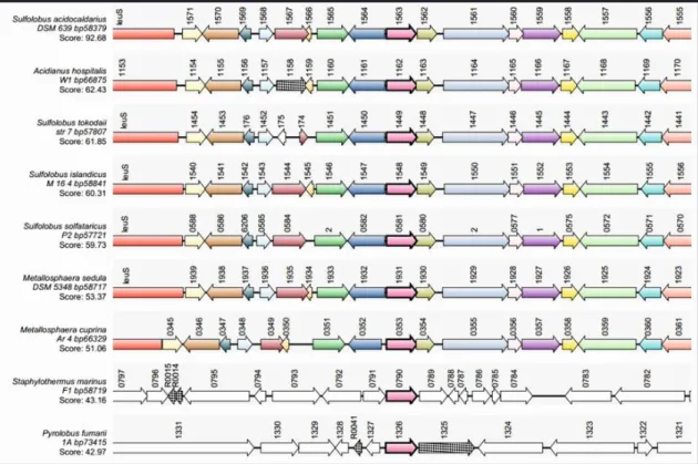
Fig. 8 saci_1563 is conserved among several Crenarchaeota, and the genes surrounding
saci_1563 are the same for the members belonging to the order Sulfolobales. Figure from Sartor, P. Master Thesis.
22
Fig. 9 The G-motifs of Saci_1563 are indicated in blue in the following order: G4 (NKGD); G5
(SAT); G1 (GYPKTGKS); G2 (T); and G3 (DTPG). G1 and G3 are also called Walker A and Walker B motifs, respectively. The isoleucine in red next to G3 corresponds to the putative catalytic amino acid that, as in HAS-GTPases, is hydrophobic.
23
Fig. 10 In order to measure the motility diameter, the diameter of the outside halo was measured
in two different orientations (Do1 and Do2). The two values were averaged to determine the outer diameter. The same was performed for the inner halo (darker). The motility radius corresponds to half the subtraction of the inner diameter from the outer diameter.
36
Fig. 11 Schematic representation of the starvation experiment. Before splitting the 200 ml culture
one sample of 2 ml and one sample of 10 ml are harvested. This corresponds to time 0 (t=0) in the experiment. The other time-points at which samples were harvested and the OD was meas-ured, in hours after the beginning of the experiment, are indicated in the rightmost part of the scheme.
xiv
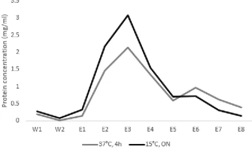
Fig. 12 Chromatographic profile of Saci_1563 purification from E. coli induced at 37°C for 4h or at 15°C overnight (ON), induced with 0.5 mM IPTG. W: washingfractions; E:elution fractions. 46
Fig. 13 Typical SDS-PAGE of Saci_1563 purified from E. coli with the set of buffers G1. The lower
bands found in the fractions most enriched with Saci_1563 were hypothesised to be degradation products of this protein.
47
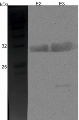
Fig. 14 Western blot performed on the elution fractions 2 and 3 of a culture of E. coli induced for 4h at 37°C with 0.1 mM IPTG. The low molecular weight band present in the E3 lane is indicative of protein degradation.
47
Fig. 15 A SDS-PAGE analysis of the fractions obtained from purification of Saci_1563 with buffers
containing 10% glycerol. B Western blot of the same fractions as in A. The bands at approximately 17 kDa appear to be degradation products of Saci_1563, both because the intensity of the low molecular weight bands match the intensity of Saci_1563 band and because those low molecular weight bands are detected by an anti-His tag antibody. L: loading; FT: flow-through; W: washing fractions; E: elution fractions.
48
Fig. 16 SDS-PAGE gel of fractions recovered from IMAC using the set of buffers G2 with A all
buffers at pH 10 or B buffers A and B at pH 8.6 and the remaining buffers at pH 10. 49
Fig. 17 Recovered fractions from protein purification with Profinia. The elution fraction was
con-centrated and separated once more by SDS-PAGE. As shown in the rightmost lane, 4 other bands besides that corresponding to Saci_1563 are present..
50
Fig. 18 A To improve the purity of the elution fraction and, simultaneously, recover the maximum
protein possible, the flow-through and W1 fractions from the purification of the cell lysate were mixed and purified. The resulting elution fraction (Elution 2) was mixed with the elution fraction from the purification of the cell lysate (Elution 1). Loading 3 was finally subjected to buffer ex-change and purified again. B After the concentration of Elution 3 only the band that corresponds to Saci_1563 is obvious. However, two very faint bands at higher molecular weights can be iden-tified.
51
Fig. 19 Increased stringency of the second washing step (100 mM imidazole) results in more
protein loss during this step, but does not result in increased purity of the elution fraction. 52
Fig. 20 Saci_1563 with a mutation in A Walker A or B Walker B region were purified using the
buffers P1. Contaminants are present in the elution fraction of both proteins, as is the case with the wild-type protein. Red arrows point to contaminant bands.
53
Fig. 21 C-terminal His10-Strep-tagged Saci_1563 overexpression from plasmid pSVA3222 in S. acidocaldarius. Apparently there are no bands at higher molecular weights. The band present in the SDS-PAGE gel could not be detected by immunoblotting with α-His antibodies.
54
Fig. 22 A Chromatography profile of two independent purifications, indicated by different line
colour. The black line corresponds to the same samples that were loaded in B. B Fractions ob-tained from gravity chromatography purification of Saci_1563 from S. acidocaldarius MW396 transformed with pSVA3233 were analysed by SDS-PAGE. C Western blot analysis of the same samples used in A with α-Strep antibody. Bands are at an approximate molecular weight of 30 kDa. FT: flow-through; W: wash fractions; E: elutions.
55
Fig. 23 Green malachite assay using inorganic phosphate. After 30 minutes at 55°C, the green
malachite assay yields a linear response in the range of approximately 0 to 50 µM of inorganic phosphate. Points are the average of three independent assays with two technical replicates each, and error bars are standard deviation.
57
Fig. 24 Phosphate release assays for Saci_1563 in the presence of GTP (200 µM) with
incuba-tion for 30 minutes at A 55°C and B 65°C. Data shown as average ± standard deviaincuba-tion of at least two independent experiments with two technical replicates each.
58
Fig. 25 A ATPase and B GTPase activity seem to be optimal at 37°C. This points at the possibility
that both detected activities are due to an E. coli contaminant rather than Saci_1563. Data shown as average ± standard deviation.
58
Fig. 26 OD value of MW396 and MW001 grown in rich medium or in starvation conditions (+
xv
Fig. 27 Expression profile of saci_1563 in MW001 grown in rich medium or in starvation condi-tions for 4 hours. Fold induction is compared to t = 0. Red lines indicate the threshold over which the transcription is different from t = 0. Data shown as average ± standard deviation. Starvation conditions seem to lead to a slight saci_1563 induction, at least when compared to MW001 grown in rich medium.
60
Fig. 28 Expression profile of flaB and flaX, whose gene products are part of the archaellum, and
arnA, the gene coding for ArnA. ArnA is a negative regulator of the archaellum during whose purification Saci_1563 was detected. Red lines indicate log2 = 2.
61
Fig. 29 Representative whole cell SDS-PAGE for cultures grown A in rich medium or B under
starvation. This analysis allowed to verify if the amount of protein loaded in each sample was approximately the same. If so, any difference between lanes in the immunoblotting analysis would be due to real phenotypic differences and not a bias due to different protein quantity.
62
Fig. 30 Immunoblotting with antibodies against ArnA for cells grown A in rich medium and B
under starvation. ArnA seems to be similarly translated for both the wild-type (MW001) and the Δsaci_1563 strain (MW396). ArnA translation seems to be indifferent to the nutrition status of the cell for the duration of the entire experiment (4 hours).
63
Fig. 31 Immunoblotting with antibodies against FlaB for cells grown A in rich medium and B
under starvation. FlaB cannot be detected when either MW001 or MW396 were grown in rich medium and its induction is similar in both strains. Purple arrow indicates the region where FlaB band is to be expected.
63
Fig. 32 Immunoblotting with antibodies against FlaX for cells grown A in rich medium and B
under starvation. FlaX can be detected mainly at t = 0 and t = 4 in A. In B FlaX can be detected in all samples with similar intensity. There are no apparent differences between MW001 and MW396.
64
Fig. 33 A Representative colonies from each of the strains whose swimming ability in soft media
was tested. B Percentage change in swimming radii for every strain relative to MW001, given as means ± standard deviation. Three biological replicates with six technical replicates each were measured. MW332 and MW351 swimming radii differ significantly from that of MW001 (p-value < 0.0001, n =18).
1
2
1. – Archaea
In the beginning a series of as yet unknown chemical events resulted in the first life forms. For 4 billion years life evolved (and still does) to adapt to the myriad of environments that can be found on our planet and by sheer chance, and thus became astonishingly diverse. This diversity has, of course, posed a great challenge to humanity ever since humans decided that classifying things could be helpful. Indeed, the first classification of organisms was based on how they could benefit or harm our species.
Later the attention was devoted to subjects that were not related to our immediate survival and, therefore, other ways of classifying organisms were devised. Carl Linnaeus (1739-1778), the father of taxonomy, tried to systematically organize and classify in a standardized fashion all living beings. Under his system the classification of organisms was defined by a given set of traits: for example, plants were classified according to their sexual morphology. After the publication of the book On the Origin of Species in 1859 it became obvious that the classification of organisms should reflect their evolutionary history. Comparative morphology and analysis of the fossil record allowed for such classification for the so called higher organisms. Unicellular organisms, on the other hand, were not amenable to have their evolutionary history assessed by such methods, and therefore establishing the phylogeny of those morphologically simple organisms was dismissed as a worthless endeavour. That changed when molecular biology techniques became available. Carl Woese (1928-2012) was the first to suggest that ribosomal RNA (rRNA), due to its broad distribution and slow rate of mutation, is an adequate molecule to study the phylogeny of the bacteria known by that time. By comparing the sequences of 16S rRNA or 18S rRNA, Carl Woese and George Fox showed in 1977 that organisms belong to one of three main groups or urkingdoms (now kingdoms): eubacteria (now Bacteria), urkaryotes (renamed Eukarya) and archaebacteria (currently Archaea) (C. R. Woese and Fox 1977). Bacteria included bacterial species that had already been studied with some degree of detail like Escherichia coli. Eukarya included all the cells that have a nucleus separated from the cytoplasm, and Archaea encompassed a little studied group of organisms thought to be bacteria that, at that time, was mainly composed of cells able to synthesise methane (i.e., methanogens). Further sequence analyses and phenotypic studies widened the gap between the three proposed groups and established each of them as a primary phylogenetic division. This led to the proposal of a new taxon called domain in 1990 (Carl R. Woese, Kandler, and Wheelis 1990). In that seminal paper Woese and collaborators suggested that there are three domains of
3
life, which were called Eucarya, Bacteria and Archaea (Fig. 1) (Carl R. Woese, Kandler, and Wheelis 1990). The suffix “bacteria” was dropped from the first suggestion (“archaebacteria”) to emphasise that these groups of organisms, despite their morphological resemblance, are as distinct from each other as they are distinct from eukaryotes.
Currently Woese’s ideas are widely accepted. However, there are still heated debates about the topology of the tree of life. One hypothesis that has garnered attention recently suggests that eukaryotes originated from within archaea. If this hypothesis, named the Eocyte model, proves to be correct, the three-domain model will have to be discarded in favour of a two-domain model in which Eukarya and Archaea form a monophyletic group (Hug et al. 2016; Spang et al. 2015). This question is intrinsically related to one of the hottest topics of the 21st century Evolutionary Biology, namely that of the origin of the Eukaryotes. Studying and understanding Archaea will be essential to understand the Evolution of organisms that are placed in the deepest branches in the Tree of Life.
Fig. 1 First tree proposed with the phylogenetic relationship between the three primary domains. The domain Archaea is here shown to be composed by two main branches, the Euryarchaeota and the Crenarchaeota. Figure adapted from Carl R. Woese, Kandler, & Wheeli.
1.1. – Archaeal diversity
Simultaneously to the proposal of the three-domain model, Woese and collaborators offered evidence for the existence of two major phyla of Archaea (Fig. 1) (Carl R. Woese, Kandler, and Wheelis 1990). Crenarchaeota is composed mainly of hyperthermophiles, while the
4
euryarchaeotes show a broader diversity, including hyperthermophiles, methanogens and halophiles (Madigan 2012). This division, and others still within Archaea, were possible to identify and confirm due to the increasing number of sequenced archaeal genomes. Although the number of sequenced archaeal genomes pales in comparison to the number of sequenced bacterial genomes, 220 complete archaeal genomes can be found in the KEGG database§ as of April of 2016. With the advent of metagenomics it became possible to study the biological diversity within environments whose inhabitants cannot be cultured in the laboratory. Sequencing technologies and metagenomics have thus allowed to expand our knowledge regarding the diversity of Archaea, and new phyla and superphyla have been proposed. Of the most recently proposed phyla, Thaumarchaeota is probably the best understood. Thaumarchaeota was previously known as “mesophilic Crenarchaeota”, but now it has been ascribed a phyla of its own (Céline Brochier-Armanet et al. 2008). This class of archaea is particularly relevant because of their widespread distribution (approximately 20% of all picoplankton cells in the sea) and because of their impact on the nitrogen cycle (Karner, DeLong, and Karl 2001; Wuchter et al. 2006). Attesting to their environmental significance, in some mesophilic environments ammonia-oxidizing archaea belonging to Thaumarchaeota outnumber their ammonia-oxidizing bacterial counterparts (Leininger et al. 2006). Other phyla have been proposed, but for some of them only few (and sometimes only one!) organisms are known and cannot be cultured in the laboratory. Besides, while candidate phyla like the Korarchaeota and the Lokiarchaeota are usually recognized as legitimate phyla, others such as the phylum Aigarchaeota are controversial (Elkins et al. 2008; Spang et al. 2015; Celine Brochier-Armanet, Forterre, and Gribaldo 2011; Forterre 2015).
1.2. – Ecology
Due to the disproportionate presence of archaea in extreme environments it was initially thought that all archaea were extremophiles. However, this is not the case. Archaea can indeed be found in places where other organisms have a hard time surviving, let alone thriving, such as solfataras, alkaline salterns and acid mine drainage (Grant et al. 1999; Baker et al. 2010; Brock et al. 1972). Nevertheless, milder environments including freshwater sediments, ocean water and soil are also
5
inhabited by archaea (Herrmann, Saunders, and Schramm 2008; Leininger et al. 2006; Karner, DeLong, and Karl 2001). Members of the domain Archaea have also been shown to be part of the human microbiome, but no pathogenic archaeon has been reported so far (Lurie-Weinberger and Gophna 2015).
New technologies and methods such as metagenomics and single cell genomics are revolutionising our understanding about the diversity of life and, by extension, its evolution. No longer restrained by our inability to grow bacteria and archaea in the laboratory, we are now in a position to start uncovering the microbial dark matter. The study of DNA in environmental samples has already offered us intriguing results, such as the Lokiarchaea – the most eukaryote-like non-eukaryote found so far –, as well as entire groups of organisms that, despite their abundance, have eluded us like the bacterial “candidate phyla radiation” (Spang et al. 2015; Hug et al. 2016). We can only expect that our knowledge on the Earth’s biodiversity continues to increase and that we are able to translate that information into answers for fundamental biological questions and into new technologies.
1.3. – Archaeal physiology and genetics
Under a microscope archaea look no different than any bacteria. For that reason archaea were only recognized as a different group of organisms when molecular methods were used and their ribosomes were found to differ from those of both bacteria and eukaryotes. However, before archaea had been recognized as such, several biochemical hints already pointed at some oddities shared by members of this group.
The lipid composition of the cell membrane of archaea is unique. While the membrane of eukaryotes and bacteria is composed of unbranched fatty acid molecules ester-linked to a 3-phosphate molecule, in archaea isoprenoid side chains are ether-linked to a sn-glycerol-1-phosphate moiety. Moreover, monolayer membranes are common in archaea. While unconventional side chains can also be found in the membrane of some bacterial species, the stereochemistry of the glycerol-phosphate found in archaea is an idiosyncrasy of this domain (De Rosa, Gambacorta, and Gliozzi 1986; Weijers et al. 2006). The cell wall of archaea also differs from that of bacteria. Peptidoglycan is not present in archaeal cell walls; in fact, only a group of
6
archaea possess a polymeric cell wall composed of pseudomurein. Instead, most species rely on a proteinaceous layer (S layer) that serves similar functions to those of the bacterial cell wall (Albers and Meyer 2011).
The metabolic diversity of Archaea matches the diversity of archaeal lifestyles. As Bacteria, Archaea can be aerobes or anaerobes, and they can obtain carbon from organic compounds or from CO2 (Madigan 2012). Interestingly, the central carbon metabolism of Archaea somehow resembles the pathways found in Eukaryotes and Bacteria, but numerous participating genes are non-homologous to their bacterial/eukaryal counterparts (Brasen et al. 2014). The gain of reducing equivalents can be accomplished by chemo-, litho- or phototrophy. Although there is no record of bona fide photosynthesis in Archaea, several halophiles belonging to Euryarchaeota have been shown to produce bacteriorhodopsin, a membrane protein that converts light to a proton gradient that can be used to synthesise ATP (Madigan 2012; Racker and Stoeckenius 1974). Methanogenesis, the biological process of methane production, is found only in some members of Euryarchaeota and is a metabolic pathway found exclusively in Archaea (Madigan 2012). Methane is one of the most potent greenhouse gases, attesting to the importance of Archaea for the composition of Earth’s atmosphere and its geochemical cycles.
The genetic information of archaeal species is commonly stored in a circular chromosome. As in Bacteria, archaeal open reading frames are commonly transcribed into polycistronic mRNA, due to the organisation of genes in operons. While bacterial chromosomes usually have a single origin of replication, archaeal chromosomes on the other hand can have multiple. In every organism DNA has to be packaged in order to fit the small cellular volume. Some archaea species use proteins similar to the eukaryotic histones to package their DNA (Reeve, Sandman, and Daniels 1997; White and Bell 2002). Others, such as members of the Crenarchaeota, use nucleoid-associated proteins that share no homology with eukaryotic histones (Driessen and Dame 2013).
Transcription in archaea is very similar to transcription in eukaryotes. Archaea use a single RNA polymerase (RNAP), similar to the eukaryotic RNAPII. The initiation of transcription requires a B recognition element (BRE) region and binding of proteins to the TATA-box (Ouhammouch 2004).
The translation process in Archaea is more complex than in Bacteria. As in Bacteria, archaeal ORFs can be transcribed into polycistronic messenger RNAs (mRNA). A typically archaeal feature is the number of mRNA molecules without a 5’ untranslated region (5’-UTR), i.e., leaderless mRNAs (Benelli and Londei 2011). These mRNAS are very uncommon in both bacteria and eukaryotes, but prevalent in Archaea. Although Shine-Dalgarno sequences can be found in archaeal mRNAs,
7
this is a rather uncommon feature, and the mechanism of initiation of translation for leaderless mRNAs remains largely unexplored. Overall, as with transcription, translation in archaea resembles more the same process in Eukarya rather than in Bacteria (Bell and Jackson 1998).
The shared evolutionary history as well as episodes of lateral gene transfer resulted in the coexistence of bacterial and eukaryotic features in archaeal cells (Garcia-Vallvé, Romeu, and Palau 2000). These shared features should not however obscure the fact that this domain is also characterised by unique traits that can only be realized to their full extent with a continuous and dedicated study of the organisms belonging to this domain.
1.4. –
Sulfolobus
as a model organism
In 1972 Brock and collaborators published the isolation and characterization of an organism that was found to be widespread in solfatara areas (Brock et al. 1972). The description stated that it was possible to isolate specimens from thermal acid soils and acidic hot springs, with pH less or equal to 3 and temperatures ranging from 65°C to 90°C. That proved to be the first description of an organism from the genus Sulfolobus, namely S. acidocaldarius (Brock et al. 1972). The first organism was isolated from the Yellowstone National Park, in the United States, but species from this genus have already been found in places as distant from each other as Japan, Italy and Iceland (Huber and Stetter 1991; Brock et al. 1972; Suzuki et al. 2002). Sulfolobus belongs to Crenarchaeota and to the Sulfolobaceae family, which is characterised for comprising thermoacidophilic organisms. Indeed, considering all the seven characterised species of Sulfolobus, the range of optimum growth temperatures is between 65°C and 83°C, and the optimum pH values have a minimum of 2, for S. acidocaldarius, and a maximum of 4, for S. yangmingensis (Brock et al. 1972; Jan et al. 1999; Albers and Siebers 2014).
More than one species of Sulfolobus is considered a model organism for the study of archaeal molecular and cell biology, but in this work the focus will be on S. acidocaldarius. This species was the first of its genus to be described. Morphologically the cells are coccoid, albeit with lobes, with a diameter ranging from 0.1 to 0.8 µm (Fig. 2). S. acidocaldarius cells are protected by a proteinaceous S-layer (Albers and Meyer 2011; Weiss 1974; Brock et al. 1972). This archaeon has different types of surface structure, such as an archaellum (functionally analogous to the flagellum of bacteria), and aap pili (Henche et al. 2012; Lassak et al. 2012; Ken F. Jarrell and Albers 2012).
8
S. acidocaldarius cells are peritrichously archaellated and the motility of this organism is mediated by the archaellum (Albers and Siebers 2014; Lassak et al. 2012).
Fig. 2 Sulfolobus acidocaldarius cells are lobed, spherical cells. Bar represents 0.5 µm. Figure from Brock et al., 1972.
S. acidocaldarius is an obligate aerobe and can grow auto- or heterotrophically (Brock et al. 1972). Autotrophic growth is accomplished in the presence of elemental sulphur and yields slower doubling times than heterotrophic growth on complex organic substrates (Shivvers and Brock 1973; Brock et al. 1972). The range of carbon sources that can be used for growth by S. acidocaldarius is more limited than the carbon sources available to other species of its genus. However, this species can grow on several amino acids, on sucrose and on dextrin (Grogan 1989). Sulfolobus is the only genus of the Crenarchaeota kingdom for which genetic manipulation techniques are available. Therefore these organisms are invaluable for research focused on Archaea in general and Crenarchaea in particular. An extensive genetic toolbox for S. acidocaldarius has been developed, and nowadays several common molecular biology techniques are available in this organism. Among them are the production of single, double and triple mutants, overexpression of proteins, gene complementation and specific gene mutation (Wagner et al. 2012; Allers and Mevarech 2005).
It comes thus as no surprise that this organism has been used extensively to probe into details of archaeal biology, such as the cell cycle, DNA-repair systems and regulation, assembly and
9
structure of surface structures like the archaellum and gene regulation (Bernander 2007; van Wolferen et al. 2016; Banerjee et al. 2015; Chaudhury et al. 2015; Lassak et al. 2013; Reimann et al. 2012).
2. – Motility
Microbial locomotion allows cells to search for favourable conditions and to escape stress, making this an important ability to survive and reproduce. Not surprisingly, several microorganisms developed the ability to do so by different means. Bacteria can swim with the aid of the flagellum, a rotating surface structure that propels the cells through liquid media. The flagellum is not a strict requirement for motility, however, since other mechanisms of locomotion are also known, e.g., twitching motility. Pseudomonas aeruginosa moves over solid surfaces by twitching motility through cycles of extension and retraction of type IV pili (Burrows 2012).
Swimming motility can also be found in some Archaea species. Though archaeal cells are propelled by a surface structure functionally (and visually) similar to the flagellum, its genetic and structural basis shares no resemblance with the flagellum. Some of the features that set the archaellum apart from the flagellum will be detailed in the next section.
2.1. – Setting the Archaellum and the Flagellum apart
As with many other characteristics of Archaea, it was soon found out that the archaellum is different from the flagellum found in bacteria. Besides the discovery that archaea can have multiple archaellins – proteins that compose the filament –, while Bacteria commonly have a single flagellin (the flagella filament protein in Bacteria), one of the first and most striking characteristics of the swimming organelle of archaea that set it apart from the flagellum was that the archaellins are glycosylated (Wieland, Paul, and Sumper 1985). At the time glycosylation was not recognized as a post-translational modification present in Bacteria, and although O-glycosylation has been found in some flagellins, N-glycosylation of archaellins remains a specific archaeal modification (Iwashkiw et al. 2013). The presence of the N-glycosylation pathway, and, in some species, N-glycosylation
10
of archaellins, is an essential requirement for the correct assembly of the archaellum (Meyer, Birich, and Albers 2015; Tripepi et al. 2012; Ding et al. 2015).
Interestingly the archaellins are produced as pre-proteins. The first 6 to 12 amino acids of archaellins are cleaved prior to assembly of the archaellum filament (Ng, Chaban, and Jarrell 2006). While flagellins are assembled without prior modification, pilins from type IV pili undergo a similar process, and the N-terminal sequence carried by both archaellins and type IV pilins is conserved (Faguy et al. 1994). These two points led to the hypotheses that the archaellum has a different mode of assembly from the flagellum and that type IV pili and the archaellum are evolutionary related. Moreover, it has been shown that FlaK/PibD – proteins that cleave the signal peptide in pre-archaellins – are homologous to prepilin peptidases that process the signal of type IV pili prepilins in Bacteria (Szabo, Albers, and Driessen 2006).
The ultrastructural details of archaella also set this organelle apart from the flagellum. The diameter of the archaellum is placed between 10 and 13 nm, approximately 10 nm narrower than that of the flagellum (Thomas, Bardy, and Jarrell 2001). The filament of the flagellum is hollow, contrary to the archaellum (Trachtenberg and Cohen-Krausz 2006). This has important implications on the mode of assembly of both structures. The flagellins travel through the hollow filament to assemble at the tip of the flagellum, as indicated in Fig. 3, but the absence of a central channel in the filament of the archaellum precludes such assembly mode. It has therefore been proposed that archaellins are added at the base of the archaellum (K. F. Jarrell, Bayley, and Kostyukova 1996). The filament of type IV pili also grows in this fashion, highlighting once more the similarities between the archaellum and type IV pili (Craig and Li 2008). While the connection between the filament and the motor complex in the flagellum is mediated by a hook, this structure is not a universal feature in Archaea (Albers and Jarrell 2015). The motor complex itself shows a different topology when observed under the microscope: differently from the flagellum, which has distinct rings at its proximal end, the archaellum exhibits instead a knob-like structure (Kalmokoff, Jarrell, and Koval 1988; Beeby et al. 2016).
The flagellum is powered by a proton-motive force or, rarely, a sodium-motive force. The archaellum has been experimentally shown for Halobacterium salinarum to be dependent on ATP instead, and so it is assumed that all archaella are powered by ATP hydrolysis rather than an electrochemical gradient (Streif et al. 2008). How ATP powers the rotation of the motor complex of this organelle remains unknown.
11
Fig. 3 Schematic representation of different microbial surface structures. Proteins that share the same colour in the archaellum and type IV pili scheme represent homologous proteins. Image from Ken F. Jarrell and Albers 2012.
Most of these findings that questioned a shared evolutionary history between the archaellum and the flagellum were made before the advent of the genomic age. With the development of sequencing technologies all those findings were confirmed, because no homologue gene required for the assembly of the flagellum was found in Archaea, giving support to the hypothesis that the archaellum and the flagellum are fundamentally different. To establish definitely the different nature of the flagellum and the archaellum, the rebranding of “archaeal flagellum” to “archaellum” was proposed in 2012 (Ken F. Jarrell and Albers 2012).
What we know about the archaellum and its regulation lags very much behind our understanding on the flagellum. The studies that allowed us to gain insight about the flagellum provided us with important knowledge about bacterial physiology, signalling pathways and genetic regulation. There is therefore a strong basis for the assumption that our knowledge about the Archaea – and, as a consequence, about life itself – will benefit greatly from studies on this yet enigmatic organelle.
2.2. – Structure of the archaellum
The archaellum is a helical filament that imparts motion to archaeal species by rotation (Trachtenberg and Cohen-Krausz 2006; Shahapure et al. 2014). The functional similarity between flagella and archaella allows us to predict that a stator and a rotor will be present in the archaellum.
12
The stator is the stationary, membrane-bound part of the flagellum, while the rotor converts chemical energy to movement (Minamino, Imada, and Namba 2008). Several components of the archaellum have been recently elucidated, but the mechanism that allows for the rotation of this structure remains elusive.
The genomic organization of the genes encoding archaellins and the other structural proteins of the archaellum (called fla accessory genes) is consistent throughout several species. The archaellin and fla accessory genes are found in the fla operon, with the fla accessory genes usually downstream of the archaellin(s) (Albers and Jarrell 2015). This operon is controlled by two promoters: a constitutive promoter that controls the expression of the fla accessory genes and an inducible promoter that drives the expression of the archaellins. The activation of the latter has been found to have polar effects on the expression of fla accessory genes as well (Lassak et al. 2012).
There are between 7 and 13 genes in the fla operon, that vary between Crenarchaeota and Euryarchaeota, as shown in Fig. 4. The fla operon of the former encodes for FlaX, a protein that is absent from the Euryarchaeota. Species belonging to the Euryarchaeota, on the other hand, can contain FlaC, D and E, which are absent from Crenarchaeota. The fla operon of S. acidocaldarius contains seven genes, and each of them has been shown to be essential for the assembly of the archaellum, and therefore for swimming behaviour (Albers and Jarrell 2015; Lassak et al. 2012).
The gene that codifies for archaellins is flaB (and flaA in some species). Some species like Halobacterium salinarum have genes coding for different archaellins, while others like S. acidocaldarius possess a single archaellin. After processing of the pre-archaellins, as mentioned in the previous section, these proteins assemble to form rigid right-handed helices that compose the filament (Szabo et al. 2007; Alam and Oesterhelt 1984; Cohen-Krausz and Trachtenberg 2002). The process that underlies archaellins assembly in a filament remains unknown, but assembly modes based on the homology between archaellins and type IV pillins have been proposed (K. F. Jarrell, Bayley, and Kostyukova 1996).
FlaX is a membrane-bound protein whose soluble domain forms an oligomeric ring with a ~300 nm diameter. This protein interacts strongly with FlaI and FlaH, and the stability of FlaX is also dependent on the presence of FlaJ. Taking all this into account, it has been proposed that FlaX is a static component of the archaellum motor, as well as a priming subunit for archaellum assembly (Banerjee et al. 2012; Banerjee et al. 2013).
13
FlaH and FlaI are the only predicted cytoplasmic proteins of the archaellum. Both proteins possess Walker A and B motifs (involved in triphosphate nucleotide binding and hydrolysis), although FlaH has a non-canonical Walker B motif. FlaI shares homology with bacterial ATPases, such as PilB and PilT, involved respectively in the assembly and disassembly of type IV pili (Craig and Li 2008; Burrows 2005). The ATPase activity of FlaI has already been demonstrated, as was that its activity increases in the presence of archaeal tetraether lipids (Ghosh et al. 2011). FlaI oligomerises in crown-like hexamers in the presence of ATP, whose conformation is dependent on the bound nucleotide (i.e., ATP or ADP) (Ghosh et al. 2011; Reindl et al. 2013). Moreover, FlaI is involved both in the assembly and the rotation of the archaellum, as evidenced by the observation that the deletion of the first 29 amino acids results in archaellated but non-motile cells (Reindl et al. 2013). Due to the odd nature of FlaH Walker B motif, this protein had been predicted to be an ATP binding protein without ATPase activity, a hypothesis that meanwhile has gained experimental support (Chaudhury et al. 2015). The interaction between FlaI and FlaH requires the latter protein
Fig. 4 Organization of the fla operon in several species of the Crenarchaeota and Euryarchaeota. Image adapted from Albers and Jarrell 2015.
14
to be ATP-bound. Moreover, cells expressing mutant FlaH with no affinity for ATP are non-archaellated, revealing the physiological importance of this interaction (Chaudhury et al. 2015). Nevertheless, the role played by this interaction in the assembly and/or rotation of the archaellum remains obscure, but a regulatory role of FlaH towards FlaI has been suggested (Albers and Jarrell 2015). FlaH and FlaI are expected to be found inside the ring formed by FlaX, as are the cytoplasmic loops of the polytopic membrane protein FlaJ. These proteins would thus constitute the motor of the archaellum, as shown in the scheme in Fig. 5. The interaction between FlaI and FlaJ, as well as the conformational changes of FlaI triggered by ATP-binding and hydrolysis events have been suggested to underlie the assembly of archaellin subunits, the movement of the archaellum, or both (Reindl et al. 2013).
FlaF is a membrane protein whose soluble domain is located in the extracellular space between the cell membrane and the S-layer, i.e., the pseudoperiplasm (Albers and Meyer 2011; Banerjee et al. 2015). FlaF dimerization is essential for cell motility, and this protein interacts with S-layer
Fig. 5 Current schematic representation of the archaellum. FlaB (B) forms the filament, which with an average of 15 nm diameter is between the diameter of the flagellum and the type IV pili. FlaX (X) forms a platform in the membrane where the other proteins of the motor complex assemble. FlaF (F) is the stator of this nanomotor, anchoring the archaellum to the S-layer, composed by two proteins - SlaA and SlaB. FlaG (G) plays an essential but currently unknown role in the archaellum. PibD is a homologue of type IV prepilin peptidases and plays an essential role by cleaving the signal peptide from prearchaellins. Image from Banerjee et al. 2015.
15
proteins, possibly by hydrophobic interactions. This interaction suggests that FlaF anchors the archaellum to the cell envelope. Therefore, current models of the archaellum (Fig. 5) place FlaF as the stator component of this nanomotor, and it has been proposed that FlaF may also play a role in the archaellum assembly by recruiting FlaB subunits, and in the maintenance of S-layer integrity during filament rotation. FlaF may be aided by FlaG, but little is known about the latter (Banerjee et al. 2015).
Structural knowledge of the components of the archaellum are a requirement for the complete understanding of this organelle. Several important developments have been made in the last few years, but much yet remains to be unravelled. Coupling of biophysical, biochemical and genetic studies will certainly allow us to understand more thoroughly this nanodevice – the smallest rotating nanomotor found in nature – and, by extension, possibly other molecular machines in the prokaryotic world.
2.3. – Archaellum regulation
The archaellum is an expensive molecular machine. Its assembly requires the expression of several proteins and ATP hydrolysis, the latter being also required to fuel archaella rotation (Lassak et al. 2012; Streif et al. 2008). For this reason this organelle is not constitutively expressed, as has been determined for some archaellated species belonging both to Euryarchaeota and Crenarchaeota (Hendrickson et al. 2008; Mukhopadhyay, Johnson, and Wolfe 2000; Lassak et al. 2012). Even when the archaellum is assembled some degree of regulation is to be expected: swimming increases the likelihood of survival if cells swim towards favourable conditions or away from harmful ones. It comes thus as no surprise that systems that bias the movement mediated by flagella or archaella have evolved. Taxes, i.e., directed movement towards an attractant or away from a repellent have been extensively studied in Bacteria and in some species of Archaea (particularly Halobacterium salinarum) (Sourjik and Wingreen 2012; Albers and Jarrell 2015). In Archaea, however, proteins that are homologous to bacterial proteins involved in chemotaxis are only found in Euryarchaeota and Thaumarchaeota (Briegel et al. 2015). In Sulfolobus spp. no chemotactic proteins are present and the switching between counterclockwise and clockwise rotation seems to be a stochastic event. The swimming behaviour of this organism is therefore essentially random, consisting on a succession of runs and tumbles (Shahapure et al. 2014; Lewus
16
and Ford 1999). During the tumbles cells stop swimming and their swimming direction is changed due to Brownian motion. Nevertheless, temperature appears to influence the random motility of S. acidocaldarius, because the swimming speed and the run times increase from 50°C to 80°C (Lewus and Ford 1999; Shahapure et al. 2014). Computer simulations predict that this behaviour could allow populations of S. acidocaldarius to move from deadly hot spots to lower temperatures in their natural environment (Lewus and Ford 1999).
The archaellum of S. acidocaldarius is not constitutively expressed, and several studies have shown that nutrient depletion in the medium acts as an environmental cue that triggers archaella expression and assembly (Lassak et al. 2012; Reimann et al. 2012; Lassak et al. 2013). Since then several regulators involved in the regulation of archaella have been described, but the mechanism through which this stimulus elicits archaella induction remains unclear.
Post-translational modifications (PTMs) are common in all domains of life, including Archaea (Eichler and Adams 2005). Protein modification by covalent attachment of chemical moieties to amino acids allow for the diversification of the chemical nature of amino acids and consequently for an increase in the complexity of the processes carried out by proteins. Protein phosphorylation has been reported in Eukaryotes, Archaea and Bacteria and it is a particularly common PTM due to the chemistry of the phosphate group (Hunter 2012; Eichler and Maupin-Furlow 2013). The first regulator of the archaellum was identified after studies showed that a forkhead-associated (FHA)-domain containing protein from S. tokodaii (S0829) was phosphorylated by an eukaryotic-like protein kinase (ePK) with specificity towards serine and threonine (Wang et al. 2010). FHA-domain containing proteins recognize phosphorylated threonine (pThr) residues, and this domain has been found in regulators such as transcription factors (Durocher and Jackson 2002). ST0829 binds specifically to the flaX promoter in a phosphorylation-dependent manner: phosphorylation of ST0829 was shown to reduce its DNA-binding activity (Duan and He 2011). This protein has also been found in S. acidocaldarius in a cluster alongside a von Willebrand (vWA) domain-containing protein – a protein domain found in proteins that form multi-protein complexes and that can be found involved in several processes such as signal transduction, cell adhesion and migration (Whittaker and Hynes 2002; Duan and He 2011). These FHA- and vWA-domain containing proteins found in S. acidocaldarius were renamed ArnA and ArnB (archaellum regulatory network), respectively, and they were found to interact with each other. Moreover, deletion mutants of either arnA or arnB, or both simultaneously, resulted in a hyperarchaellated, hypermotile phenotype, with increased FlaB and FlaX expression. ArnA and ArnB were found to be constitutively expressed in
17
the exponential and stationary phases, and the increase in FlaB and FlaX expression was only achieved after induction by starvation, pointing at the need of a positive regulator to induce archaella expression (Reimann et al. 2012). Curiously the transcripts of flaB and flaX were not found to differ much in ΔarnA and ΔarnB compared with the wild-type MW001, indicating a post-transcriptional regulatory role for these two negative regulators of the archaellum; this finding was not expected due to the specific protein-DNA interaction found for S. tokodaii protein ST0829 (Reimann et al. 2012; Duan and He 2011). However, similarly to ST0829, both ArnA and ArnB can be phosphorylated. Two eukaryotic-like protein kinases (ePKs) – ArnC (Saci_1193) and ArnD (Saci_1694) – were found to be involved in the phosphorylation of ArnA and ArnB. (Albers and Jarrell 2015; Reimann et al. 2012). The physiological significance of this phosphorylation is yet to be determined.
The positive regulator(s) were found in two genes that flank the archaellum operon in S. acidocaldarius. The gene products of saci_1180 and saci_1171 were renamed ArnR and ArnR1, respectively, according to the same acronym used for ArnA and B, and both were found to comprise a helix-turn-helix (HTH) N-terminal domain and two C-terminal transmembrane domains. Interjacent to both domains a putative sensing domain and a possible signal-transducing HAMP domain were found. While the HTH of both proteins showed a very high degree of similarity, the other domains were relatively distinct, leading to the hypothesis that both regulators act on the same DNA motif in response to different signals (Lassak et al. 2013). ArnR and ArnR1 have been proposed to be membrane-bound transcriptional activators that act on flaB promoter. ArnR1 expression is constitutive regardless of the presence or absence of nutrients in the medium; ArnR expression, on the other hand, is starvation-induced and appears to be a pre-requisite for FlaB accumulation (Lassak et al. 2013). A positive feedback loop or activation of expression by ArnA were checked as to whether they could be responsible for the ArnR induction under starvation conditions, but neither was found to be responsible. This led to the hypothesis that another regulator acting at a higher hierarchical level is necessary. The mechanism of activation of ArnR and ArnR1 is unknown, as are the mechanisms of signal perception and transduction; however, it has been proposed that these proteins sense alterations on the membrane potential and/or composition, as these properties have been shown to depend on the energetic state of the cell in some bacterial species (Lassak et al. 2013; Mukamolova et al. 1995; Smigielski, Wallace, and Marshall 1989). The archaellum regulation by ArnR1 is possibly dependent on the phosphorylation state of ArnR1, since this protein has been found to be phosphorylated in S. acidocaldarius.
18
Moreover, both arnR and arnR1 were found to be up-regulated in the Δsaci_pp2a strain. Saci-PP2A is a phosphatase with activity towards phosphorylated (p) Ser, pThr and pTyr, and one of the two phosphatases annotated in S. acidocaldarius genome (Reimann et al. 2013).
Besides the archaellum regulatory network genes that have already been identified, other cellular processes are intertwined with the archaellum regulation. For example, Saci_0446 (AbfR1), a regulator protein involved in biofilm production, has been shown to bind to and activate the flaB promoter (Orell et al. 2013). A relationship between the expression of different surface structures has to be considered as well. A deletion mutant that lacks the Aap pilus has been found to be hyperarchaellated and, adding to that, AbfR1 also binds to the promoter of aapA genes, possibly repressing them (Lassak et al. 2012; Orell et al. 2013). Moreover, overexpression of ArnA leads both to archaellum repression and to hyperpiliation, putting once more in evidence the connection between archaella and Aap pili (Reimann et al. 2012). A representation of the archaellum operon and the influence of the already known regulators is presented in Fig. 6.
The currently identified participants on the archaellum regulatory network are not enough to explain the regulation of the archaellum in a holistic manner: other components that act at the same or other hierarchical levels of the already identified regulators must exist.
Fig. 6 Summary of the currently known regulators of archaella expression. ArnR/R1 are membrane-bound activators, and ArnR expression itself is stimulated in starvation conditions, pointing at the existence of another unknown regulator. ArnA and ArnB are negative regulators of the archaellum that can be phos-phorylated by ePKs and dephosphos-phorylated by phosphatases like PP2A, which has been shown to influence the archaellum expression. AbfR1 is a biofilm regulator that activates the FlaB promoter. Image from Albers and Jarrell, 2015.
19
In a pull-down experiment it was also found that the putative GTP-binding protein Saci_1563 co-purified with ArnA (Julia Reimann and Sonja-V. Albers, personal correspondence). Given that GTP-binding proteins are implicated in several processes of cell signalling, it was hypothesised that Saci_1563 could have some role in the regulation of expression of the archaellum (Kjeldgaard, Nyborg, and Clark 1996). In the next section an overview about GTP-binding proteins and Saci_1563 in particular will be provided.
2.4. – The GTPase superfamily of proteins
Nucleoside triphosphates and their di- and mono- derivatives are essential for life as we know it, as nucleotides are the building blocks of DNA and RNA. Despite their role in information storage and processing, nucleoside triphosphates, particularly adenosine and guanosine triphosphate (ATP and GTP, respectively) also have an important role in cell metabolism. While ATP is the major source of energy, allowing enzymes to catalyse reactions and molecular motors to convert chemical energy into movement, GTP is instead implicated usually in regulatory functions (Wittinghofer and Vetter 2011).
P-loop NTPases are a group of proteins that bind to and hydrolyse nucleotides. This group includes, among others, proteins that bind and hydrolyse GTP in a specific manner, usually in order to regulate cellular processes. Due to the GTPase activity of these proteins the superclass of proteins to each they belong is rightfully called the superfamily of GTPases. Proteins belonging to this class exist in all three domains of life and, in the case of humans, are necessary for processes like vision and olfaction, and mutated versions of GTPases (like KRAS) are implicated in carcinogenesis (Leipe et al. 2002; Pylayeva-Gupta, Grabocka, and Bar-Sagi 2011; Lehninger, Nelson, and Cox 2005). GTPases can be further divided in two classes: the TRAFAC (translation factors) and SIMIBI (signal recognition particle, MinD and BioD). GTPases belonging to the TRAFAC superfamily are involved, among others, in the regulation of translation, signal transduction and motility. SIMIBI proteins, on the other hand, contain signal-recognition-associated GTPases and other proteins with kinase or phosphate transferase activity. From now on this section will only concern GTPases belonging to the TRAFAC class (Verstraeten et al. 2011).
GTPase proteins adopt cyclically one of three possible conformational states: i) the GTP bound state, in which the protein is said to be activated; ii) the GDP bound state, made possible by the
20
hydrolysis of GTP; and iii) a transient empty state in which the protein is able to bind again to a GTP molecule. The GTP and GDP bound states are frequently called the “on” and “off” states of the GTPase, respectively. The hydrolysis of GTP and the turnover of GDP for a GTP molecule can be intrinsically slow processes which are, for this reason, commonly aided respectively by GTPase-activating proteins (GAP) and guanine exchange factors (GEF) (Bourne, Sanders, and McCormick 1991; Vetter and Wittinghofer 2001).
GTPases are molecular switches characterized by four to five motifs that contact the nucleotide, designated G1 to G5 regions (Bourne, Sanders, and McCormick 1991). Regions G1 to G5 form the GTP-binding domain of GTPases and are usually highly conserved among GTPases. The core domain of G-proteins is structurally conserved as well, containing usually 5 α-helixes and 6 β-strands. The G1 region, also called the P-loop or Walker A, is present both in ATP and GTPases. This region has a conserved GX4GK(S/T) that establishes bonds with the α- and β- phosphates of the nucleotide. The G2 region has a conserved threonine residue that is coordinated with a Mg2+ ion that is essential for GTPase activity. The Mg2+ ion also binds oxygens of β- and γ-phosphates of GTP. The loop that contains the conserved threonine usually undergoes structural changes according to the bound nucleotide (i.e., GTP or GDP). This nucleotide-dependent changes are the reason why this region is also called Switch I. G3 region, also known as the Walker B motif, has a conserved glycine residue typically in the context hhhDX2G. Possibly due to the interaction with the amide proton of the invariant glycine to the γ-phosphate of GTP, the conformation of this region is, similarly to G2 region, nucleotide-dependent. For this reason G3 is also known as Switch II. The conformational change induced by GTP hydrolysis can be seen in Fig. 7 for Ras (Paduch, Jeleń and Otlewski 2001). Effectors, i.e., molecules that interact specifically with the GTP-bound form of the GTPase, interact with the Switch I and II regions of GTPases (Vetter and Wittinghofer 2001). In the human protein Ras, a protein commonly used as the prototype for small GTPases, a glutamine residue adjacent to G3 is associated with the catalysis of GTP hydrolysis. Other proteins have been shown to have rather a substitution of the equivalent glutamine residue by a hydrophobic amino acid. These proteins, termed HAS-GTPases, retain their activity despite the amino acid replacement (Bourne, Sanders, and McCormick 1991; Mishra et al. 2005). G4 region contains amino acids that interact with the guanine ring and that are responsible for the specificity of the GTPase towards GTP. This region usually contains the sequence [NT]KxD. Finally, the G5 region is the least conserved region of them all, although the sequences SA[KL] and SAL seem to be conserved in