Daniela Correia de Melo
Departamento de Conservação e Restauro Mestrado em Conservação e Restauro
Fungal stains on paper:
Melanins produced by fungi
Dissertação para obtenção do Grau de Mestre em Conservação e Restauro
Orientador: Professora Doutora Maria Filomena Macedo
Co-orientador: Doutora Sílvia Sequeira
Co-orientador: Professor Doutor João Lopes
Monte de Caparica
Daniela Correia de Melo
Department of Conservation and Restoration
Master degree in Conservation and Restoration
Fungal stains on paper:
Melanins produced by fungi
Dissertation presented at Faculdade de Ciências e Tecnologia, Universidade Nova de
Lisboa, in fulfilment of the requirements for the Master degree in Conservation and
Restoration
Supervisor: Maria Filomena Macedo Dinis
Co-supervisor: Sílvia Sequeira
Co-supervisor: João Lopes
Monte de Caparica
Fungal stains on paper: Melanins produced by fungi
Copyright © Daniela Correia de Melo, Faculdade de Ciências e Tecnologia, Universidade NOVA de Lisboa
Acknowledgments
This master’s thesis was developed at the Conservation and Restoration Department, FCT,
Universidade NOVA de Lisboa (DCR, FCT-UNL) and at VICARTE Research Unit
(UID/EAT/00729/2013), UNL. This work was supported by CleanART research project
(PTDC/EPH-PAT/0224/2014), financed by national funds through FCT/MCTES (PIDDAC).
Being included in a project of such dimensions was so enriching in terms of knowledge, team
cooperation and so many other aspects. I couldn’t express enough my gratitude to everyone
who contributed to this work and its achievements, and hope that it will be helpful for the many
upcoming goals.
Writing this thesis was not an easy task, as it always isn’t for all things that are really worth it.
Nevertheless, it wouldn’t be possible if not by all the professors, colleagues, friends and
family’s support, knowledge, love, strength and most of all, their mighty patience.
To my supervisor Prof. Mª Filomena Macedo, for all the readiness and willingness to help
whenever necessary, for all the guidance and constant positive encouragement and for all the
patience as a continuous psychology office.
To my co-supervisor Dr. Sílvia Sequeira, who has been one of my examples, for all the
patience, support and for having the courage and the will of starting her guidance work with
me, allowing me to become a better professional and person.
To my co-supervisor Prof. João Lopes, for the helpful suggestions, valuable advice and
readiness to help in whatever necessary.
To all the people who made available the respective techniques and instruments and for all
the support in analyzing the results: Prof. Márcia Vilarigues (DCR, FCT-UNL), for the FTIR
analyses; Prof. João Lopes (Faculty of Pharmacy, Univ. of Lisbon), for the analysis of FTIR
results; Dr. Miguel Silva (DCR, FCT-UNL), for the Raman analyses; Prof. Marta Corvo and
Tiago Paiva (CENIMAT, FCT-UNL), for the UV-Vis and SSNMR analysis and discussion; Prof.
José Paulo Sampaio (DCV, FCT-UNL), for access to the autoclave. I deeply thank the
willingness to make this work possible.
To Ana Maria, our holy savior, for always keeping us sane during these dramatic seasons and
always solving whatever necessary, whenever necessary.
To all my colleagues who became loving and supportive friends after this journey together:
Susana Pimentel, my Nilo’s princess; Soraia Teixeira, my little redhead; Joana Amaral, my
awesome Felix, Mafalda Santiago, my dearest dwarf; Artur Neves, my favorite lady and tech
support, for all the “coco”-guidance; Amanda Verdasca, my forever roomie; all of my VICARTE
Vera Gomes. One could not ask for a better and more unite class. Also to my favorite Dra.,
Tatiana Vitorino, for all the patience and help, especially in this final mile; I couldn’t have done
it without your support, when you could use some yourself. There is a place in heaven for a
special Dra.. And to Vanessa Otero, for the helpful suggestions throughout this work and all
the unconditional encouragement through every step of the way.
To my little college pilgrims, Ana Catarina Faia, Ana Melo, Joana Tomás and Samuel for
allowing me to guide you in this journey and for many times helping me in mine with supportive
words and true friendship. Specially to my eldest, Faia, who has accompanied me since my
second year in late nights working to get to where I am today.
To my family during this masters, my college godfather and lifetime neighbor, Marco Aurora,
and my borrowed cousin Raquel for all the support and guidance, even before I started this
path; my college godmother and nearly mother indeed, Daniela Pinto; my couple fathers,
Tomás Cabrita and Ricardo Gomes; “7 e derivados”; and the whole “Xaxa” crew. For all the
love, support, friendship and mostly everything that is expected of familiar unconditional love.
To my Azorean sisters, Joana Santos and Juliana Silva, who have always had my back no
matter what, this was no exception; I deeply thank you for supporting me since we were barely
walking and for not stopping ever since. And to all my dearest Azoreans who ever support me
and helped me distract from the master thesis, whether from Porto, Coimbra, Lisbon or
Terceira. You make my home everywhere we are.
To my boyfriend, Caio Lourenço, with who I’ve grown most as a person throughout all these
masters’ years. There is no road with no bumps, and in ours you helped me with the rougher
ones. Words could never explain how much I appreciate all the extreme patience and
encouragement many times needed in this never ending journey.
Finally, to all my family. I couldn’t have gotten any better and bigger if I were to choose, so that
an entire page wouldn’t be sufficient to thank you all. Especially, to my little brother, Alexandre
Melo, who walked me through this year, first hand, and for all the other years of companionship
since birth; and to my parents, my bedrocks; my mother for always being my best friend,
adviser and most supportive person in the world; my father, for always pushing me to be my
best and for passing me this stubborn personality, that always enables me to do whatever I
can to make for the best I can; and to my grandfather, for having passed it on to him.
I really hope this work will make you all proud and reaches your expectations. If not, I apologize,
but, looking back, I don’t think I would’ve made it any different and with no better support team
Abstract
Books, prints, drawings, watercolours, engravings as well as all other works of art based on
paper comprise a great portion of our cultural heritage. Therefore, its preservation is a matter
of great concern.
Paper can be deteriorated due to physical, chemical and biological agents. Fungi are among
the most common biodeteriogens affecting paper-based collections, causing severe material
and information losses.
This work focused on fungal stains on paper which are often coloured. These interfere with the
readability of the artefacts diminishing theirartistic and monetary value. Up to now, there is still
no definitive answer for this problem. The successful cleaning of fungal stains from paper is a
mandatory conservation task, considered a priority by paper conservators. However, most of
the authors refer the stain colour and patterns but they do not indicate the colourant or
colourants (or chemical compound) responsible for the stain.
Black stains on paper are of major concern because, not only they are very frequent, as well
as its dark colour leads to a great loss of visibility. Therefore, this work focused primarily in the
extraction and characterization of fungal melanins from three different species: Aspergillus niger, Chaetomium globosum and Cladosporium cladosporioides, known to be responsible for black staining on paper and melanin production.
UV-Vis, µ-FTIR and µ-Raman analyses were carried out for all three fungi melanin extracts.
UV-Vis, µ-FTIR and µ-Raman results show that, after extraction and purification, purified
melanin samples were obtained from the three-fungal species. Moreover, SSNMR allowed to
characterize A. niger’s melanin as a L-DOPA type melanin, and Cl. cladosporioides as a DHN type melanin, by comparison with the synthetic L-DOPA melanin (Sigma-Aldrich) and the
literature.
This will allow for a colourant-specific testing of newly developed cleaning methods,
considering the base structure of the polymer (melanin) to be removed from the paper.
Resumo
Livros, impressões, desenhos, aguarelas, gravuras, bem como todas as outras obras de arte
em papel compreendem uma grande porção da nossa herança cultural. Sendo assim, a sua
preservação é de elevada importância.
O papel pode ser deteriorado devido a agentes físicos, químicos ou biológicos. Os fungos são
os principais microrganismos responsáveis pela biodeterioração de coleções em papel,
provocando graves perdas de material e de informação.
Este trabalho focou-se em manchas provocadas por fungos em papel. Estas manchas
coloridas interferem com a leitura das obras, diminuindo o seu valor artístico e monetário. Até
então, não há uma solução definitiva para este problema. A limpeza destas manchas em papel
é uma tarefa indispensável, considerada uma prioridade pelos conservadores. No entanto, a
maioria dos autores refere a cor da mancha, mas não indica os colorantes (ou compostos
químicos) responsáveis pela mancha.
As manchas negras em papel são de maior preocupação, pois, não só são extremamente
frequentes, como a sua cor leva a grande perda de visibilidade. Sendo assim, o presente
trabalho focou-se primeiramente na extração e caracterização de melaninas produzidas por
três espécies de fungos: Aspergillus niger, Chaetomium globosum e Cladosporium
cladosporioides, reconhecidos como responsáveis pela produção de manchas negras em papel, associadas a melaninas.
As melaninas produzidas pelas três espécies de fungos foram extraídas e purificadas. Os
resultados das análises de UV-Vis, µ-FTIR e µ-Raman, realizadas para todas as melaninas
demonstraram que foi possível obter melaninas purificadas. Além disso, as análises de
SSNMR permitiram caracterizar a melanina do A. niger como sendo do tipo L-DOPA e a
melanina do Cl. Cladosporioides como sendo DHN melanina, através de comparações com a
melanina sintética L-DOPA (Sigma-Aldrich) e a literatura.
Isto permitirá testes específicos de novos métodos de limpeza desenvolvidos, considerando
a estrutura base do polímero (melanina) a ser removido do papel.
Index of Contents
Index of Figures
... viii
Index of Tables
... x
Symbols and Abbreviations
... xi
1. General Introduction
... 1
1.1. Objectives ... 2
2. Colourants produced by fungi colonizing paper: an overview
... 3
2.1. Filamentous fungi ... 3
2.2. Colourants produced by fungi ... 3
2.2.1. Polyketide colourants ... 4
2.2.2. Carotenoids ... 5
2.3. Fungal stains on paper versus Colourants produced by fungi ... 5
3. Extraction and characterization of melanins produced by fungi
... 8
3.1. Materials and Methods ... 10
3.1.1. Extraction and purification of melanins from fungal biomass ... 10
3.2. Results and Discussion - Characterization of melanins ... 11
Conclusions
... 21
References
... 23
I. Appendix I
... 36
II. Appendix II
... 36
III. Appendix III
... 37
IV. Appendix IV
... 54
V. Appendix V
... 55
VI. Appendix VI
... 56
Index of Figures
Figure 1.1. Three examples of different stains on paper. A - Black, brown and purple fungal
stains on an archival document (AHU-DGLAB, Portugal); B - Foxing stains on an etching
(private collection)[11]; C – Black and brown stains on an etching – CleanART case study
(private collection). ... 1
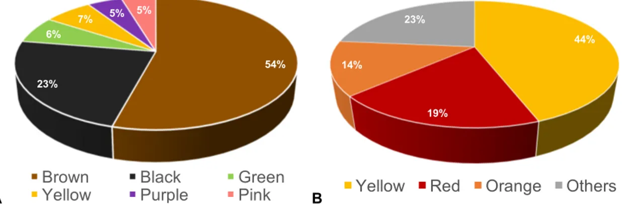
Figure 2.1. A. Percentage of the stains’ colours reported in the paper conservation literature. B. Colours of the colourants identified in the chemical/food/pharmaceutical literature. ... 6
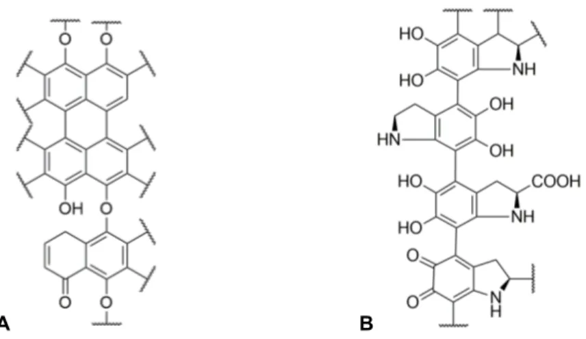
Figure 3.1. Proposed chemical structure for the DHN-melanin polymer (A) and for the L-DOPA-melanin polymer (B). ... 8
Figure 3.2. A. niger colonies on PDA (Potato Dextrose Agar medium), 14 days, 25°C, (A) front and (B) reverse. Petri dish = 90mm. A. niger (C) conidiophore, (D) conidia, bars = 50 μm. ... 9
Figure 3.3. Ch. globosum colonies on MEA (Malt Extract Agar medium) (A) front and (B) reverse, 14 days, 25°C. Petri dish = 90mm. Ch. globosum (C) perithecia and (D) ascospores (bars = 100 μm). ... 9
Figure 3.4. Cl. cladosporioides colonies on MEA, (A) front and (B) reverse, 14 days, 25°C. Petri dish = 90mm. Cl. cladosporioides (C) conidiophore and (D) conidia (bars = 50 μm). ... 10
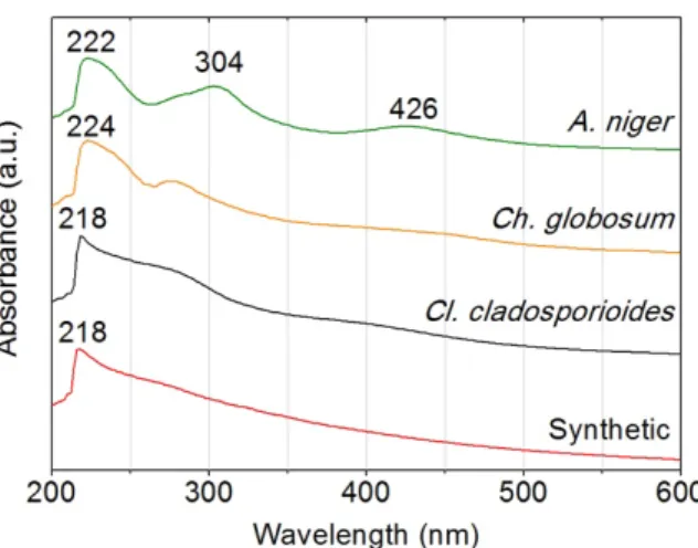
Figure 3.5. UV-Vis spectra of the purified melanins. ... 11
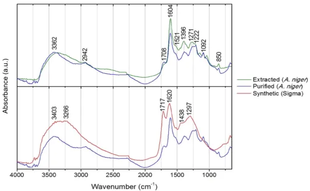
Figure 3.6. µ-FTIR spectra of the extracted and purified melanins from Aspergillus niger in comparison with synthetic melanin from Sigma. ... 12
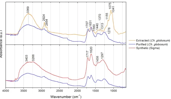
Figure 3.7. µ-FTIR spectra of the extracted and purified melanins from Chaetomium globosum in comparison with synthetic melanin from Sigma. ... 13
Figure 3.8. µ-FTIR spectra of the extracted and purified melanins from Cladosporium cladosporioides in comparison with synthetic melanin from Sigma. ... 13
Figure 3.9. μ-Raman spectra from extracted melanins: Aspergillus niger (a), Chaetomium globosum (b), Cladosporium cladosporioides (c) and synthetic melanin (d) from Sigma Aldrich. ... 17
Figure 3.10. Cl. cladosporioides 13C CP/MAS spectrum ... 18
Figure 3.11. A. niger13C CP/MAS spectrum ... 18
Figure 3.12. Sigma-Aldrich synthetic melanin 13C CP/MAS spectrum ... 19
Figure I.1. Life cycle of fungi where both the teleomorphic and anamorphic states are observed and, thus, both ascospores and conidia are produced [12]. Terms used for describing parts of conidiophores are discriminated. Scale bar = 10 μm [63]. ... 36
Figure IV.1. Diagram of the experimental procedure for the extraction of melanin from fungal
biomass from Aspergillus niger, Cladosporium cladosporioides and Chaetomium
globosum.. ... 54 Figure V.1. Infrared spectra from extracted melanins: Aspergillus niger (a), Chaetomium globosum (b), Cladosporium cladosporioides (c) and synthetic melanin (d) from Sigma Aldrich. ... 55
Figure V.2. Chemical structure of chitin (left) [146]. µ-FTIR spectra of A. niger, Ch. globosum
Index of Tables
Table 3.1. FTIR band wavenumbers (cm-1) and assignments for Ch. globosum and Cl. cladosporioides; n=streching mode; d=bending mode. ... 15
Table 3.2. FTIR band wavenumbers (cm-1) and assignments for A. niger; n=streching mode.
... 16
Table 3.3. Raman band wavenumbers (cm-1) and assignments for A. niger, Ch. globosum, Cl.
cladosporioides and synthetic melanins; s=strong, m=medium, w=weak, n=streching
mode. ... 17
Table 3.4. Characteristic Chemical shift regions in 13C CP/MAS Spectra. ... 19
Table III.1. Case studies reviewed in this work together with the type of paper, type of study,
location and respective reference are presented chronologically organized. Information
regarding if the paper was already colonized or if it was inoculated under laboratorial
conditions, is also given. ... 37
Table III.2. Fungal genera or specie found staining on paper together with their stain colour,
colourants, fungal identification method and respective references. (C – Culture; MB –
Molecular Biology) ... 39
Table III.3. Fungi known to produce melanins and to colonize paper. ... 42
Table III.4. Azaphilone colourants chemical structure (base structure in bold), colour and the
fungi responsible for its production. Note that only the genera that are also found on paper
are reported here. ... 43
Table III.5. Quinone, HAQN and Naphthoquinone colorants chemical structure (base structure
in bold), colour and the fungi responsible for its production. Note that only the genera that
are also found on paper are reported here. ... 45
Table III.6. Other colorants chemical structure, colour and the fungi responsible for its
production. Note that only the genera that are also found on paper are reported here. .. 48
Table III.7. Carotenoid colorants chemical structure, colour and the fungi responsible for its
production. Note that only the genera that are also found on paper are reported here. .. 53
Table V.1. FTIR band wavenumbers (cm-1) and proposed assignments for chitin in melanin
extractions for A. niger, Ch. globosum and Cl. cladosporioides; n=streching mode; d=bending mode [146,147]. ... 55
Symbols and Abbreviations
DCR Department of Conservation and Restoration
FTIR Fourier Transform Infrared Spectroscopy SSNMR Solid StateNuclear Magnetic Resonance UV-Vis Ultraviolet-Visible Spectroscopy
DHN type melanin
L-DOPA type melanin MEA Malt Extract Agar
PDA Potato Dextrose Agar
1. General Introduction
Books, documents, maps and works of art on paper are the carriers of a precious heritage left
by our ancestors. These special items are of great significance since they ensure the link
between past, present and future by sending a set of values from one generation to another
[1]. To ensure the passage of such legacy to future generations, knowing how to preserve
paper materials is a matter of the utmost importance.
Paper can be deteriorated due to physical, chemical and biological agents. This material is
particularly susceptible to biodeterioration processes due to its organic composition and
hygroscopicity [2]. Biodeterioration is any unwanted alteration in a material caused by the vital
activities of organisms [3], namely microorganisms, such as bacteria, fungi or lichens [4].
The main microorganisms that deteriorate paper based collections in musea, archives, and
libraries all over the world are filamentous fungi [1,5,6]. Filamentous fungi growing on paper
induce several chemical and physical decomposing processes, due to the excretion of
metabolic substances that react with the substrate, and the development of fungal structures
that alter the structure of the paper [7,8]. The excreted metabolites include colourants,
enzymes (cellulases and proteases),organic and inorganic acids, chelating agents and other
biochemical substances [8]. The excreted substances and the fungal structures themselves
are often coloured [9] and interfere with the readability of the artefacts, diminishing their value.
Accordingly to the literature, fungal stains on paper present a great variety of colours from
black to brown, red, yellow and purple stains and can ultimately deem the document
unreadable [7,10]. Figure 1.1 presents examples of different kinds of fungal stains. These
stains may migrate through successive pages, causing irreversible damage.
A B C
Figure 1.1. Three examples of different stains on paper. A - Black, brown and purple fungal stains on
an archival document (AHU-DGLAB, Portugal); B - Foxing stains on an etching (private collection)[11];
C – Black and brown stains on an etching – CleanART case study (private collection).
According to a recent survey [6], in the perspective of paper conservators, fungal stain removal
is one of the major topics that needs further research in the area of paper biodeterioration. To
better achieve a successful removal of the stains, knowing their chemical composition is
essential.
1.1. Objectives
Currently, fungal stain removal from paper documents and artworks is still very problematic,
since there is no known method that does not damage the substrate. A clear understanding
on the composition of such stains is essential for their successful and safe removal; however,
information on this area of knowledge is currently very scarce.
In order to create a solution and give a response to this lack of effective methods, the present
work is included in a starting project, CleanART, which envisioned a new methodology for
cleaning fungal stains in paper. The project activities encompass four phases: identification of
the colourants causing paper stains and the respective fungal flora; synthesis of innovative
compounds to remove fungal stains; testing the effect of the developed compounds on the
properties of different papers and media; and testing their effectiveness on real case studies
using antique documents and artworks or fac símilies.
The present work is part of the first phase of this project and is mainly focused on the extraction
and characterization of colourants produced by fungi, which were formerly identified as stain
causing species. This study will contribute to identifying which compounds should be targeted
and removed from the paper structure; and will allow for a colourant-specific testing of the
newly developed cleaning methods.
To achieve this main goal, it was necessary to:
• Review the literature regarding fungal stains on paper and the respective fungal
colourants; select the fungal species to carry out the extractions; analyse and
characterize the extracted colourants.
This work focused primarily in fungal melanins, since these are the most damaging colourants
for paper documents and artwork, not only due to their dark colour and frequent occurrence,
2. Colourants produced by fungi colonizing paper: an overview
2.1. Filamentous fungi
Filamentous fungi belonging to the Ascomycota phylum are the main microorganisms
deteriorating paper-based collections worldwide, being mainly responsible for the appearance
of different colour patches with biological origin on paper.
In their vegetative stage, fungi consist of a tangle of slender, thread-like hyphae, whereas the
reproductive stage is usually more obvious [8]. In their reproductive stage, both teleomorphic
(sexual) and anamorphic (asexual) states may be observed, according to species (Appendix
I). In the large majority of the cases, “conidium” is the term used to describe ascomycetes’
asexual spores [12]. Adhesion to the substrate is very important, since it is strictly linked to the
ability to transform it. Fungi attach directly by hyphae [13], acting like a root for a better
adherence and nutrient absorption, while the reproductive structures like spores allow wide
distribution and increase fungal resistance to adversity [8].
Fungal spores can be easily carried by air movements, and eventually settle in the dust [14],
which is deposited in all kind of materials, including paper documents. Its high resistance to
adverse conditions allows them to remain in a latent state until relative humidity and
temperature conditions favours germination [13], putting archival materials at risk.
2.2. Colourants produced by fungi
Fungi can produce organic colourants during their development. These are characteristic of
different species, but the colour of fungal stains arises not only from the chemical composition
of colourants, but also from many other factors, such as the chemical composition of the
substrate; presence of metallic trace elements (e.g. iron); nutrient availability; acidity/alkalinity
of the medium; presence of other microbial species; and environmental conditions [13].
Fungal colourants are composed of complex chemical substances that are formed during
metabolic processes; these colourants may be encrusted in spores, present in mycelium (the
mass of filaments constituting the body of the fungus), or secreted to a substrate such as paper
[15]. The release of colourants on the substrate or the presence of coloured microorganisms
causes the appearance of different colour stains or patches on many works of art [13].
Some colourants are enzyme inhibitors or antibiotics [13], but the main biological functions of
fungal colourants are related to light harvesting and processing, photo-protection, as well as
absorption and neutralization of protons that could potentially damage fungal cellular structures
[16]. Melanins, for instance, have had several biological functions attributed to them, such as:
protection against radiation (e.g. UV), enzymatic lysis, high temperatures, or oxidizing agents;
metals; action as a virulence factor; or capacity of increasing resistance to fungicides [17].
Besides, such as in flowers, the striking colouration of fungi can help in their dispersion, since
conidia could easily adhere to any animal attracted and touching the mycelial surface [18].
Biosynthetically, most colourants produced by fungi are polyketide-based [19]. Representative
classes may include structures such as azaphilones, anthraquinones, hydroxyanthraquinones,
naphthoquinones and others, each exhibiting an array of colour hues [20]. Colourants from
fungi can be broadly classified chemically as polyketides and carotenoids [20].
2.2.1. Polyketide colourants
Fungal polyketides are natural products that include fungal melanins, and other colourants.
These are one of the largest and most structurally diverse classes of naturally occurring
compounds, ranging from simple aromatic metabolites to complex macrocyclic lactones [21].
2.2.1.1. Melanins
Melanins are polymers formed by oxidative polymerization of phenolic or indolic compounds
[22]. In some instances, the polymer subunits have been discovered; still, the exact
arrangement of these subunits in the polymer remains unknown [23]. They have resisted
atomic-level structural examination due to their insolubility and amorphous organization [24].
Microscopic studies show an overall granular structure.
In fungi, melanin granules are localized in the cell wall where they are likely cross-linked to
polysaccharides [23], being very complex since they also contain intra-granular proteins [25].
Often the resulting colourants are brown or black in colour [22,23]. In fungi, the two most
important types are DHN-melanin (named for one of the pathway intermediates,
1,8-dihydroxynaphthalene – 1,8-DHN) and DOPA-melanin (named for one of the precursors,
L-3,4- dihydroxyphenylalanine) [22] (Appendix II). In the Ascomycota fungi, where most of the
fungi found in archives are represented [8], melanin colourants are generally synthesized from
the pentaketide pathway in which 1,8-DHN is the immediate precursor of the polymer [26].
Due to their chemical and physical endurance, the safe removal of melanized fungal stains
from paper is very problematic [27], making them the hardest colourants to remove from paper
substrates.
2.2.1.2. Azaphilones
Azaphilones are polyketide derivates that can be defined as a structurally diverse class of
fungal secondary metabolites [19,21], with a highly oxygenated pyranoquinone bicyclic core,
usually known as isochromene, and a quaternary carbon centre [21]. Azaphilones can be
coloured or uncoloured, and, when coloured, are responsible for the bright yellow, red or green
including genera Aspergillus, Penicillium, Chaetomium, among others[21], which may colonize
paper causing the appearance of coloured patches.
2.2.1.3. Quinones
Quinones are a class of organic compounds derived from aromatic compounds by conversion
of an even number of –CH= groups into –C(=O)– groups with any necessary rearrangement
of double bonds, resulting in a fully conjugated cyclic dione structure [28]. Numerous quinones
biosynthesized by the polyketide pathway have been isolated from fungi [29], and display an
array of colours, like yellow, orange, or red, according to the position of the keto groups [30].
Hydroxyanthraquinoid (HAQN) colourants, derivatives of quinones, are widespread in nature
and have also been found abundantly in microorganisms, particularly in filamentous fungi
belonging to the genera Penicillium and Aspergillus , with different colour hues [31].
Naphthoquinone colourants are also derivatives of quinones, very common in fungi and have
been the subject of many studies regarding their chemical structure, biosynthesis and
biological significance [30].
2.2.2. Carotenoids
Since fungi are non-photosynthetic organisms, carotenoids are not as widespread as they are
in plants, where they play an important role in the photosynthetic process. Nevertheless,
carotene hydrocarbons have been found in several fungi [18,29], and are widely accepted as
protecting agents against oxidative stress [18]. Containing an aliphatic polyene chain usually
composed of eight isoprene units that include light-absorbing conjugated double bonds,
carotenoids provide characteristic yellow, orange or reddish colours [18].
2.3. Fungal stains on paper versus Colourants produced by fungi
A review of the literature from the paper conservation area dating from 1970 to 2015, was
compiled in 26 studies focused on the identification of fungal species from stains on
biodeteriorated paper (Appendix III, Table III.1). Accordingly to this review, fungal stains can
be divided into three main groups: various colour stains; melanised stains (dark brown or black
stains caused by melanin) and foxing (small and round-shaped spots with reddish or
yellowish-brown colour resulting from the ageing and/or oxidation of fungal residues).
After gathering all the information from the available literature, 25 different fungal genera were
found to cause staining on paper (Appendix III, Table III.2). The fungi that are most frequently
identified on paper belong to the genera Aspergillus (29%) and Penicillium (13%).
The most common stains found on paper and associated with fungal activity were brown stains,
followed by black and yellow to greenish tones (Fig. 2.1.A). The brownish colour is most
frequently associated with the foxing phenomena described in the literature [32]. Black colour
percentage (around 13%) of the authors associate a coloured stain with specific colourants
produced by fungi. However, no analyses are actually carried out in these studies to confirm
the presence of such colourants.
Therefore, a research outside the scope of paper conservation was necessary to understand
which chemical structures could be causing this damage, namely the staining of paper by fungi.
There is a great variety of colourants produced by fungi, but not all of them colonize paper. In
the literature, several fungal colourants were mentioned and characterized, being used in the
food, chemical and pharmaceutical industries. However, not all of them were detected on
colonized paper, according to a previously carried out review [11].
From the colourants produced by fungi that colonize paper, circa 80 different compounds were
gathered, from literature between 1934 and 2016, and are presented in tables (Appendix III,
Tables III.3-III.7) regarding their chemical structure, colour and producing fungal species.
The majority of these colourants were polyketides (about 96%) and only 4% were carotenoids.
There were only three different carotenoids identified: Neurosporaxanthin, β-carotene and
Sporopollenins. The latter are oxidative polymers of carotenoids and carotenoid esters [35].
From the 96% of polyketides reviewed on the literature, about 20% were azaphilones and 33%
were quinones. The remaining 46% were attributed to other types of colourants, that didn’t
belong to the chemical classes defined initially. Additionally, at least 9 species of fungi
identified on paper are known to produce melanins. Melanin was taken into account as a single
colourant, although it can be produced by two biosynthetic pathways: DHN and DOPA.
The majority of colourants produced by fungi in the chemical/food/pharmaceutical literature
have a yellow colour (44%), followed by red (19%) and orange (14%) (Fig.2.1. B). Many times,
the colour is a mixture of hues (e.g. red-brown for Tritisporin [36]). In the paper conservation
literature, the most common stains produced by fungi on paper substrates are coloured in
brown or black (Figure 2.1. A), having no clear correspondence with the bright colours studied
in the chemical/food/pharmaceutical areas (Figure 2.1-B).
A B
Figure 2.1. A. Percentage of the stains’ colours reported in the paper conservation literature. B. Colours of the colourants identified in the chemical/food/pharmaceutical literature.
54% 23%
6%
7% 5% 5%
Brown Black Green
Yellow Purple Pink
44%
19% 14%
23%
However, these data must be considered at light of the research area they belong to, since red
and yellow hues are appealing colours and have been the most extensively used food
colourants [20], for instance. Besides, the colours found on stained paper may be a
combination of the colourants produced by fungi present on paper. When extracting the
colourant from Aspergillus melleus, it was verified that the brown solid contained viomellein –
reddish-brown (11%), xanthomegnin – orange (50%), and viopurpurin – purple-black (1%) [37].
Also, stains’ colour produced by fungi on paper may as well be a result of the colourants’
degradation in the given medium, causing its darkening. For instance, sporopollenin is a brown
product of oxidative polymerization of yellow b-carotene [31].
Besides, many of the identified colourants are biosynthetic precursors of others, so depending
on the development stage, a different colour may be observed. This is the case of '4C-labeled
red averufin, the orange versiconal hemiacetal acetate, and the pink-orange versicolourin A,
which are converted to the yellow sterigmatocystin by Aspergillus versicolour [38]. This is also
the case of dopachrome and melanochrome, the pink and purple intermediates in the formation
of the melanin colourant in Aspergillus nidulans by the L-DOPA pathway [26].
Finally, different colours may often be due to the modification of the same colourant, depending
on different reactions with the substrate. For instance, variation in copper levels among
commercial fungal culture media (e.g. potato dextrose agar) correspond to variable colouration
in multiple fungal species, including Stachybotrys atra and Cladosporium herbarum [39].
Overall, there is no doubt that colourants produced by fungi are a serious problem to
conservators, especially as the dominant species that produce these colourants belong to the
most common genera identified on paper substrates, namely Aspergillus and Penicillium. Each
colourant, with different properties, may require diverse removal methods. Therefore, it is of
the utmost importance that developments are carried out in this field of knowledge, so that
targeted removal techniques can be developed without damaging our cultural records on
paper.
The present study is an important advance in terms of knowledge, since almost no information
was published about the colourants produced by fungi on paper. By relating the information
acquired in different areas, such as the food industry, with the information already acquired in
the conservation area, we are one step closer to define which colourants may be present in
specific stains on the paper substrate, which will have an impact on the conservation strategy.
By knowing which colourants to target, new and safer methods can be developed. Likewise, it
is also relevant to consider whether the fungi produce colourants within their structure or as
excreted coloured products, which determines the extent of staining – whether a simple
removal of fungi is sufficient, or its products are linked to the paper structure, which carries
3. Extraction and characterization of melanins produced by fungi
According to the litterature, there are virtually no studies regarding the chemical structures of
the colourants that fungi produce on paper substrates. To get enlightenment on this subject,
melanins produced by fungi were selected as primary colourants for the initial phase of the
CleanART project, where the extraction and characterization of colourants produced by fungi
is intended. Melanins were chosen because they cause much of the black and brown staining
on paper objects, existing either as components of the fungal structures or as excreted
polymers attached to the paper fibres [27]. Removal of melanins without damaging works of
art is a problematic issue, since these polymers have a great chemical endurance [17].
After decades of investigation, the exact molecular structure of these colourants remains a
mistery. This is due to several challenging features of the system, including almost complete
insolubility in all solvents, an amorphous particulate character, and extreme molecular
heterogeneity. A proposed chemical structure for these polymers’ (DHN-melanin and
L-DOPA-melanin) molecular arrangement is given in Fig. 3.1, based on the literature [52–55, for e.g.],
where several models have been proposed. The main, if not the only, similarity between the
various models is the presence of the monomers linked by covalent bonds, where these
primary units are in different states of oxidation. The spatial arrangement of these units in the
biopolymer structure may be quite variable, providing the material with increased
heterogeneity.
A B
Figure 3.1. Proposed chemical structure for the DHN-melanin polymer (A) and for the L-DOPA-melanin polymer (B).
For the characterization of fungal melanins, three species known to produce this colourant on
paper [11,15,22,23,30,33,44] were selected as case studies: Aspergillus niger, Chaetomium
globosum and Cladosporium cladosporioides. The characteristics of the three-selected
Aspergillus niger
Aspergillus niger is a very common member of the Aspergilli [29]. It’s a filamentous ascomycete
[45], that produces a white mycelium before forming black conidia. Mature colonies appear
grey/ greenish-black (Fig.3.2.A) [29]. Its reverse is usually yellow (Fig.3.2.B) [46].
A B C D
Figure 3.2. A. niger colonies on PDA (Potato Dextrose Agar medium), 14 days, 25°C, (A) front and (B)
reverse. Petri dish = 90mm.A. niger (C) conidiophore, (D) conidia, bars = 50 μm.
A. niger’s brown Aspergillin has been the subject of biochemical characterization, and was
assumed to be a polymer composed of DHN melanin and a hexahydroxyl pentacyclic quinoid
(HPQ) compound [47]. Consequently, one of the main characteristics of A. niger is the
production of black or dark brown conidia resulting from combination of these dark brown
melanins with hexahydroxyl pentacyclic quinoid green colourants [44].
Although most Ascomycetes produce melanin through the DHN pathway, recent studies
suggest that some Aspergillus spp. produce L-DOPA melanin, A. niger included. According to
the literature [48] melanin synthesis by A. niger was inhibited by kojic acid and tropolone,
indicating a L-DOPA melanin pathway.
Chaetomium globosum
Chaetomium globosum belongs to the phylum Ascomycota [12]. Ascomata are superficial,
greenish olivaceous or slightly dark olivaceous. Terminal hairs are abundant and brown and
the ascospores are olivaceous and brown when mature (Fig. 3.3. A) [49]. Orange exudates
diffusing into the medium are noticeable on its reverse (Fig. 3.3. B) [49].
Ch. globosum is a known melanin producer and, according to the literature [50], this
biosynthesis is via the DHN pathway; as, when tricyclazole, an inhibitor of the DHN pathway,
was added to plates, it resulted in an absence of pigmentation.
A B C D
Cladosporium cladosporioides
Cladosporium cladosporioides belongs to one of the largest genera of dematiaceous
hyphomycetes [51]. Colonies are rapidly growing, olive-gray to olive-brown or black (Fig. 3.4.
A, B) [52].
A B C D
Figure 3.4. Cl. cladosporioides colonies on MEA, (A) front and (B) reverse, 14 days, 25°C. Petri dish = 90mm. Cl. cladosporioides (C) conidiophore and (D) conidia (bars = 50 μm).
This fungus has already been frequently isolated from paper materials [53] and has been
pointed out as a DHN-melanin producer [51], since when adding tricyclazole, a specific inhibitor
of DHN-melanin synthesis, growth and colouration of colonies were affected.
3.1. Materials and Methods
A. niger was obtained from the mycological collection of Instituto Nacional de Saúde Doutor
Ricardo Jorge (Lisbon, Portugal). Ch. globosum and Cl. cladosporioides were isolated from
fungal stains on an engraving and identified by Molecular Biology at Universidade de Coimbra
(Coimbra, Portugal).
Fungal cultures were grown in three culture media: a) Potato Dextrose Agar (PDA) - Solid
medium, b) Malt Extract Agar (MEA) – Solid medium and c) Malt Extract (ME) – Liquid medium.
PDA was used for maintenance of the cultures. Liquid ME allowed for an easier extraction of
colourants from A. niger and Cl. cladosporioides by filtration. Solid MEA had to be used for Ch.
globosum, since it favoured its sporulation, and the melanin to be extrated was located in those
reproductive structures (spores).
3.1.1. Extraction and purification of melanins from fungal biomass
This experimental protocol was adapted from the literature [48]. Twenty Petri dishes with Ch.
globosum cultures on MEA were incubated for one month in the dark, at 25ºC inside a drying
oven, after which the cultures were collected and boiled in distilled water and filtered.
One-month old cultures of A. niger and Cl. cladosporioides in ME were also filtered. The individual
fungal masses obtained were separately crushed with 2M NaOH (pH 10.5) and allowed to
stand for 48 h. After a second filtration to remove fungal debris, the resulting filtrates were
precipitated with 2M HCl to pH 2.5. These were incubated overnight at room temperature and
using 6M HCl at 100ºC for 2h to remove carbohydrates and proteins. They were then treated
with organic solvents: chloroform, ethyl acetate and ethanol to remove lipids. The resulting
precipitates were dried at room temperature, dissolved in 2M NaOH and filtered. The collected
solutions were precipitated with 6M HCl (kept overnight), washed with distilled water and
allowed to dry at room temperature (Appendix IV). These precipitates were considered to be
purified melanin and were used for further analyses.
3.2. Results and Discussion - Characterization of melanins
UV-Vis analyses
Melanin samples were dissolved in a NaOH 0.1 M solution and scanned from 200 to 600 nm
with 2 nm intervals on a T90+ spectrometer from PG Instruments. Before starting all
measurements, a baseline was made with the solvent. Since the apparatus has a double
beam, all measurements were made against a reference cell which had solvent, thus
discounting any possible fluctuations in absorption which may have other origins as physical
differences in the cells used.
The UV-Vis absorption spectrum of a given
colourant, melanins in this case, is a graphical
representation of the absorbed radiation as a
function of the wavelength (λ) [16]. The spectra
obtained for the purified fungal melanins and
synthetic melanin showed a more pronounced
band at the UV region (Fig. 3.5). These appear
to be atypical in organic chromophores and are
probably the result of a structure that presents
considerable heterogeneity and disorder.
In the melanin samples, the monomeric units from DHN-melanin and L-DOPA-melanin, as well
as units resulting from the oxidation of these monomers, are predominant in their structure
(Fig. 3.1). The presence of this variety of species in the composition of the melanin polymers
leads to an overlapping of energy levels, with a greater number of electronic transitions along
the electromagnetic spectrum and, consequently, an extension of the absorption spectrum. On
the other hand, the aromaticity of the units that make up the melanin structure favours efficient
electronic delocalization, which contributes to the stabilization of the polymer and,
consequently, to an increase in its photo-protective capacity [54]. The result is typical of the
absorption profile of these type of aromatic compounds, such as melanin, which absorb
strongly in the UV region and decrease progressively as the wavelength is increased [55]. This
is in agreement with the photoprotection function of melanin suggested in fungi [25].
The nature of the purified colourants from A. niger, Ch. globosum and Cl. cladosporioides were
therefore confirmed by their spectral profiles, since among biological colourants, only melanins
absorb throughout the entire visible region. Since the colours we observe are the result of the
selective light absorption process, if all wavelengths of light are absorbed and none reflected,
we recognize it as black. This characteristic is responsible for the dark colour of these
colourants, since we perceive no colour [55]. Recent reports mention a characteristic
absorption peak was observed at 217 nm with a small shoulder around 260-280 nm, which is
similar to synthetic melanin, and suggests the presence of phenol groups [48,56].
For the Aspergillus niger’s spectrum, two distinct maxima are observed at 304 and 426 nm,
respectively. These maxima can be assigned to absorption of light by the residual protein
fragments which are not completely removed during the isolation and purification procedure
[25]. Besides, according to the literature [47], A. niger’s melanin is linked to a green colourant,
which could be the reason of the absorbance in the violet region (426 nm).
µ-FTIR analyses
Micro-FTIR analyses were performed on a Nicolet Nexus spectrophotometer interfaced with a
Continuμm microscope with a MCT-A detector cooled by liquid nitrogen. The spectra were
collected in transmission mode, with a spatial resolution of 50–100 µm, optical resolution of
4 cm−1 and 128 co-added scans, using a thermo-diamond anvil compression cell.
FTIR spectra were carried out and studied to confirm if the extracted and purified colourants
were melanins. The obtained spectra are shown in Figs. 3.6-3.8.
Figure 3.6. µ-FTIR spectra of the extracted and purified melanins from Aspergillus niger in comparison
with synthetic melanin from Sigma.
Ab
s
o
rb
a
n
c
e
(a
.u
Figure 3.7. µ-FTIR spectra of the extracted and purified melanins from Chaetomium globosum in comparison with synthetic melanin from Sigma.
Figure 3.8. µ-FTIR spectra of the extracted and purified melanins from Cladosporium cladosporioides
in comparison with synthetic melanin from Sigma.
Firstly, spectra were acquired for the extracted melanins, prior purification, to assess if this
would be sufficient. Melanins are typically localized in cell walls where they are most likely
cross-linked to polysaccharides, mainly chitin [56] (Appendix V, Fig. V.2) which is a major
constituent of the fungal cell wall [29]. Accordingly, many of the characteristics bands of fungal
chitin were observed in the acquired spectra (Appendix V, Fig. V.1), showing that the extraction
Ab
s
o
rb
a
n
c
e
(a
.u
.)
Ab
s
o
rb
a
n
c
e
(a
.u
procedure alone is not sufficient for the isolation from other cell wall constituents, especially
for Ch. globosum and Cl. cladosporioides, for which the FTIR spectra are almost coincident
with their mycelia’s FTIR spectrum (Appendix V, Fig. V.2). A proposed assignment for the chitin
bands present in the spectra of the extracted melanins is given in Table V.1 (Appendix V).
Following purification of the extracted melanins, new FTIR spectra were acquired. These were
compared with Sigma’s synthetic melanin spectrum in order to verify their nature, and also with
the previously obtained spectra of the extracted melanins, to confirm their differences and a
successful purification (Fig. 3.6, 3.7 and 3.8).
Although early researchers on melanin structure used UV and visible spectroscopy, as well as
IR spectra, to distinguish between melanins from different sources, these methods are now
generally regarded to be of dubious value [17]. However, it is possible to access if the extracted
and purified samples are in fact from melanin, by comparison with the synthetic melanin from
Sigma-Aldrich (DOPA-melanin). As previously mentioned, there are two distinct pathways for
fungal melanin formation. Ch. globosum and Cl. cladosporioides are reported to produce
DHN-type melanins (Fig. 3.1.A), whereas A. niger is reported to produce L-DOPA-melanin (Fig.
3.1.B), each of which having different precursors (1,8-DHN) for DHN-melanin and
dihydroxyindoles for L-DOPA-melanin).
The presence of carboxyl groups in the melanins’ structures, as well as aromatic rings and
phenolic groups, is evidenced by bands in the obtained spectra, characteristic of these
polymers’ molecular arrangement (Fig.3.1). Purified melanin spectra are very similar to the
spectrum of synthetic melanin, having common absorption peaks: the broad bands in the
3600-3200 cm-1 region, attributed to OH stretching, further indicating the presence of phenolic and
carboxylic groups in the melanin structure [51,56,57]; the clear peaks at 1720-1700 cm-1,
corresponding to the oscillations of C-O groups from acids, esters or ketones [56]; a strong
absorption at 1650–1620 cm-1 attributed to stretching vibrations of aromatic C=C or C=O
groups [48,51], which is typical of a conjugated quinoid structure and is believed to be
important for the identification of melanin [48]; in case of L-DOPA melanins, such as the
synthetic melanin from Sigma, it may be indicative of the compounds containing carbonyl
groups conjugated with a benzene ring, corresponding to the typical quinone structure of
DOPA-melanin [51]; an absorption band at 1440-1370 cm-1, indicating nitrogen content (C-N
bending) [56], and indolic NH stretches [57], as DOPA melanins, in the case of synthetic DOPA
melanin, and phenolic COH bends in both melanins; and the peak around 1250 cm-1 that
corresponds to C-OH stretching or angular deformation of O-H [51,56].
Comparing the purified melanins from Ch. globosum and Cl. cladosporioides with the extracted
melanin prior purification, a great difference is confirmed between the different samples. Chitin
aliphatic nCH2; nCH3, which could indicate a remaining contamination of these cell wall
carbohydrates [48,56,57].
A proposed assignment for the specific peaks of these melanins (Ch. globosum/Cl.
cladosporioides) is given (Table 3.1), regarding the chemical structure from
dihydroxynaphthalene (DHN)-melanin, formed by oxidative polymerization of 1,8-DHN (Fig.
3.1. A). The similarity between these melanins’ spectra supports the literature, since both these
fungi are reported to produce DHN-type-melanins.
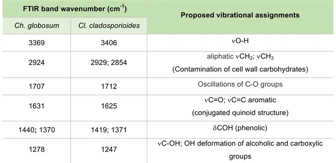
Table 3.1. FTIR band wavenumbers (cm-1) and assignments for Ch. globosum and Cl. cladosporioides;
n=streching mode; d=bending mode.
FTIR band wavenumber (cm-1)
Proposed vibrational assignments
Ch. globosum Cl. cladosporioides
3369 3406 nO-H
2924 2929; 2854 aliphaticnCH2; nCH3
(Contamination of cell wall carbohydrates)
1707 1712 Oscillations of C-O groups
1631 1625 nC=O; nC=C aromatic
(conjugated quinoid structure)
1440; 1370 1419; 1371 dCOH (phenolic)
1278 1247 nC-OH; OH deformation of alcoholic and carboxylic groups
Absorption peaks from IR spectrum obtained from A. niger’s purified melanin are nearly
coincidental with the one from its extracted melanin (Fig. 3.7). Its extraction was easier than
for Ch. globosum and Cl. cladosporioides, showing that without a purification process, the
extracted melanin was already quite pure when compared with the extractions carried out for
the other species. This could be associated with cellular location of the biologically synthesized
melanin. A. niger’s melanin is located in its black conidiospores which are rather abundant
[44], whereas Cl. cladosporioides’ is located in the mycelia and conidia [51] and Ch. globosum
in the perithecia, with conidia not generally produced [46]. Ch. globosum was in fact the hardest
fungal melanin to extract, requiring larger amounts of mycelia in order to obtain a sufficient
amount of sample for the analyses.
Comparing A. niger’s to Sigma’s and other fungal melanins, characteristic peaks are also
observed as previously described. However, differences are noticed particularly in the carbonyl
and indolic regions, which is in agreement with reports from literature regarding the
absorption regions are observed with defined peaks: 1521 cm-1 attributed to N-H stretching
and bending [51,56]; and 1092 cm-1 attributed to C-O stretching, to the aromatic ring C-H
stretching and characteristic of C-N elongation, which may derive from the pyrrole units that
make up the L-DOPA melanin structure [48,51], reported to be produced by this fungal specie.
Moreover, the weak absorption peaks at 800-600 cm-1 could indicate some positions of
aromatic rings were substituted and the conjugated system with low amount of aromatic
hydrogen content was formed [57].
A proposed assignment for the specific peaks A. niger’s purified melanin is given in Table 3.2,
regarding the proposed chemical structure of the L-DOPA-melanin polymer (Fig. 3.1. B).
Table 3.2. FTIR band wavenumbers (cm-1) and assignments for A. niger; n=streching mode.
FTIR band
wavenumber (cm-1) Proposed vibrational assignments
3362 nN-H2; nO-H
2942 AliphaticnCH2; nCH3
nNH
1708 Oscillations of C-O groups
1604 nC=O; nC=C aromatic (carbonyl groups conjugated benzene ring – typical structure of DOPA melanin); nCOO
1396
nCN (strong suggestive of a substantial amount of indole groups in the
structure of the colourant – typical structure of DOPA melanin)
nNH (indolic)
nCOH (phenolic)
1271; 1222 nC-OH; OH deformation of alcoholic and carboxylic groups
1092 nC-O; aromatic ring C-H;
850 Some positions of aromatic rings were substituted and the conjugated system with low amount of aromatic hydrogen content was formed.
μ-Raman analyses
Raman Microscopy was carried out using a Labram 300 Jobin Yvon spectrometer, equipped
17 mW HeNe laser operating at 632.8 nm. Melanin samples were analyzed with a solid state
laser operating at 532 nm. Spectra were recorded as an extended scan. The laser beam was
focused either with a 50x or a 100x Olympus objective lens. The laser power at the samples’
surface was varied with the aid of a set of neutral density filters (optical densities 0.6, 1 and 2),
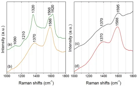
The obtained spectra were dominated by two intense and broad peaks: a higher intensity band
at circa 1580 cm-1 and a second band located at around 1370 cm-1 (Fig. 3.9).
Figure 3.9. μ-Raman spectra from extracted melanins: Aspergillus niger (a), Chaetomium globosum (b), Cladosporium cladosporioides (c) and synthetic melanin (d) from Sigma Aldrich.
A broadband background is noticeable due to auto fluorescence emission properties of
melanin. It has been proved [58], using multiple wavelengths and a variety of melanin sources,
that the two prominent peaks observed in melanins are indeed due to inelastic Raman
scattering rather than fluorescence or other nonlinear processes. These two peaks can be
interpreted as originating from the in-plane stretching of the aromatic rings and the linear
stretching of the C-C bonds within the rings, along with some contributions from the C-H
vibrations in the methyl and methylene groups.
Other bands or shoulders of lower intensity that appear in the spectrum of A. niger can be
attributed to the non-pigment residues of fungi. For instance, lipid bands at 1210 and 1080
cm−1 that can be both assigned to C–C skeletal stretching.
However, the actual assignment of the two main melanin bands is not entirely agreed upon
between authors, but it is most likely that the broad features of melanin are composed of many
overlapping Raman bands because of the vibrations of the constituent monomers [59].
Table 3.3. Raman band wavenumbers (cm-1) and assignments for A. niger, Ch. globosum, Cl.
cladosporioides and synthetic melanins; s=strong, m=medium, w=weak, n=streching mode.
Raman band wavenumber (cm-1) Proposed vibrational assignments
A. niger Ch. globosum Cl. cladosporioides Synthetic (Sigma)
1566 (s)
1620 (s) 1588 (s) 1595 (s) 1588 (s) nCCH aromatic
Solid-State NMR analyses
Samples were packed into 4 mm zirconium rotors and sealed with Kel-F caps. On selected
experiments an insert was packed with the sample and placed inside the rotor.
13
C CP/MAS spectra was obtained on an Avance III WB spectrometer operating at 7.2 T,
corresponding to a 300 MHz Larmor frequency for 1H and 75 MHz for 13C.
Magic angle spinning rate was 5-10 KHz. The cross-polarization was performed applying a
variable spin-lock sequence RAMP-CP/MAS with a contact time 1.2 s and a recycle delay of 2
s. 5000 points were sampled and accumulated during 4000-15000 transients.
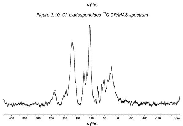
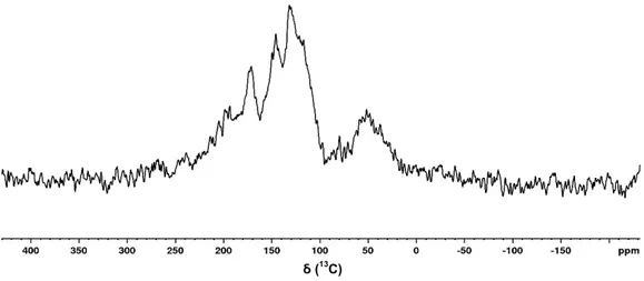
The 13C CP/MAS spectra of the melanin samples revealed differences in chemical shift
regions, maximum of those bands and in linewidth of the peaks (Fig. 3.10-3.12).
Figure 3.10. Cl. cladosporioides 13C CP/MAS spectrum
Figure 3.11. A. niger 13C CP/MAS spectrum. *Spinning Side Bands.
ẟ (13C)
ẟ (13C)
Figure 3.12. Sigma-Aldrich synthetic melanin 13C CP/MAS spectrum
Unfortunately, it was only possible to obtain sufficient melanin sample for two of the selected
species, namely Cl. cladosporioides (reported to produce DHN-type melanin) and A. niger
(reported to produce L-DOPA-type melanin). This is a solid state analyse, so a major amount
of sample must be sparred to do so. Characteristic chemical shift regions for the spectra indeed
obtained are given in Table 3.4.
Table 3.4. Characteristic Chemical shift regions in 13C CP/MAS Spectra.
Sample Carbonyl Aromatic Aliphatic
Cq CH
A. niger 215-150 (Carbonyl + Cq) 140-88 84-0
Cl. cladosporioides 190-165 165-150 144-100 81-9
Sigma-Aldrich 185-163 163-137 140-90 70-12
Note: Chemical shifts are reported in ppm; CH represents protonated carbons and Cq represents non-protonated carbons.
Analyzing the obtained spectra, the samples obtained are predominantly aromatic, with phenol
groups, having some fragments oxidized to carbonyls, which is consistent with the type of
structures expected from the consulted literature and with the previously carried out analyzes,
also showing carbonyl, phenolic and aromatic groups.
The synthetic melanin sample from Sigma-Aldrich is known to be from L-Tyrosine origin,
oxidized in DMSO with H2O2. Comparing that spectrum with the one from the A. niger sample
and the reported 13C chemical shifts for tyrosine derived compounds in Table VI.1 (Appendix
VI), the melanin production pathway in A. niger can be assigned to a L-DOPA pathway
(Appendix II), which is in agreement with the reports from literature [48] and the previously
carried out analyses - namely indolic vibrations in the IR spectrum, indicating Nitrogen content
(correspondent to the typical structure of the L-DOPA-melanin).
Regarding characteristic regions, the aromatic and carbonyl regions are different between A.
niger’s and synthetic melanin, which can be attributed to a lower oxidation of the monomers
[60] in the synthetic melanin, resulting in fewer signals in the carbonyl region. The linewidth is
also different, with A. niger‘s melanin having much sharper peaks. This broadening effect is
caused, in the synthetic melanin, by either a higher molecular weight that decreases the
molecule correlation time and makes the NMR signal decay faster, or to a higher crosslinking
in the polymer which has the same effect as the size of the polymer, by restricting the molecular
motion inside the polymer structure.
The molecular weight (MW) difference between natural and synthetic melanin in literature is
conflicting, with reports of similar MW regardless of the source, and of higher size in the natural
produced melanins [61]. The natural polymer being of higher size than the synthetic polymer
does not fit the experimental data, as it would lead to sharper peaks in the synthetic sample
rather than in the A. niger’s melanin. Therefore, the synthetic sample should have a more
crosslinked structure than the produced by A. niger.
Cl. cladosporioides melanin sample has a similar peak width to the Sigma-Aldrich sample, also
indicating a very crosslinked or a higher molecular weight polymer than A. niger’s.
Cl. cladosporioides melanin is reported in literature to be of 1,8-DHN pathway [62]. Comparing
the observed 13C chemical shift with model compounds present in Table VI.1 (in Appendix VI)
from literature, it is likely to be of the DHN pathway as the observed signals are in better
agreement with the naphthalene derived structures. At 160 ppm the naphthalene phenol
groups fit the one of the observed shifts in the Cl. cladosporioides melanin, as the 140 ppm
shift from the catechol ring found in L-DOPA fits the shifts in the synthetic Sigma melanin and
the A. niger sample.
Without further analyses, it is only possible to ascertain that, regarding the observed
differences between the carbonyl and aromatic regions of the obtained spectra, the melanins
produced by the two species analyzed, namely A. niger and Cl. cladosporioides, are clearly
from two different biosynthetic pathways. This is in agreement with the initially consulted
literature and with the other analyses carried out. Also, melanin sample produced by A. niger
is considerably more crystalline and organized than Cl. cladosporioides’ melanin, which is
Conclusions
The successful cleaning of fungal stains from paper is considered a priority by paper
conservators, but data from the literature review show that, despite the relevance of this
subject, there is still a lack of information since most of the authors refer only the stain colour
and patterns (e.g. black, purple, etc.) but they do not indicate the colourant or colourants (or
chemical compound) responsible for the stain or the colorimetric parameters. Therefore, the
present work was of the utmost importance in linking the knowledge between the conservation
literature concerning fungal stains on paper and the remaining literature (from e.g. food,
chemical and pharmaceutical industries) concerning fungal colourants production.
The paper conservation literature shows that the most common stains produced by fungi on
paper substrates were brown or black, having no clear correspondence with the bright colours
(e.g. red and yellow) reported by studies from other areas of knowledge. However, stains’
colour produced by fungi on paper may be a result of the colourants’ degradation in the given
medium, causing its darkening, or a convergence of different colourants produced by the same,
or different species, present in the fungal stains.
It was assessed that these fungal stains on paper are in no doubt a serious problem to
conservators, particularly because the two most common fungal genera producing these
colourants are the same as the ones identified on paper substrates: Aspergillus spp. and
Penicillium spp.
Each colourant, with different properties, may require distinct removal methods. Thanks to the
present work, innovative cleaning techniques targeting the most concerning type of stain for
paper conservators, namely black stains caused by melanised fungi, will be addressed and
tested specifically for these molecules.
The extraction and characterization of fungal melanins produced by fungi isolated from paper
was a stepping stone in the current investigation concerning which colourants may be present
in a given stain, in order to develop new reliable and safer methods for these stains’ removal.
A successful melanin extraction of the three selected fungal species was obtained according
to the UV-Vis, µ-FTIR and µ-Raman data, showing a predominantly aromatic structure with
carbonyl and phenolic groups for all samples, with a special difference noticed in the IR
spectrum of the sample obtained from A. niger – namely nitrogen content, indicating indolic
groups, typical of the L-DOPA melanin structure.
For the two species where enough melanin sample was obtained to carry out SSNMR
analyses, a proposed monomer type melanin was given, namely DHN type melanin for Cl.
cladosporioides and L-DOPA melanin for A. niger. These analyses were consistent with the
![Figure 1.1. Three examples of different stains on paper. A - Black, brown and purple fungal stains on an archival document (AHU-DGLAB, Portugal); B - Foxing stains on an etching (private collection)[11];](https://thumb-eu.123doks.com/thumbv2/123dok_br/16623600.740298/14.892.114.788.768.954/figure-examples-different-archival-document-portugal-foxing-collection.webp)