Acta Cir. Bras. 2018;33(12):1078-1086 DOI: http://dx.doi.org/10.1590/s0102-865020180120000005
Marcelo Campos Moraes AmatoI, Bruno César AprileII, Cézar Augusto de OliveiraIII, Vinicius Marques CarneiroIV, Ricardo Santos de OliveiraV
Experimental model for transforaminal endoscopic spine
1Abstract
Purpose: To validate the porcine spine as a model for learning and practicing transforaminal percutaneous endoscopic lumbar procedures (TF-PELP).
Methods: TF-PELP was performed in three porcine cadaver lumbar spine levels. Anatomical features of the current cadaver were compared to human and porcine spines. Performance and documentation of endoscopic procedures were described.
Results: This study shows that this representative animal model reflects anatomical characteristics of the human spine. Transforaminal approaches were successfully completed. Although lower disc heights make disc puncture more difficult, the outside-in technique is feasible and more useful to identify anatomical parameters and to practice different surgical steps and maneuvers.
Conclusion: This is an effective and representative model for learning and practicing this procedure. Difficulties of the procedure, as well as the differences compared to the human spine, were described.
Key words: Discectomy, Percutaneous. Endoscopy. Intervertebral Disc Displacement. Lumbar Vertebrae. Learning Curve. Models, Animal.
IPhD, Amato - Instituto de Medicina Avançada, Sao Paulo-SP, Brazil. Conception and design of the study; acquisition, analysis and interpretation of data; technical procedures; manuscript preparation and writing; final approval.
IIMD, Amato - Instituto de Medicina Avançada, Sao Paulo-SP, Brazil. Conception and design of the study, acquisition of data, technical procedures, critical revision.
IIIMD, Amato - Instituto de Medicina Avançada, Sao Paulo-SP, Brazil. Technical procedures, acquisition of data.
IVMD, Division of Neurosurgery and Pediatric Neurosurgery, Department of Surgery and Anatomy, Hospital Universitário, Ribeirao Preto Medical School, Universidade de São Paulo (USP), Ribeirao Preto-SP, Brazil. Substantive scientific and intellectual contributions to the study, critical revision.
axis. The length from the occiput to the end of the sacrum was of 84cm.
Antero-posterior (AP) and lateral
fluoroscopic images were obtained from cervical, thoracic and lumbar spine of the
porcine model. Anatomical dimensions of
the lumbar vertebrae were measured and
compared to the average measures of human
and porcine cadavers studied by other
authors6,9-12.
Relevant anatomical features of
the lumbar vertebrae of the porcine spine was described. As well as performance and documentation of several transforaminal
endoscopic procedures in order to determine
whether the porcine lumbar spine can be a representative model for the practice of such
procedures.
TF-PELP was performed in three lumbar
spine levels (L3L4, L4L5 and L5L6) on a single porcine cadaver,
This procedure entails the use of a
thin tubular device that contains the optical system and working channel, that is introduced through a stab incision. After positioning and marking, a dorsal lateral needle puncture is
targeted to the disc, a wire is then inserted
inside the needle to guide the obturator through the disc. The working sleeve is passed over the obturator, which is removed to insert
the endoscope. A mono portal technique is
standard and surgery is performed under constant saline irrigation13-16. After the
transforaminal approach under fluoroscopic control and endoscopic visualization, anatomical structures were identified and
■
Introduction
Spine endoscopy is used for a variety of surgical procedures such as discetomy, corpectomy, biopsy and tumor resection1,2. Transforaminal percutaneous endoscopic
lumbar procedures (TF-PELP) are minimally invasive surgeries for disc herniations and
foraminal stenosis. Although performed for
more than twenty years, the continuous
development of the surgical apparatus
and indications for different types of disc herniations recently increased its popularity3,4.
Although the use of cadaver spines are
considered gold standard for spine surgery education, the availability of human cadaver material is very limited in many regions5,6.
Porcine spines are frequently used as an alternative model to human specimens for
in vivo and in vitro experiments involving spinal
fusion and instrumentation techniques1,6-9.
Although extensive biomechanical research has been done with animal spines, anatomical studies are still scarce6,9 and no previous study considered their use for this spine endoscopic technique.
The goal of this descriptive study was
to determine whether the porcine spine can
be a representative model for learning and practicing TF-PELP.
■
Methods
All procedures performed in studies involving animals were in accordance with the
Tables 1 and 2 show anatomical
dimensions of the current porcine lumbar
spine, as well as the dimensions of human and
porcine lumbar spines from other studies. Relevant considerations of the
procedures realized on the porcine cadaver
and comparison to actual transforaminal
endoscopic surgeries are described below. The surgical steps required real-time
anteroposterior (AP) and/or lateral view in the C-Arm.
Table 1 - Average CT measurement of the Busscher et al.6 study on the porcine and human lumbar
vertebrae, Dath et al.9 anatomical measurements of the porcine lumbar vertebrae, Panjabi et al.11
human vertebrae dimensions and the current porcine model fluoroscopic measurement of Vertebral Body Height anterior (VBHa), Vertebral Body Height central (VBHc), Vertebral Body Height posterior (VBHp).
VBH VBHa VBHc VBHp
Human Human Porcine Human Porcine Human Porcine
Table 2 - Average CT measurement of the Busscher et al.6 study on the porcine lumbar Interverterbral
Disc Height (IDH), MRI measurement of human IDH from the Abdollah et al.12 study and current
porcine model fluoroscopic measurement of important parameters for an TF-PELP: Intervertebral Disc Height central (IDHc), Intervertebral Disc Height medial pedicular line (IDHmp), Intervertebral Disc Height posterior (IDHp), Pedicle Height (PedH), Intervertebral Foramen Height (IVFH) and Intervertebral Foramen Depth (IVFD). The IDHc was measured in the AP as well as in the lateral fluoroscopic view. IDHmp was an average of left and right measurements. PedH and IVFH were measured in the AP view and IVFD in the lateral view. The data form the Abdollah study considered measurement of disc height using a combination of Dabbs and Hurxthals of 43 human spine MRIs with disc in different grades of degeneration6.
IDH (Buscher
et al)
Human IDH (Abdollah
et al)
IDHc model
AP
IDHmp model
AP
IDHc model lateral
IDHp model lateral
PedH
left PedH right IVFH left IVFH right
IVFD
L1L2 2.7 (0.5) 4.1 2.6 4.0 2.8 12.3 13.2 19.8 17.1 9.9
L2L3 2.9 (0.4) 4.0 3.1 4.3 2.5 15.9 15.5 16.9 16.2 10.0
L3L4 2.6 (0.8) 4.4 3.1 4.9 2.1 17.2 16.1 15.8 15.9 8.9
L4L5 2.7 (0.9) 10.5 (2.0) 5.4 3.3 5.0 2.5 19.7 15.2 14.6 19.2 8.2
L5L6
(L5S1) 3.0 (0.8) 9.8 (2.4) 4.9 3.2 4.5 2.0 18.4 18.0 12.4 13.3 7.8
L6S1 2.9 (0.9) 3.6 2.5 3.4 3.3 16.6 17.9 NA NA 6.8
First step
Positioning and marking (Figure 1A-E)
A guide wire was positioned over the spinous process line in the anteroposterior (AP) view to allow drawing a line over the dorsum of the porcine model (Figure 1A). Then the guide
wire was positioned across the middle disc line
(Figure 1C) and the facets line (Figure 1D) to draw security lines over the postero-lateral
aspect of the dorsum. The last two lines are
Figure 1 - A,B. AP Fluoroscopic views for midline and disc line marking. C,D. Lateral views for spinous process
and facets lines marking. E. Corresponding marks on the model dorsum.
Needle insertion into the disc (Figure 2A-B)
The target point used for needle
insertion was the same as used for humans:
medial pedicular line in the AP view and
posterior vertebral or posterior disc line in the
lateral view.
The insertion of a 18G needle into the disc was possible in L3L4 and L4L5, although very difficult due to lower disc heights in the porcine lumbar spine when compared to human discs (Table 1). In L5L6, disc puncture was not possible, but it did not prevent the surgeon to continue the TF-PELP. For these steps it is recommended to start on real-time AP view and shift to lateral view as the disc or any other structure is felt, always preventing
that the needle goes further than the medial
pedicular line. If the trajectory must be corrected, shift back to AP view to start over; after disc puncture, the following steps can be performed on the lateral view. In the discs
where the needle was inserted, it was possible
to inject a small amount of methylene blue,
not greater than 1ml.
Guide wire, obturator, working-sheet and
endoscope (Figure 2C-F)
The guide wire is than inserted inside the
cannulated needle without further difficulties
in L3L4 and L4L5. Except for the L5L6, where
the needle stayed in the posterior aspect of the vertebral body, adjacent to the disc, the guide-wire was forced inside the bone to allow
the next steps of the procedure. The following
Figure 2 - Fluoroscopic views of the procedure. Insertion of the needle (A, B), guide wire. (C), obturator. (D),
working canula (E), and withdrawal of the obturator (F). Documentation of instruments inside the disc at the
end of the procedure on AP (G) and lateral (H) views.
Second step
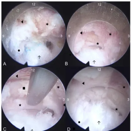
Endoscopic view and anatomic identification In this second step, fluoroscopy is only used to check position of the instruments and verify anatomic parameters. After insertion of the endoscope and cleaning of the operatory field with radiofrequency ablation, it was possible to identify the same structures as in human procedures: the blue stained disc, the superior articular process (SAP) of the inferior vertebrae and the emerging nerve root (Figure 3A). Additionally it was possible to identify the inferior articular process (IAP) of the superior
SAP drilling and exposition of the dural sac Using an oval cutting burr and a Kerrison punch, it was possible to partially drill out the
SAP and IAP to expose the descending root within the dural sac (Figure 3B-C).
Figure 3 - Endoscopic views of the procedure. 12 o’clock is midline (right side), 3 is caudal, 6 is lateral (left side) and 9 is cranial. A) ◄ inferior laminae and articular process ✱ superior articular process; ● emerging nerve
root; ↑ annulus fibrosus (blue stained); B) after drilling the SAP and IAP:◄ inferior laminae and articular
process ✱ superior articular process; ↑ annulus fibrosus (blue stained); ★ dural sac; C) closer midline view:
◄ inferior laminae and articular process ✱ superior articular process; ↑ annulus fibrosus (blue stained) ★
dural sac; ■ yellow ligament; D) after slight rotation of the endoscope, a closer superior view, cranial is at 11
and midline is at 2 o’clock: ◄ inferior laminae and articular process ✱ superior articular process; ↑ annulus
fibrosus (blue stained); ★ dural sac; ● emerging nerve root.
■
Discussion
Although endoscopic spinal surgery is becoming more popular, the lack of proper training often leads to poor clinical results, discouraging the use of this undoubtedly effective technique.
As previously described, TF-PELP has a
very steep learning curve. Lee and Lee17 show
a significant reduction in operating time after treating their first 17 patients, additionally Kafadar et al.18 show a significant higher
number of reoperations in their first 8 months
experience with this technique. Therefore,
along with proper indication of the procedure, education of the surgeon is essential.
Due to limited availability of human cadaver spines, particularly in Brazil, surgeons are being forced to travel abroad to participate in available cadaver hand-on courses to learn or improve such techniques, or may even be skipping this important step of the learning
process.
As well as several other anatomical
studies of the porcine spine, this study shows that this representative animal model reflects anatomical characteristics of the human
spine6. Busscher et al.6 used 6 four
month-old domestic Landrace pigs with an average weight of 40kg for a fine anatomical study based on CT scans. Dath et al.9 used 18 to
80kg, and they macroscopically dissected the vertebrae to perform their measurements. Although different methods were used,
their studies considered larger samples and detailed anatomical measurements that were
important to evaluate whether our study would be possible to replicate. The current model is bigger than those used by Busscher
et al.6 and smaller than those used by Dath
et al.9, however the main characteristics and
proportions are maintained. It is interesting to note that the IVFH has the same behavior as in humans, higher in the upper lumbar spine and increasingly smaller as going caudally. Although the IDH is a bit greater when compared to Busscher samples, it is still much smaller
when compared to humans. The human spine
demands relatively larger caudal vertebral bodies to balance the higher longitudinal loads, in opposition to the quadruped spine. Also the greater range of motion of the lumbar human spine demands adaptable joints. These are probably some of the explanations for the smaller intervertebral discs heights observed in the porcine, which can be up to four times smaller than human disc heights in the lumbar region as shown in Table 2 and described by
Busscher et al.6.
Additionally the lumbar Intervertebral
foramen as height as 12.4 to 19.8mm allow
enough space for the endoscope introduction. The IVFD however (6.8 to 10.0mm) limits the introduction of the endoscope inside
the foramen, and with the correct use of its
denomination, the transforaminal approach. The lower disc heights also make the disc puncture more difficult. However, training
■
Conclusions
The porcine spine is an effective, easily reproducible and representative model for learning and practicing transforaminal percutaneous endoscopic lumbar procedures. Although the described anatomical differences should be known, they do not interfere in learning and practicing all steps of TF-PELP in
the porcine model.
■
References
1. Bozkus H, Crawford NR, Chamberlain RH, Valenzuela TD, Espinoza A, Yüksel Z, Dickman CA. Comparative anatomy of the porcine
and human thoracic spines with reference to thoracoscopic surgical techniques. Surg
Endosc. 2005;19(12):1652-65. doi: 10.1007/
s00464-005-0159-9.
2. Caputy A, Starr J, Riedel C. Video-assisted endoscopic spinal surgery: thoracoscopic discectomy. Acta Neurochir (Wien). 1995;134(3-4):196-9. PMID: 8748781. 3. Nie H, Zeng J, Song Y, Chen G, Wang X, Li Z,
Jiang H, Kong Q. Percutaneous endoscopic lumbar discectomy for L5-S1 disc herniation
via an interlaminar approach versus a
transforaminal approach: A prospective
randomized controlled study with
2-year follow up. Spine (Phila Pa 1976). 2016;41 Suppl 19:B30-7. doi: 10.1097/
BRS.0000000000001810.
4. Choi KC, Park CK. Percutaneous endoscopic lumbar discectomy for L5-S1 disc herniation: consideration of the relation between the iliac crest and L5-S1 disc. Pain Physician. 2016;19(2):E301-8. PMID: 26815257.
comparison of large animal models for the
human lumbar spine. Spine (Phila Pa 1976). 2002;27(8):E200-6. PMID: 11935119.
8. Goel VK, Panjabi MM, Patwardhan AG, Dooris AP, Serhan H, American Society for Testing and Materials. Test protocols for evaluation of spinal implants. J Bone Joint Surg Am. 2006;88 Suppl 2:103-9. doi: 10.2106/JBJS.E.01363.
9. Dath R, Ebinesan AD, Porter KM, Miles AW. Anatomical measurements of porcine lumbar vertebrae. Clin Biomech (Bristol, Avon). 2007;22(5):607-13. doi: 10.1016/j. clinbiomech.2007.01.014.
10. Kumar N, Kukreti S, Ishaque M, Mulholland R. Anatomy of deer spine and its
comparison to the human spine. Anat Rec.
2000;260(2):189-203. PMID: 10993955. 11. Panjabi MM, Goel V, Oxland T, Takata K,
Duranceau J, Krag M, Price M. Human lumbar vertebrae. Quantitative three-dimensional anatomy. Spine (Phila Pa 1976). 1992;17(3):299-306. PMID: 1566168.
12. Abdollah V, Parent EC, Battié MC. Reliability and validity of lumbar disc height quantification methods using magnetic
resonance images. Biomed Tech (Berl). 2018
doi: 10.1515/bmt-2017-0086.
13. Ahn Y, Kim CH, Lee JH, Lee SH, Kim JS. Radiation exposure to the surgeon during percutaneous endoscopic lumbar discectomy: A prospective study. Spine (Phila Pa 1976). 2013;38(7):617-25. doi: 10.1097/BRS.0b013e318275ca58.
14. Yeung AT, Tsou PM. Posterolateral
endoscopic excision for lumbar disc herniation: Surgical technique, outcome, and complications in 307 consecutive cases. Spine (Phila Pa 1976). 2002;27(7):722-31. PMID: 11923665.
15. Ahn Y, Lee SH, Park WM, Lee HY, Shin SW, Kang HY. Percutaneous endoscopic lumbar discectomy for recurrent disc herniation: Surgical technique, outcome, and prognostic factors of 43 consecutive cases. Spine (Phila Pa 1976). 2004;29(16):E326-32. PMID:
15303041.
16. Birkenmaier C, Komp M, Leu HF, Wegener B, Ruetten S. The current state of endoscopic disc surgery: Review of controlled studies
comparing full-endoscopic procedures for
disc herniations to standard procedures. Pain Physician. 2013;16(4):335-44. PMID:
23877449.
17. Kafadar A, Kahraman S, Akbörü M.
Percutaneous endoscopic transforaminal
lumbar discectomy: a critical appraisal. Minim Invasive Neurosurg. 2006;49(2):74-9. doi: 10.1055/s-2006-932184.
18. Lee DY, Lee SH. Learning curve for
percutaneous endoscopic lumbar
discectomy. Neurol Med Chir (Tokyo). 2008;48(9):383-8. PMID: 18812679.
■
Acknowledgements
To Dr. Salvador Amato and Mr. Timothy Coyne for their technical assistance.
Correspondence:
Marcelo Campos Moraes Amato
Avenida Brasil, 2283
01431001 São Paulo - SP Brasil
Tels.: (55 11)5053-2222 / 99937-2565
Received: Aug 03, 2018 Review: Oct 08, 2018 Accepted: Nov 05, 2018
Conflict of interest: none Financial source: none
1Research performed at Division of Neurosur