December 2012
Tânia Raquel Martins dos Santos
Undergraduate in Biology
Genetic characterization of Portuguese
Fasciola hepatica
isolates
Dissertation presented to obtain the Master Degree in
Molecular, Genetics and Biomedicine
Supervisor: Prof.ª Doutora Maria Manuela Calado
Professora Auxiliar
Instituto de Higiene e Medicina Tropical, Universidade Nova de Lisboa
Co-supervisor: Doutora Ana Júlia Pinto Fonseca Sieuve Afonso
Investigadora Auxiliar
Instituto de Higiene e Medicina Tropical, Universidade Nova de Lisboa
Members of the Jury:
President: Prof.ª Doutora Paula Maria Mendes Bernardo Gonçalves
Principal Examiner: Doutora Fernanda Henriques de Jesus Rosa
Tânia Raquel Martins dos Santos
Undergraduate in Biology
Genetic characterization of Portuguese
Fasciola hepatica
isolates
Dissertation presented to obtain the Master Degree in
Molecular, Genetics and Biomedicine
Supervisor: Prof.ª Doutora Maria Manuela Calado
Professora Auxiliar
Instituto de Higiene e Medicina Tropical, Universidade Nova de Lisboa
Co-supervisor: Doutora Ana Júlia Pinto Fonseca Sieuve Afonso
Investigadora Auxiliar
Instituto de Higiene e Medicina Tropical, Universidade Nova de Lisboa
December 2012
Members of the Jury:
President: Prof.ª Doutora Paula Maria Mendes Bernardo Gonçalves
Principal Examiner: Doutora Fernanda Henriques de Jesus Rosa
Genetic characterization of Portuguese Fasciola hepatica isolates
Copyright Tânia Raquel Martins dos Santos, FCT/UNL, UNL
Part of the results discussed in this dissertation was presented in the following communications:
R. Santos, M. Calado, J. Sampaio, C. Ferreira, A. Afonso and S. Belo. Contribution to the genetic characterization of Fasciola hepatica populations in Portugal. XXXVII Portuguese Genetic
Conference, Lisbon, Portugal, May 28th-30th 2012 [poster communication]
R. Santos, M. Calado, J. Sampaio, C. Ferreira, A. Afonso and S. Belo. Contribution to the genetic characterization of Fasciola hepatica populations in Portugal. Arquivos Portugueses das Ciências
ACKNOWLEDGEMENTS
I want to express my gratitude to all that supported me in the realization of this Master and dedicate to them several words in my native language:
Em primeiro lugar quero agradecer à Professor Doutora Silvana Belo por tudo o que fez para permitir que este projecto fosse avante e por me ter recebido tão bem na sua equipa.
Quero também agradecer à Professora Doutora Manuela Calado por toda a ajuda prestada, pelos conhecimentos partilhados, pela paciência que teve comigo e por todo o tempo que me disponibilizou. A sua orientação e supervisão foram fundamentais para levar este trabalho a bom porto. Muito obrigada!
Agradeço à Doutora Ana Afonso por todo o apoio e ajuda que me deu, pelos ensinamentos que me transmitiu e pela disponibilidade que sempre teve comigo. A nível pessoal tenho de agradecer pelas suas palavras amigas e pelos conselhos que me deu ao longo destes anos, sem ela nada disto seria possível. Muito obrigada!
À Mestre Cátia Ferreira agradeço a ajuda prestada nas primeiras etapas deste trabalho. Os seus conhecimentos foram essencias à realização deste trabalho. Agradeço toda a disponibilidade, todas as dúvidas esclarecidas e todos os artigos emprestados.
À Técnica Especialista Isabel Clemente agradeço o carinho, os ensinamentos e as palavras amigas.
Ao Professor Doutor José Paulo Sampaio agradeço por ter coordenado o Mestrado de forma tão eficiente e dedicada e pela prontidão na ajuda prestada.
Agradeço a todos os meus professores que me abriram os olhos para o mundo da ciência e do desconhecido. Sem eles não teria percebido que o caminho da investigação é sempre o mais divertido. Às minhas gurias, fantásticas colegas de Mestrado, agradeço o companheirismo, o carinho e a amizade que agora nos une.
Agradeço a todos os meus amigos, por estarem sempre lá, sem pedir nada em troca. Por estarem presentes, mesmo quando eu me ausentava por meses a fio. Pelas palavras de encorajamento e apoio reforço positivo. Por todos os momentos de diversão e pelas gargalhadas. Mas, principalmente, pelo carinho, respeito e confiança demonstrados ao longo dos anos. Obrigada!
Ao Timon, muito obrigada por toda a ajuda fornecida ao longo deste trabalho e pela disponibilidade e paciência demonstradas. Obrigadão! Espero estar à altura de retribuir.
Quero agradecer à minha família o amor e apoio incondicional que sempre me deram. São eles que dão sentido a todos os meus projectos de vida.
Quero também agradecer ao meu mano, à Susana e à Elsa, por serem os melhores irmãos do mundo, por cuidarem de mim, por serem seres humanos fantásticos e, especialmente, por serem a imagem do que eu queria ser quando fosse grande.
Ao meu pai agradeço por toda a confiança que sempre depositou em mim e por permitir que eu seguisse o meu caminho, com toda a liberdade.
À minha mãe quero agradecer por tudo o que sou. É o meu maior exemplo de amor, moral, coragem, preserverança, esforço e espírito de sacrifício. Agradeço por nunca me ter deixado sozinha em momento algum e por ser o meu porto de abrigo. Agradeço sobretudo por ser para mim muito mais do que o que eu mereço.
Quero ainda agradecer ao Splinter por ser extraordinário. Por cuidar de mim, dar força e por me puxar para o lado bom, sempre. Por acompanhar esta viagem como se fosse sua. Por fazer deste planeta um lugar muito mais bonito para se viver. Obrigada! Muito obrigada!
ABSTRACT
Fasciola hepatica is a parasitic trematode with debilitating and socio-economically devastating
effects. At present near to 600 million animals and 2.4 million people in the entire world suffer from fascioliasis. Genetic characterization is of the utmost importance to an efficient epidemiologic control of helminth infections. In the present study we aimed to provide the first insights into the genetic variability of F. hepatica in Portugal. 47 isolates from different hosts (cattle and sheep) and
geographical locations (Beja, Castelo Branco, Coimbra, Évora, Faro, Leiria, Lisboa, Portalegre, Santarém and Setúbal) were analyzed through Random Amplified Polymorphic DNA-Polymerase Chain Reaction (RAPD-PCR), Restriction Fragment Length Polymorphism (RFLP) and sequencing of NADH dehydrogenase subunit 1 (nad1) gene, cytochrome c oxidase subunit 1 (cox1) gene and
Internal Transcribed Spacers (ITS) region. RAPD-PCR and RFLP patterns were similar for all the analyzed samples, despite their host and geographical origin. Nucleotide sequencing revealed low levels of genetic diversity within Portuguese isolates and no direct correlation was observed between haplotype and geographical location or host. Phylogenetic analysis revealed a high similarity within samples from Mediterranean countries, such as Portugal, Spain, Tunisia, Algeria and Egypt, possibly due to livestock import/export trade between these countries. Moreover, Portugal presents a low risk of fascioliasis drug-resistance.
RESUMO
A Fasciola hepatica é um parasita tremátode com efeitos devastadores na saúde e a nível
socio-económico. Actualmente, perto de 600 milhões de animais e 2.4 milhões de pessoas em todo o mundo sofrem de fasciolíase. A caracterização genética é de extrema importância para um controlo epidemiológico eficiente de infecções helmínticas. Neste trabalho pretendemos iniciar o estudo sobre a variabilidade genética de F. hepatica em Portugal. 47 isolados de F. hepatica provenientes de
diferentes hospedeiros (bovinos e ovinos) e localizações geográficas (Beja, Castelo Branco, Coimbra, Évora, Faro, Leiria, Lisboa, Portalegre, Santarém e Setúbal) foram analisados através de Random Amplified Polymorphic DNA-Polymerase Chain Reaction (RAPD-PCR), Restriction Fragment Length Polymorphism (RFLP) e sequenciação do gene codificante da subunidade I da desidrogenase NADH (nad1), do gene codificante da subunidade I do citocromo C oxidase (cox1) e da região que
comprime os Internal Transcribed Spacers (ITS). Os padrões de RAPD-PCR e de RFLP foram semelhantes em todas as amostras analisadas, independentemente do hospedeiro e da região de origem. A sequenciação nucleotídica revelou baixos níveis de diversidade genética nos isolados portugueses e não foi observada nenhuma correlação entre o haplótipo e a localização geográfica ou o hospedeiro. A análise filogenética revelou elevada semelhança nas amostras vindas de países mediterrânicos, como Portugal, Espanha, Tunísia, Argélia e Egipto, provavelmente devido ao comércio de animais entre estes países. Os nossos dados levam-nos ainda a crer que Portugal apresenta um baixo risco de resistência a fármacos contra a fasciolíase.
TABLE OF CONTENTS
ACKNOWLEDGEMENTS ... vii
ABSTRACT ... ix
RESUMO ... xi
TABLE OF CONTENTS ... xiii
INDEX OF FIGURES ... xv
INDEX OF TABLES ...xvii
ABBREVIATIONS ...xix
Chapter I. INTRODUCTION ...1
1. General Aspects ...1
2. Fasciola hepatica ...3
2.1. Taxonomy ...3
2.2. Life Cycle ...3
2.3. Morphology...7
2.4. Genetics ...9
2.5. Geographical Distribution ... 10
2.6. Intermediate Hosts ... 11
2.7. Primary Hosts ... 12
2.8. Ecology ... 13
3. Fascioliasis ... 15
3.1. Historical Background ... 15
3.2. Sources of Infection and Transmission ... 17
3.3. Fascioliasis in Portugal ... 18
3.4. Fascioliasis in the World... 19
3.4.1. Human Prevalence ... 20
3.4.2. Animal Prevalence ... 22
3.5. Pathology ... 23
3.5.1. Clinical Manifestations in Man ... 23
3.5.2. Clinical Manifestations in Animals ... 24
3.6. Diagnosis ... 25
3.7. Treatment ... 26
3.8. Prevention and Control ... 27
4. The Importance of Genetic Characterization ... 28
Chapter II. MATERIALS AND METHODS ... 33
1. Sample Collection ... 33
2. Nuclear and Mitochondrial DNA Extraction ... 33
3. Random Amplified Polymorphic DNA-PCR (RAPD-PCR) ... 34
4. Polymerase Chain Reaction Amplification of cox1 Gene ... 35
5. Polymerase Chain Reaction Amplification of nad1Gene ... 36
6. Polymerase Chain Reaction Amplification of ITS Region ... 37
7. Restriction Fragment Length Polymorphism (RFLP) ... 38
8. Purification of Polymerase Chain Reaction Products ... 39
9. Sequencing ... 39
10. Analysis of Nucleotide Sequence Data and Phylogenetic Inference ... 39
Chapter III. RESULTS ... 43
1. Sample Collection ... 43
2. Nuclear and Mitochondrial DNA Extraction ... 44
3. Random Amplified Polymorphic DNA-PCR (RAPD-PCR) ... 44
4. PCR Amplification of the Cytochrome C Oxidase Subunit 1 (cox1) Gene ... 46
5. PCR Amplification of NADH Dehydrogenase Subunit 1 (nad1) Gene ... 47
6. PCR Amplification of the Internal Transcribed Spacers (ITS) Region ... 48
7. Restriction Fragment Length Polymorphism (RFLP) ... 48
8. Purification of the PCR Products ... 53
9. Sequencing ... 53
10. Analysis of Nucleotide Sequence Data ... 53
11. Phylogenetic Analysis ... 57
Chapter IV. DISCUSSION ... 63
Main Conclusions ... 68
Future Perspectives ... 68
INDEX OF FIGURES
Fig. I.1. Adult specimen of Fasciola hepatica ... 2
Fig. I.2. Fasciola hepatica life cycle including the stages egg (1.a and 1.b); miracidium (2), sporocyst (3), redia (4), cercaria (5), metaceraria (6), juvenile (7) and adult (8) ... 4
Fig. I.3.Fasciola hepatica life cycle. Assexual reproduction with sporocyst, redia and cercaria stages, inside intermediate host, in detail ... 5
Fig. I.4.Fasciola hepatica adult morphology. A: Schematic draw of F. hepatica internal structures, B: Optical microcopy image of F. hepatica ... 8
Fig. I.5.Fasciola hepatica egg (A), miracidium (B) and cercaria (C) ... 8
Fig. I.6.Fasciola hepatica worldwide distribution. ... 10
Fig. I.7. Galba truncatula adult specimen (A) and details of its sheel (B) ... 12
Fig. II.1. Gene segment of ribosomal DNA that contains 18S, 5.8S, and 28S tracts and the internal transcribed spacers (ITS) 1 and 2. ... 37
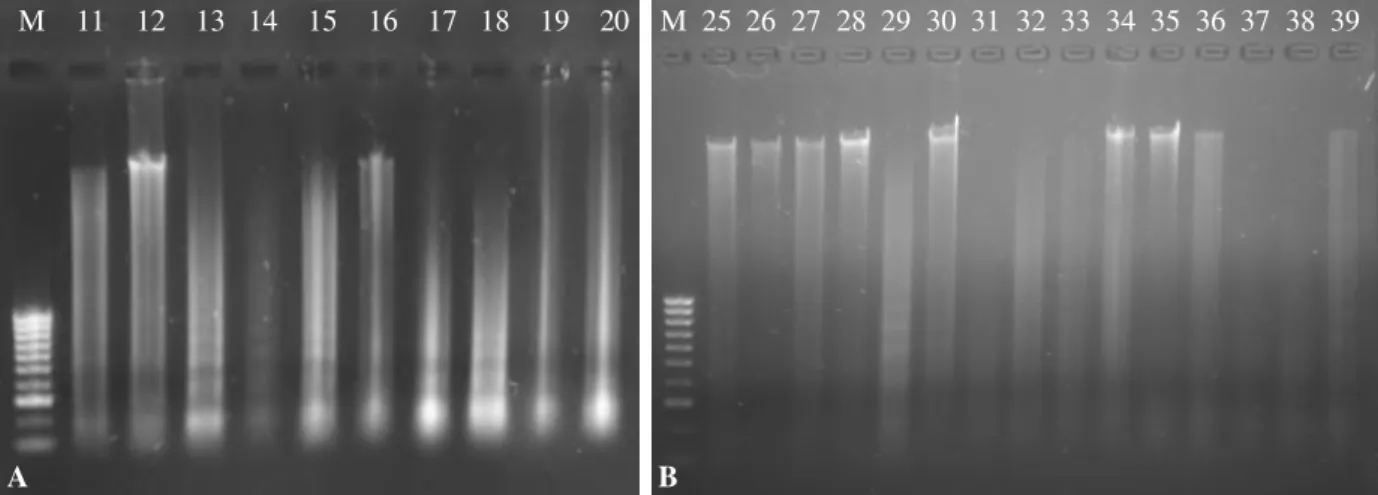
Fig. III.1. Agarose gel electrophoresis of DNA extraction from isolates 11 - 20 (A) and 25 - 39 (B) . 44 Fig. III.2. Agarose gel electrophoresis of primer AP9 RAPD-PCR products ... 44
Fig. III.3. Agarose gel electrophoresis of primer AP2 RAPD-PCR products ... 45
Fig III.4. Agarose gel electrophoresis of primer OSA9 RAPD-PCR products... 45
Fig III.5. Agarose gel electrophoresis of primer OSA10 RAPD-PCR products ... 45
Fig III.6. Agarose gel electrophoresis of primer OSA11 RAPD-PCR products ... 46
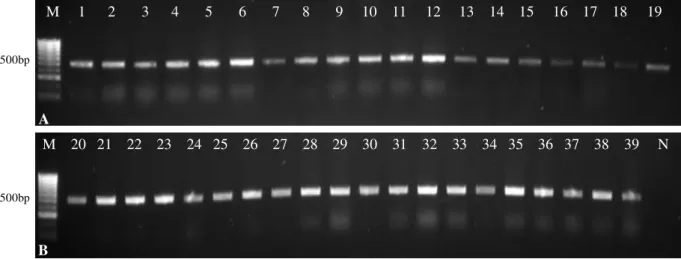
Fig III.7. Agarose gel electrophoresis of the cox1 gene PCR product from isolates 1 - 19 (A) and 20 - 39 (B) ... 46
Fig III.8. Agarose gel electrophoresis of the cox1 gene PCR product from isolates 40 – 47. ... 47
Fig III.9. Agarose gel electrophoresis of the nad1 gene PCR product from isolates 1 – 19 (A) and 20 - 39 (B) ... 47
Fig III.10. Agarose gel electrophoresis of the nad11 gene PCR product from isolates 40 - 47. ... 47
Fig III.12. Agarose gel electrophoresis of the ITS region PCR product from isolates 40 - 47 ... 48
Fig III.13. Agarose gel electrophoresis of the cox1 gene PCR product after digestion with HinfI. ... 49
Fig III.14. Agarose gel electrophoresis of the cox1 gene PCR product after digestion with AluI ... 49
Fig III.15. Agarose gel electrophoresis of the nad1 gene PCR product after digestion with HinfI ... 50
Fig III.16. Agarose gel electrophoresis of the ITS region PCR product after digestion with AluI A: Isolates 1, 6, 9, 14, 20, 21, 27, 31, 34 and 37. B: Isolates 40 - 47 ... 50
Fig III.17. Agarose gel electrophoresis of the ITS region PCR product after digestion with HinfI A: Isolates 2, 5, 11, 13 and 18. B: Isolates 40 - 47. ... 51
Fig III.18. Agarose gel electrophoresis of the ITS region PCR product after digestion with RsaI A: Isolates 1, 6, 9, 14, 20, 21, 27, 31, 34 and 37. B: Isolates 40 - 47 ... 51
Fig III.19. Agarose gel electrophoresis of the ITS region, cox1 gene and nad1 gene PCR product after purification. A: Isolates 1, 3, 6, 8, 10, 12, 14, 15, 19, 20, 21, 22, 26, 28, 29, 32, 33, 35 and 37 cox1 gene PCR products. B: Isolates 38, 42 and 46 cox1 gene PCR products, isolates 4, 6, 12, 13, 17, 21, 26, 30, 35, 38, 42 and 44 nad1 gene PCR product and isolates 7, 12 and 25 ITS region PCR products ... 53
Fig III.20. Map of Portugal with correspondence between regions and haplotypes from cox1gene, nad1 gene and ITS region ... 56
Fig III.21. The cox1 gene phylogenetic tree representing the relationships among Fasciola hepatica isolates from differente geographical locations ... 58
Fig III.22. The nad1 gene phylogenetic tree representing the relationships among Fasciola hepatica isolates from different geographical locations ... 59
INDEX OF TABLES
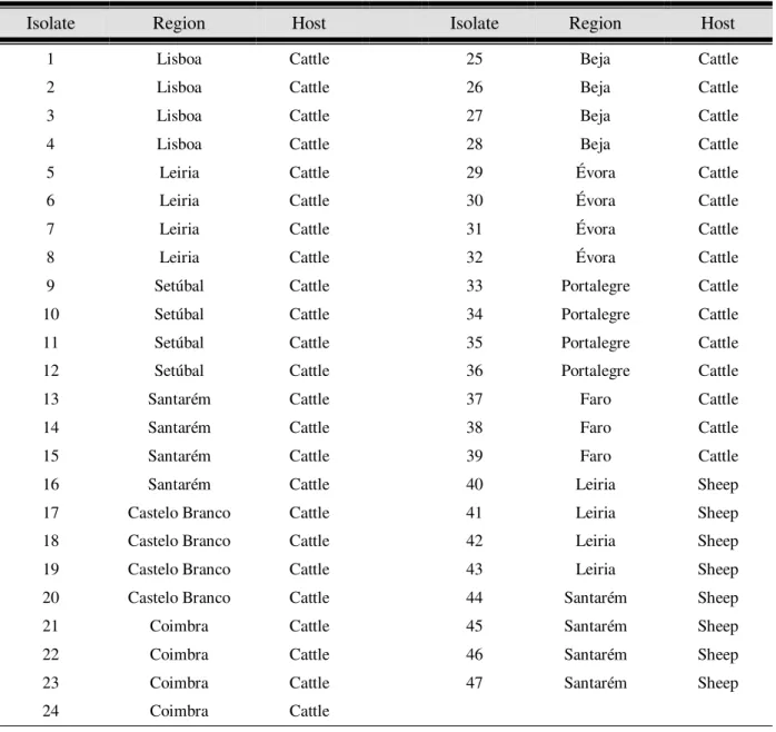
Table II.1.Nucleotide sequences and G + C content of the primers used in the RAPD-PCR assays ... 35 Table II.2. Recognition sites of the restriction endonucleases used in the RFLP assays ... 38 Table III.1. Fasciola hepatica isolates according to their geographical and host origin ... 43 Table III.2. Representative diagram of the PCR and RFLP patterns obtained for all Fasciola hepatica
isolates before and after digestion of cox1, nad1 and the ITS region with endonucleases HinfI, AluI
andRsaI ... 52 Table III.3. Comparison of the cox1 gene sequences of Fasciola hepatica isolates from different hosts
and geographical regions in Portugal and respective haplotypes ... 54 Table III.4. Comparison of the nad1 gene sequences of Fasciola hepatica isolates from different hosts
and geographical regions in Portugal and respective haplotypes ... 55 Table III.5. Comparison of the ITS region sequences of Fasciola hepatica isolates from different
hosts and geographical regions in Portugal and respective haplotypes... 55 Table III.6. Number of analyzed isolates, average number of nucleotide differences, nucleotide diversity, number of existent haplotypes and haplotype diversity for cox1 gene, nad1 gene and ITS
ABBREVIATIONS
π Nucleotide diversity
ATP Adenosine Triphosphate
bp Base pairs
BSA Bovine Serum Albumine
CTAB Hexadecyltrimethylammonium Bromide
cox1 Cytochrome c oxidase subunit 1
dATP Deoxyadenosine Triphosphate
dCTP Deoxycytidine Triphosphate
dGTP Deoxyaguanosine Triphosphate
DNA Deoxyribonucleic Acid
Dna SPTM DNA Sequence Polymorphism
dTTP Deoxythymidine Triphosphate
EDTA Ethylenediamine Tetraacetic Acid
ELISA Enzyme-Linked Immunosorbent Assay
EtBr Ethidium bromide
F. gigantica Fasciola gigantica
F. hepatica Fasciola hepatica
Fig Figure
G + C Guanine + Cytosine
G. truncatula Galba truncatula
G. cubensis Galba cubensis
H Haplotype
HCL Hydrochloric acid
Hd Haplotype diversity
ITS Internal Transcribed Spacers
K Average number of nucleotide differences
KCl Potassium chloride
MEGATM Molecular Evolutionary Genetics Analysis MgCl2 Magnesium chloride
NaCl Sodium chloride
nad1 NADH dehydrogenase subunit 1
NADH Nicotinamide Adenine Dinucleotide Hydride
NCBI National Center for Biotechnology Information
PCR Polymerase Chain Reaction
RAPD Random Amplified Polymorphic DNA
rDNA Ribosomal DNA
RFLP Restriction Fragment Length Polymorphism
RNA Ribonucleic Acid
rpm Rotations per minute
TAE Tris-acetate + EDTA buffer
TE Tris-HCL + EDTA buffer
Chapter I. INTRODUCTION
1. General Aspects
Fascioliasis, or fasciolosis, is one of the most prevalent helminth infections of ruminants in the world causing significant morbidity and mortality and considerable socioeconomic problems (Okewole et al., 2000).
The disease is caused by digenean trematodes, commonly referred to as liver flukes. The two etiological agents of fascioliasis are Fasciola hepatica (Linnaeus, 1758) and Fasciola gigantica
(Cobbold, 1885). The former species has a world-wide distribution, mainly in temperate climates and the latter species exists predominantly in tropical areas (Mas-Coma et al., 2005).
Animals become infected by ingesting encysted metacercariae attached to aquatic or semi-aquatic plants. Evidence also indicates that infection may occur by drinking water from ponds and sloughs contaminated with floating metacercariae. Once ingested, the metacercariae excyst, migrate through the intestinal wall to the body cavity and then to the liver. The young flukes migrating in the hepatic parenchyma eventually locate a bile duct and complete their development to the adult stage (Troncy, 1989). When the hermaphrodite fluke reaches sexual maturity it begins to lay eggs that reach the small intestine and are excreted with the stools to continue the life cycle in a freshwater snail from the family Lymnaeidae, its intermediate host in which the parasite reproduces assexually (Maher et al., 1999).
The fluke devastates the liver while dwelling in the bile ducts and gall bladder. The infection can result in biliary cirrhosis, sclerosing cholangitis associated with destructive jaundice, liver abscesses and other serious hepatic and ectopic clinical manifestations (Price et al., 1993).
Parasites from the genus Fasciola infect mainly sheep, goat, cattle and occasionally affect
humans, thus considered as a zoonotic infection (Andrews, 1999; Okewole et al., 2000; Savioli et al.,
1999).
Other animals that may be affected include horses and pigs. Recently was also reported that llamas in South America, camels in Africa and marsupials in Australia may be acting as reservoir hosts in these regions (Mas-Coma et al., 2005).
Fascioliasis causes significant economic loss, as valued by animal productivity, estimated at approximately 3.2 billion US dollars per annum to the global agricultural community with 600 million animals infected (Mas-Coma, 2005; McManus and Dalton, 2006; Spithill et al. 1997).
Apart from its veterinary and economic importance throughout the world, fascioliasis has recently been shown to be a widespread zoonosis affecting people from the entire world (Mas-Coma et al., 1999a). Accordingly to the World Health Organization (WHO), at present there are nearly 2.4
million people, from 61 countries, that suffer from fascioliasis, but the number of people subjected to the risk of infection is 180 million worldwide (Keiser and Untzinger, 2005; WHO, 2007). Therefore, fascioliasis is no longer considered only as a secondary zoonotic disease but is now recognized as an emerging or re-emerging human disease in several countries (Mas-Coma, 2004; Mas-Coma et al.,
1999a).
Fasciola hepatica is a physically and economically devastating parasitic trematode whose rise
in recent years has been attributed to climate change. Climate has an impact on the free-living stages of the parasite and its main intermediate host Galba truncatula, with the interactions between rainfall
and temperature having the greatest influence on transmission efficacy (Fox et al., 2006).
Due to its zoonotic signification and economic importance the treatment and prevention of fascioliasis is of major importance.
Several control methods against Fasciola hepatica are available and can either be used
independently or as a combination of two or more of them, for example: reduction of the number of intermediate snail hosts, water treatment against metacercariae and chemotherapy against the adult parasites (Savioli et al., 1999; WHO, 2007).
However, for an efficient eradication of fascioliasis it is extremely important to understand the biology of this parasite. Genetic characterization of Fasciola hepatica populations is of major
importance for a well-organized management of this disease. Control programs should consider the genetic diversity of the parasite as being the primary cause of resistance to antihelmintics and fast adaptation to climate changes.
2.
Fasciola hepatica
2.1. Taxonomy
According to Lofty et al. (2008) and the National Center for Biotechnology Information
(NCBI) (2012a) the taxonomic classification of Fasciola hepatica is presented as follows:
Phylum: Platyhelminthes
Subphylum: Neodermata
Class: Trematoda
Subclass: Digenea
Superorder: Anepitheliocystida
Order: Echinostomida
Suborder: Echiostomata
Superfamily: Echinostomatoidea
Family: Fasciolidae
Genus: Fasciola
Specie: Fasciola hepatica (Linnaeus, 1758)
2.2. Life Cycle
Fasciola hepatica was among the first digenetic trematodes to have its live cycle completely
elucidated, one which represents a typical example of a digenean life cycle, with miracidial and cercarial stages. The life cycle is heteroxenic and complex involving several phases and two hosts: a mammalian definitive host and an amphibious snail acting as the intermediate host (Andrews, 1999).
There may be some variations in the Fasciola life cycle, particularly within different definitive
hosts, but the main factors affecting the life cycle tend to be the requirement of suitable temperature and sufficient moisture (Andrews, 1999).
Like all trematodes, with the exception of the genus Schistosoma, Fasciola spp. is
hermaphrodite. Self-fertilization is the most common form of sexual reproduction. However, in stress conditions cross-fertilization can occur (Fletcher et al., 2004). The presence of assexually reproducing
diploid and triploid Fasciola in Asia suggests that abnormal spermatogenesis and parthenogenic
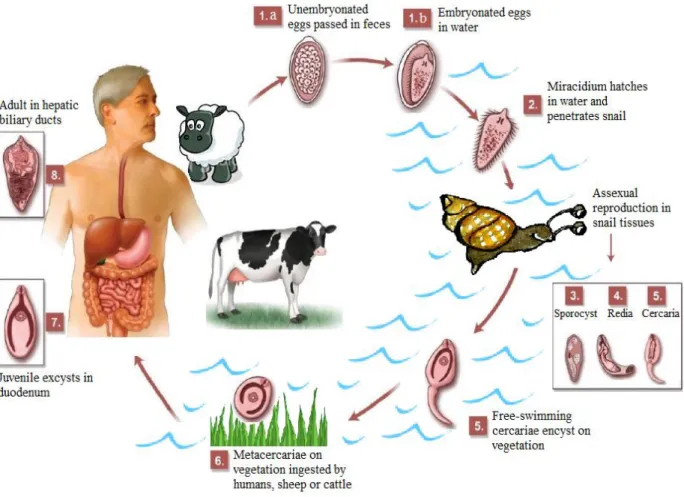
Fig. I.2. Schematic representation of Fasciola hepatica life cycle including its several phases:
(1) egg, (2) miracidium, (3) sporocyst, (4) redia, (5) cercaria, (6) metaceraria, (7) juvenile and (8) adult.
Imagem do ciclo de vida – fazer referências no texto
Fig. I.2.Fasciola hepatica life cycle including the stages egg (1.a and 1.b); miracidium (2), sporocyst (3), redia
(4), cercaria (5), metaceraria (6), juvenile (7) and adult (8). Adapted from Dusak et al., (2012).
(1) Egg:
The oval-shaped eggs are laid by the adult parasite in the bile duct system of the definitive host and pass into the host alimentary tract, specifically the duodenum, with the bile. The eggs then leave the host through the faeces. Eggs have a thin shell and at this stage they are still not embryonated. Despite their thin shell, eggs are structurally strong (Andrews, 1999).
Embryonic maturation and development occurs when the eggs are released in fresh water and is regulated mainly by temperature, moisture and light, but also by the chemical characteristics of the water. Eggs can withstand temperatures from 0ºC to 37ºC, but the optimum temperature for egg development is between 15ºC to 25ºC and it generally takes 9 to 15 days before the eggs hatch to release the fully developed and motile miracidium (Togerson and Claxton, 1999).
(2) Miracidium:
The miracidium is the first free-living larval stage of F. hepatica and is a non-feeding
facilitated by the secretion of proteolytic enzymes (Simpkin et al., 1980; Smythe and Halton, 1983).
The need to find a suitable host to penetrate is an urgent one. Miracidia that fail to do so generally die within 24 hours (Andrews 1999; Smythe and Halton, 1983).
(3)Sporocyst:
After penetrating the snail, specifically inside the muscle tissue, the miracidium loses its cilia and metamorphoses into a rounded sporocyst, which migrates into the snail’s digestive gland (Smythe and Halton, 1983).
(4) Redia:
The sporocyst contains a fixed number of germ balls. Mother sporocysts reproduce by asexual mitotic divisions giving rise to the first generation of daughter rediae. The daughter rediae of the first generation free themselves by breaking through the body wall of the mother sporocyst, which subsequently dies. The daughter rediae then migrate to the snail’s hepatopancreas (Graczyk and Fried, 1999). Usually every sporocyst gives rise to 5 to 8 motile rediae. Under adverse conditions, the rediae delay their development and give rise to a second generation of rediae, also by mitotic divisions. However, in adequate conditions, the germ cells in the brood chamber of the rediae develop into cercariae (Andrews, 1999; Smythe and Halton, 1983).
Fig. I.3. Fasciola hepatica life cycle. Assexual reproduction with sporocyst, redia and cercaria stages, inside
intermediate host, in detail. Adapted from Roberts and Janovy (2009).
(5) Cercaria:
(6) Metacercaria:
After emerging from the snail, the cercaria attaches to various objects such as submerged blades of grass or other aquatic vegetation like watercress. The tail falls away and the cercarial body secretes a four-layered cyst covering from cystogenous glands located on the lateral regions of the body. The formation of the cyst wall may take up to two days. The metacercaria (the encysted and resistant form of the cercariae) is the infective form to the definitive host (Andrews, 1999).
One miracidium hatching from an egg can produce up to 4,000 infective cysts (metacercariae) due to the vegetative multiplication (mitotic division) at the sporocyst and redia stages. Metacercariae are able to survive for up to a year in appropriate conditions, like high humidity and cool temperatures, but show reduced survivability at increased temperature and in dry conditions (Andrews, 1999; Suhardono et al., 2006).
(7) Juvenile:
Upon being swallowed, along with the contaminated vegetation, by the definitive host, the metacercarial cysts enter the small intestine and are stimulated to excyst, releasing the juvenile parasite. The excystement, which occurs in the duodenum, is stimulated by high carbon dioxide concentrations, reducing conditions and temperature at 39°C (Andrews, 1999).
The emergence of the parasite is also stimulated by the presence of bile, bile salts and gastric juices (Mulcahy et al., 1999; Sukhdeo and Mettrick, 1986). Newly excysted juveniles penetrate the
intestinal wall and enter the peritoneal cavity within 24 hours. From there, they migrate directly to the liver over a period of approximately five days. The immature flukes (also referred to as adeloscaria) then penetrate the liver tissues and migrate throught the liver parenchyma consuming liver cells and blood for about six weeks until they find the bile ducts (Andrews, 1999).
(8) Adult:
After about four weeks in the bile ducts, the flukes reach sexual maturity, generally within 3 months of the initial infection. Flukes attach to the bile duct wall using their suckers, with their spines securing them in place. This abrades the host epithelia and ruptures blood vessels, providing the parasite with additional nutrition (Dawes, 1963).
Adult F. hepatica can survive for many years in the livers of infected hosts: one to two years
in cattle or as long as 20 years in sheep (Andrews, 1999). Occasionally ectopic infections can occur, with flukes located in almost any organ (Mas-Coma et al., 1999b; Nithiuthai et al., 2004; Rim et al.,
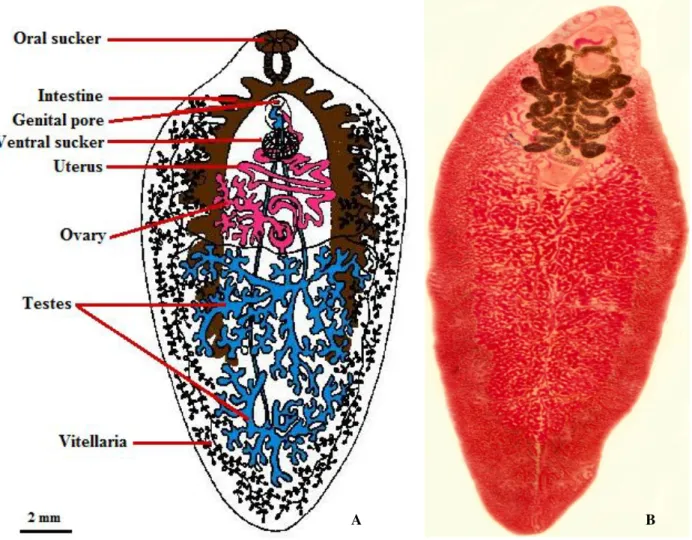
2.3. Morphology
The clade Digenea comprises a large group of species with medical and economic importance. As endoparasites of vertebrates, they present several structural adaptations for parasitism: various penetration glands or glands for cyst material production, organs for adhesion, such as suckers, and increased reproductive capacity. Digeneans also have an incomplete digestive system, with the mouth at the anterior end, and a well-developed reproductive system. The excretory and nervous systems are very simple and the sense organs are poorly developed (Hickman et al., 2004).
The hermaphroditic adult Fasciola hepatica has a flat leaf-like body and an outer tegument
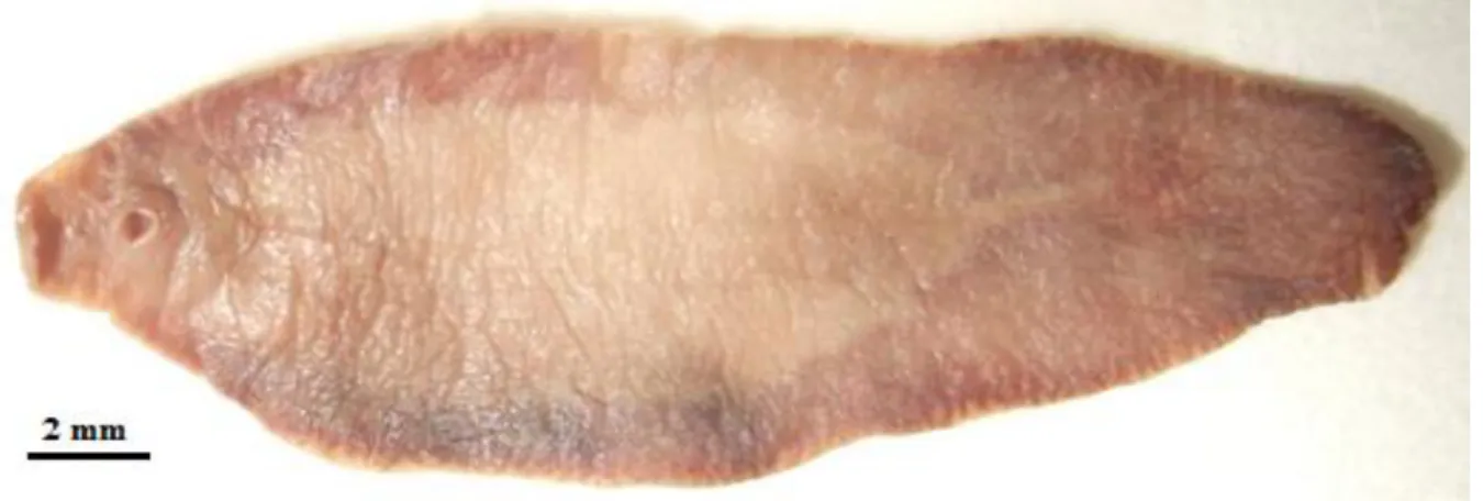
covered with tiny spines. They generally reach 20 to 30 mm and 8 to 15 mm in length and width, respectively, what makes them one of the largest digeneans to parasite humans (Fairweather et al.,1999; Valero et al., 2005).In heavy infections specimens are usually smaller (Muller, 2002).
The fluke has an anterior elongation, known as a cephalic cone, on which the oral and ventral suckers are located. The oral sucker has a diameter of 1.0 mm and the ventral sucker, which lies close behind it, has one of about 1.6 mm (Muller, 2002).
The intestine of the adult parasite is highly branched, with numerous diverticulae extending from the anterior to the posterior of the body(Fairweather et al.,1999; Mas-Coma, 2004).
Female reproductive organs are present near the ventral sucker and male reproductive organs are present near the center of the body. The pair of testes, highly branched, is located in the posterior half of the body, while the dense ovary is located just above the testes and is linked to a short convoluted uterus that opens into a genital pore above the ventral sucker. The vitellaria glands are highly dispersed and branched in the lateral and posterior region of the body (Fairweather et al.,1999;
Mas-Coma, 2004).
Eggsof Fasciolahepatica (Fig. I.5.A) are broadly ellipsoidal, very large (130-150 μm long by
60-90 μm wide) and have a yellowish brown shell with an operculum. The embryonic cells are rather indistinct (Valero et al., 2002).
The miracidium (Fig. I.5.B) has a conical shaped body covered with cilia and may be up to 130 μm by 30 μm (Mas-Coma, 2004).
The cercaria (Fig. I.5.C) has two suckers, a rounded body measuring between 250-350 μm long and a long thin unbranched tail measuring approximately 500 μm long (Fairweather et al.,1999;
Fig. I.4.Fasciola hepatica adult morphology. A: Schematic draw of F. hepatica internal structures B: Optical
microcopy image of F. hepatica. Picture A adapted from Masaba, C. (2010) and picture B by Raquel Santos.
Fig. I.5.Fasciola hepatica egg (A), miracidium (B) and cercaria (C). Pictures by Buckelew, T. (2007).
B A
2.4. Genetics
The diploid chromosome number of Fasciola hepatica is 20 (2n=20) consisting of five pairs of
submetacentrics, four pairs of subtelocentrics and one pair of telocentric chromosomes. Observations on gametogenesis demonstrated that after the two successive meiotic divisions both sperm and ovum show haploidy (Terasaki et al., 2000; Yin and Ye, 1990).
However, in Asia it is not unusual to find, along with the diploids (2n=20), triploid (2n=30) and mixoploid (with diploid and triploid cells) populations of F. hepatica (Terasaki et al., 2000).
Despite the socioeconomic impact of fascioliasis, presently there is no large-scale nuclear Deoxyribonucleic Acid (DNA) sequencing project on Fasciola hepatica (Jefferies et al., 2001).
However, in 2001 the complete nucleotide sequence and gene organization of the mitochondrial genome was determined. It comprises 14 462 base pairs (bp), contains 12 protein-encoding, 2 ribosomal and 22 transfer Ribonucleic Acid (RNA) genes and is the second complete flatworm (and the first trematode) mitochondrial sequence to be described in detail. The gene arrangement resembles that of some other trematodes (Le et al., 2001).
In the NCBI (2012b) Gene Browser we can find several of these mitochondrial genes, like the genes encoding for Cytochrome c oxidase, Nicotinamide Adenine Dinucleotide Hydride (NADH)
dehydrogenase, Cytochrome b oxidase and Adenosine Triphosphate (ATP) synthase proteins.
Although genomic sequence datasets for Fasciola species are scant, a recent study on the
transcriptome of adult Fasciola hepatica was able to give many insights into the genome organization
of this parasite. The average Guanine + Cytosine (G + C) content of the predicted coding sequences is 47.0%. The number of predicted genes expressed by the adult Fasciola hepatica is 23 447. The
estimated number of expressed proteins is 44 597 and a significant proportion of the predicted proteins, 3 804 (8.5%), is conserved across the eukaryotic organisms (Young et al., 2010).
Based on their function, proteins can be grouped into 3 major groups: biological process, molecular function and cellular component (Young et al., 2010).
The predicted proteins assigned to the group “biological process”, 8 761 (19.64%), are predominantly associated with metabolic processes, cellular processes and biological regulation processes. The predicted proteins assigned to the group “molecular function”, 16 679 (37.40%), are predominantly associated with binding processes, catalytic activities and transporter activities. Finally, the predicted proteins assigned to the group cellular component, 4 264 (9.56%), are predominantly associated with the membrane, cytoplasm, organelles and macromolecular complexes (Young et al.,
2.5. Geographical Distribution
Unlike Fasciola gigantica that occurs mainly in tropical areas such as Africa, the Middle East,
Eastern Europe and south and eastern Asia, Fasciola hepatica has a worldwide distribution and has
been reported in all continents except Antarctica (Andrews, 1999).
Due to the colonizing ability of the intermediate hosts and the parasites’ ability to infect a large range of primary hosts, F. hepatica has succeeded in expanding from the European original
geographical area and nowadays it is considered very cosmopolitan in its distribution and can be found in almost all temperate regions (Andrews, 1999; Mas-Coma and Bargues, 1997).
Fasciola hepatica is present in a very wide diversity of environments. This parasite is unique
in being able to survive in areas from below the sea level, as in the Gilan province, besides the Caspian Sea, in Iran, up to the very high altitude, as in the Andean Altiplanos and valleys of Bolivia, Peru and Venezuela (Ashrafi et al., 2007; Mas-Coma et al., 2003).
This huge adaptability to different environments has made of F. hepatica the vector-borne
parasite presenting the widest latitudinal, longitudinal and altitudinal distribution known (Mas-Coma
et al., 2003).
Infections with F. hepatica have been reported in North America: Canada, United States of
America and Mexico; Central America: Puerto Rico, Jamaica and Cuba; South America: Bolivia, Peru, Ecuador, Uruguay, Argentina, Chile, Brazil, Venezuela and Colombia; Europe: France, Spain, Portugal, the former Soviet Union, Turkey, United Kingdom, Ireland, Switzerland, Italy, Netherlands, Germany, Austria and Poland; Africa: Egypt, Kenya, Morocco, Algeria, Tunisia, Lybia, Ethiopia, Tanzania, Zimbabwe, Zambia and South Africa; Asia: Russia, Iran, Japan, Koreas, Vietnam, Thailand, Iraq, China, India, Nepal, Kazakhstan and Mongolia; Oceania: Australia and New Zealand (Esteban et al., 1998; Mas-Coma et al., 1999b; Mas-Coma et al., 2005; Yilmaz and Godekmerdan, 2004).
Fig. I.6. Fasciola hepatica worldwide distribution. Countries painted green have low to medium animal
2.6. Intermediate Hosts
The geographical distribution of trematode species is depending on the distribution of their intermediate hosts. Fasciola hepatica has a wide range of intermediate hosts and usually different
regions of the globe have different snail species has intermediate hosts (Graczyk and Fried, 1999). The intermediate hosts are amphibious snails of the family Lymnaeidae. Since traditional
morphological classification is difficult on the family Lymnaeidae, molecular methods are being used
to study the phylogenetic relationships among this family (Bargues and Mas-Coma, 1997).
In the past, most of the species in question were classified under the genus Lymnaea, but a
number of these have been reassigned to other genera, including Galba, Fossaria, Pseudosuccinea,
and Stagnicola (Acha and Szyfres, 2003).
Lymnaeidae is a taxonomic family of small to large thin-shelled and air-breathing freshwater
snails that have an elongate, dextral and ovoidal shell with a large opening and a simple lip. Lymnaieds are cosmopolitan, with members on all continents and even some islands. In common with other freshwater pulmonates they feed on algae and organic detritus and are hermaphroditic (Kohl, 2012).
Lymnaeids have a single pair of sensory tentacles on the head and an eye at the base of each tentacle. Like all pulmonates, they have a space within their mantle, lined with vascularized membrane, which is used for gas exchange. Most species maintain a bubble of air in the cavity, and occasionally refresh it at the surface of the water (Myers et al., 2012).
The most common intermediate host for F. hepatica is Galba truncatula (Muller, 1774),
formerly known as Lymnaea truncatula. It is the main intermediate host in Africa, Asia and Europe
(Graczyk and Fried, 1999).
Along with G. truncatula, it is not unusual to find Austropeplea viridis (Quoy and Gaimard,
1832) and Austropeplea ollula (Gould, 1859) as intermediate hosts in Asia, Pseudosuccinea columella
(Say, 1817) in Africa and Stagnicola fuscus (Pfeiffer, 1821) and Stagnicola palutris (Müller,1774 ) in
Europe (Bargues et al., 1997; Correa et al., 2010; Mas-Coma et al., 2001).
Fascioliasis has a European origin but succeeded in expanding to the rest of the world. In regions of the New World, where the disease was introduced, different varieties of snails, which may not be morphologically distinct from G. truncatula, play a very important role as Fasciola hepatica
intermediate hosts (Mas-Coma and Bargues, 1997; Mas-Coma et al., 2003). These include the snails Galba cubensis (Pfeiffer, 1839), Galba viatrix (d'Orbigny, 1835), Lymnaea diaphana (King, 1830)
and Pseudosuccinea columella (Say, 1817) in South America; Fossaria bulimoides (Lea, 1841), Hinkleyia caperata (Say, 1829), Lymnaea humilis (Say, 1822) G. cubensis and P. columella, in North
2003; Bargues and Mas-Coma, 1997; Bargues et al., 1997; Bargues et al., 2001; Bargues et al., 2003;
Correa et al., 2010).
Recently, G. truncatula has been reported not only in the old world, but also in South
America, especially in hyperendemic areas for fascioliasis, such as the Andean Altiplano (Bargues et al., 1997; Mas-Coma et al., 2001).
Interestingly, the most important intermediate snail host of Fasciola gigantica, Radix auricularia (Linnaeus, 1758), is resistant to F. hepatica while F. gigantica does not appear to be
capable of developing in G. truncatula (Boray, 1985; Mas-Coma et al., 2005).
Fig. I.7. Galba truncatula adult specimen (A) and details of its sheel (B). Pictures A and B by Mrkvicka, A.
(2002) and Biggs, J. (2008), respectively.
2.7. Primary Hosts
Fasciola hepatica has a wide range of primary, or definitive, hosts. Domestic herbivorous
mammals, such as Ovis aries (sheep), Capra aegagrus (goats) and Bos primigenius (cattle) are
considered the predominant primary hosts. Because of its prevalence in sheep, Fasciola hepatica is
commonly known as sheep liver fluke (Mas-Coma et al., 2005).
It has been estimated that a sheep with a mild subclinical infection can contaminate a pasture with more than 500 000 Fasciola eggs a day, and one with a moderate infection can shed 2.5 to 3
million eggs a day. Sheep are followed in importance by cattle, but their release of Fasciola eggs is
much lower (Acha and Szyfres, 2003).
F.hepatica has shown an ability to expand from infecting farm animals to parasiting wild and
exotic animals all over the world (Mas-Coma, 1998; Mas-Coma, 2004).
Sometimes, even monkeys and humans can become hosts of this parasite (Mas-Coma, 2004). Many species of domestic and wild herbivores can serve as definitive hosts. However, studies suggest that some of these latter animals are only temporary hosts and cannot maintain the cycle by
themselves for a long time. Therefore, they do not contaminate pastures to any significant extent (Acha and Szyfres, 2003).
In Europe, ruminants are very important for disease transmission, but in the rest of the world other animals may play this role. While natural parasitization by F. hepatica in Sus scrofa domesticus
(domestic pigs) and Sus scrofa (wild boars) occurs only occasionally in Europe, pig fascioliasis is
common in other geographical areas, such as Africa and South America (Mas-Coma et al., 2003).
In tropical countries, like Bolivia, some authors have observed that Eqqus africanus (donkeys)
and Eqqus ferus (horses) can act as reservoirs of F. hepatica and contribute to the spread of
fascioliasis (Mas-Coma et al., 1998).
In Australia, Fasciola hepatica was found in farmed Dromaius novaehollandiae (emus) and a
study on the prevalence of F. hepatica infection in native mammals revealed that half of the examined
marsupials were infected with this parasite (Spratt and President, 1981 and Vaughan et al., 1997)
Chronic F. hepatica infection has also been discovered in farmed and wild Rhea americana
(greater rheas) and in Bubalus bubalis (buffalo), in Brazil (Marques and Scroferneker, 2003; Soares et al., 2007).
In 1996, a veterinary study reported the presence of Fasciola hepatica in Argentinian Lama glama (llamas). One of the animals had 62 adult parasites in its liver (Cafrune et al., 1996).
During an 18-year period (1981-1998) Fasciola hepatica was repeatedly found in Belorussian
wild animals, such as Castor fiber (beavers), Lepus timidus (hares), Capreolus capreolus (roe deers), Alces alces (elks), Lutra lutra (otters) and Meles meles (badgers) (Shimalov and Shimalov, 2000).
In Corsica, there are regions in which humans become contaminated but the normal definitive hosts (livestock) are not even present. In those places Rattus rattus (peridomestic rat) has proven to be
the main reservoir host (Mas-Coma et al, 1988; Mas-Coma et al, 2003).
A similar phenomenon has been detected in France, where Myocastor Coypus (nutria), a
recently introduced rodent, plays a major role in disease transmission to humans (Menart et al, 2001).
Furthermore, infection has also been reported in different species of the families Leporidae (rabbits) and Camelidae (camelids) (Mas-Coma, 2004).
2.8. Ecology
Fasciola hepatica is dependent on a consistent set of suitable environmental conditions to
survive. They require acceptable moisture and temperature conditions both for the intermediate and primary hosts and for their own growth and development (Mas-Coma, 2004).
Adequate temperature, between 10-25ºC, is fundamental for the surviving of Fasciola eggs,
for the development of the free living stages and for the formation of metacercarial cysts. In fact, the growth of F. hepatica in warmer environments, between 17-25ºC, reduces the total amount of time
required to complete the fluke development (Andrews, 1999).
Moisture is necessary for the development of fluke eggs in dung, for penetration of the free living stages into the intermediate snail host and to enable migration of cercariae, which is shed from snails, to form cysts on grass (Andrews, 1999).
F. hepatica is found in almost all temperate regions where sheep, goats, cattle and other
ruminants are raised because, virtually, all these areas have sufficient humidity and adequate temperature conditions, at least during part of the year, to sustain a snail population (Mas-Coma, 2004).
Therefore, the ecology of Fasciola hepatica is strictly linked to the ecology of its intermediate
host. Along with the presence of water and appropriate temperatures; physiographic characteristics, soil composition, and climatic factors are of major importance in the surviving and reproduction rate of lymnaeids and, hence, in the presence of F. hepatica (Myers et al., 2012).
Specimens of the family Lymnaeidae can be found in pasturelands in widely diverse settings
throughout the world, from Andean valleys at elevations of over 3 700 meters to sea level flatlands (Mas-Coma et al., 2003).
Lymnaeids are able to adapt to very wide and extreme physical and chemical conditions and to water bodies with a large range of aquatic vegetation (Mas-Coma, 2004).
From the ecologic standpoint, the habitat of Lymnaeidae can be divided into two broad types:
primary foci, or reservoirs, and areas of dissemination:
The primary foci are located in permanently wet environments such as streams, lakes, lagoons, or canals. Snails are usually found in stagnant water bodies or near the banks, where water flows slowly. They begin to lay their eggs in springtime when temperatures rise above 10°C and continue to do so as long as the thmperature remains above this level. Warmer temperatures allow faster growth of the eggs (Acha and Szyfres, 2003; Chapuis et al., 2007).
Since new snails begin to lay eggs at 3 weeks of age, they can produce up to three generations in a single season as long as they have enough water (Acha and Szyfres, 2003).
During unusually dry and hot summers, many snails may die, but a few of them estivate and resume their development when the temperature falls and moist conditions return. Many of them also may die during very cold winters, but some go into hibernation and resume their development when temperatures once again rise above 10°C. The snails that manage to survive dry conditions, heat, and cold are the seeds for the next season’s crop of snails (Luzün-Peña et al., 1994).
Temperature above 10°C is a key factor in the epidemiology of fascioliasis because when it is any colder Fasciola eggs fail to develop, snails do not reproduce, sporocyst, redia and cercaria stages
The adaptation on lymnaeids to permanent water bodies makes transmission throughout the year possible, as observed in southern Europe and the Mediterranean islands (Valero et al., 1998).
Areas of dissemination are characterized by the alternation of flooding and droughts, and have large concentrations of Lymnaeids. Snails may reach these areas directly from original foci carried by
rising waters, or they may be reactivated after estivation during long dry spells. Seasonal foci of this kind turn pastures into enzootic areas in which serious outbreaks can occur (Acha and Szyfres, 2003; Chapuis et al., 2007).
As with many parasites there is a distinct seasonal pattern in fascioliasis outbreaks, with two key periods of infection, summer and winter (Altizer et al., 2006).
Fasciola eggs transmitted by infected animals in springtime and early summer develop inside
the snails and produce cercariae and metacercariae until the end of summer. The animals that ingest them begin to show signs of the disease at the end of autumn and during winter; this is considered a summer outbreak and occurs mostly in warm temperate regions (Fox et al., 2011; Goodall et al.,
1991).
Eggs excreted during unfavorable winters do not develop until suitable conditions are encountered. In early spring, the eggs commence their development and the metacercariae from this cycle appear at the end of spring or in early summer. When ingested by animals, these metacercariae produce symptoms in summer and autumn; this is a winter outbreak and occurs mostly in cool temperate regions (Fox et al., 2011; Goodall et al., 1991).
In sub-tropical and tropical regions disease outbreaks can occur all year round (Togerson and Claxton, 1999).
In conclusion, the climatic factors like rainfall and air temperature are decisive for the development of Fasciola hepatica, mainly because they have a very strong impact in the life cycle of
its intermediate host. A temperature range of 10-25ºC and high levels of moisture are the primary determinants of transmission efficiency (Fox et al., 2011)
3. Fascioliasis
3.1. Historical Background
Fasciola is an ancient parasite coexisting with man and animal back to approximately 3 500
BC. Fasciola eggs have been found in human coprolites from populations of the Stone Age, living at
the end of the Mesolithic period, 5 000 to 5 100 years ago and the Neolithic, a period marked by the domestication of animals and the development of agriculture (Aspöck et al., 1999; Bouchet, 1997;
Interestingly, liver fluke eggs have been found in many palaeoparasitological studies performed in Europe but never in coprolites from the New World, which clearly indicates that fascioliasis in the Americas is a relatively recent introduction (Gonçalves et al., 2003)
The earliest reference to fascioliasis in literature is contained in the Black Book of Chirk,
published around 1200, in which disease in sheep in mentioned (Froyd, 1969).
Fasciola hepatica was first observed in France by Jean de Brie, in 1379. De Brie made
mention of the disease “liver rot” in sheep but he didn’t considered the fluke as being the etiologic agent of the disease, instead he thought it was a consequence of the liver being affected by toxic substances produced by certain plants eaten by the sheep. His observations have made F. hepatica the
first trematode to be recognized (Andrews, 1999).
In the sixteenth century, fascioliasis appeared in the form of an epidemic throughout Europe, with the worst hit areas being the Netherlands and Germany. However the first sketch of an adult
Fasciola was made only in 1668, by Francesco Redi. By showing that parasites lay eggs, Redi
destroyed the false doctrine of spontaneous generation. However it was not until the end of the 18th century that reference was made to the intermediate stages of F. hepatica life cycle (Mufti, 2011).
The earliest insight into the pathology of “liver rot” was made in 1755 by Frank Nicholls, a physician that reported calcification of the bile ducts in bullocks infected with F hepatica. In 1758 Fasciola hepatica was identified by Linnaeus. "Fasciola" in Latin means fillet or small bandage and
"hepatica" means of the liver (Mufti, 2011).
Müller, in 1773, observed several different kind of cercariae swimming in water but could not correlate them with intermediate stages of helminthes. In 1803, Johann Zeder reported observing the hatching of eggs from a number of different species of trematode and the escape of a ciliated embryo, miracidium, into the water. Christian Nitzsch, in 1807, followed with the first account of cercariae encysting. The next observation of relevance was made in 1818 by Ludwig Bojanus who discovered the redial stages of trematodes, but unfortunately he did not work with F. hepatica. Unlike his
predecessors, Bojanus noted the resemblance between rediae, cercariae and adult flukes (Andrews, 1999).
Despite all these advances, it was only by the middle of the 19th century that most of the individual parts of the life cycle were brought together. In 1842 Johannes Steenstrup published his work On the Alternation of Generations where he fitted the theory to various forms of life, including
trematodes. Adolphus von La Valette St George, in 1855, demonstrated, by feeding-experiments, that certain encysted cercariae from water snails developed into sexually mature flukes in birds and Guido Wagener, two years after, observed the penetration of miracidia into snails and the subsequent development of rediae (Andrews, 1999).
In 1875 the German helminthologist David Weinland was the first person to suspect that larval stages of liver fluke occurred in Lymnaea truncatula. He conjectured that cercariae encysted on grass
1892, successfully infected several herbivorous animals by adding metacercariae to their food, confirming that mammals acquire adult fluke by ingestion of metacercariae. The final piece of the jigsaw was added by Dimitry Sinitsin in 1914, who proved that young flukes in the rabbit, after liberation from their cysts in the small intestine, penetrated the wall of the gut and migrated to the liver via the peritoneal cavity (Andrews, 1999).
3.2. Sources of Infection and Transmission
Mammals become infected with Fasciola hepatica when grazing in pastures with
contaminated vegetation. Infection occurs through the ingestion of metacercarial cysts located on the plants surface. Water has also been cited as a source of infection through direct drinking of cysts on water surface (Mas-Coma et al., 2005).
Humans become infected through ingestion of aquatic plants contaminated with metacercariae and direct drinking or contamination of food or utensils with cysts on water surface (Mas-Coma et al.,
2005).
Experimental evidence in mice and pigs also suggests that people who consume raw dishes prepared from fresh livers infected with immature flukes can also become infected (Taira et al., 1997).
Metacercariae infectivity is dependent upon storage time, being lower when cysts are older. Moreover, metacercariae viability and infectivity do not show differences between isolates from different reservoir species. Thus, flukes from secondary reservoirs, such as pigs and monkeys, involve the same potential risk of infection as those from sheep and cattle (Valero and Mas-Coma, 2000).
There are several contamination sources for human infection: - Ingestion of wild freshwater plants
Most human reports are related to watercress. However, the general term watercress includes different aquatic species, such as common watercress, Nasturtium officinale (common watercress) and N. silveris and Roripa amphibian (wild watercress). Wild watercress has been reported as the main
source of human infection in areas where fascioliasis in domestic animals is highly endemic. Other wild aquatic vegetables reported as vehicles of human infection are Taraxacum officinale (dandelion), Valerianella olitora(lamb’s lettuce) and Mentha viridis (spearmint) and Mimulus glabratus (roundleaf
monkeyflower) (Esteban et al., 1997a; Mas-Coma and Bargues, 1997; Mas-Coma et al., 1999a).
- Ingestion of cultivated freshwater plants
- Ingestion of wild and cultivated terrestrial plants
The amphibious characteristics of intermediate species, such as Galba truncatula, and the long
survival capacity and dryness resistance of metacercariae explain the human contamination by consumption of plants collected in dry habitats with temporary water bodies and plants collected in plantations of non-aquatic vegetables frequently irrigated (Mas-Coma et al., 1999a).
Eruca sativa (arugula), Lactuca sativa (lettuce), Allium porrum (leek), Portulaca oleracea
(purslane) and other contaminated plants that are eaten raw can be a dangerous source of Fasciola hepatica infection (El-Sayed et al., 1997; Motawea et al., 2001).
Thanks to the transport of vegetables, both aquatic and terrestrial, from rural endemic zones to cities, plants carrying metacercariae can be sold in non-controled city markets giving rise to urban infections (Mas-Coma et al., 1999a).
- Drinking of contaminated water
Consumption of natural water in often cited as a human infection source. In 1996, a water analysis conducted on a river crossing Tambillo, in the Bolivian Altiplano, reported that there were up to seven metacercariae in only half a litre of water. Moreover, of all the metacercariae collected in this study, about 13% were floating on water (Barges et al., 1996).
In the Egyptian locality of Tiba, where a prevalence of 18% was initially found, human infection has markedly decreased after the construction and utilization of so-called “washing-units”, in which the water is appropriately filtered (Esteban et al., 2003).
- Washing of kitchen utensils or other utensils with contaminated water
Washing with metacercariae-carrying water may be the source of inadvertent infections. In Egypt, women usually wash kitchen utensils and clothes at irrigation canals where lymnaeids and livestock are present. Infection through ingestion of food containing metacercariae can be a serious health problem in the Nile Delta region (Cadel et al., 1996; Curtale et al., 2003).
- Ingestion of raw livers
In 1997, Taira and collaborators conducted a study where mice and piglets were given fresh livers infected with immature flukes. At necropsy, they recovered several live flukes and observed hemorrhagic and granulomatous lesions, typical of those caused by active migration of early immature flukes. The results suggest that humans consuming raw liver or semi-cooked liver dishes prepared from fresh livers infected with immature flukes may also become infected (Taira et al., 1997).
3.3. Fascioliasis in Portugal
In Portugal, Fasciola hepatica can be found almost across the entire country. Reports of
The climate in Portugal favors the spread of F. hepatica. The mild temperatures and humidity
allow for the healthy development of the eggs, and the wetness in winter months promotes the abundance of its intermediate host, Galba truncatula. F. hepatica exhibits a preference towards the
areas surrounding the great rivers such as Tejo, Douro, Minho and Vouga. It is also present in certain microclimates in the Alentejo, Beiras and Algarve regions (Grácio, 1985; Rombert et al., 1991).
F. hepatica primarily affects sheep, followed by cattle, swine and goats. In a smaller scale, it
also affects horses, mules, donkeys and rodents (Ferreira and Oliveira, 1960).
Studies on animal fascioliasis in Portugal are not plentiful and most data are gathered in slaughterhouses. The first studies were conducted by Silva Leitão in 1950 and showed a high presence of bovine fascioliasis in the northern regions of the country (Leitão, 1965).
Sousa studied the presence of F. hepatica in wild animals such as boar and deer in the Tapada
Nacional de Mafra, by observing lesions and adult forms of the parasite in the animals’ liver. An increase in the prevalence of the parasite between 1992 and 2000 was reported (Sousa, 2001).
In a comparative analysis based on data from the Divisions of Veterinarian Intervention of Viseu, Coimbra, Aveiro and Leiria it was shown that most cattle with livers rejected due to fascioliasis hailed from Aveiro, followed by Coimbra (Conceição, 2001).
In Portugal, man is an accidental host and reports of fascioliasis in man are somewhat scant. However, it is known that human fascioliasis is particularly present in the northern regions of Portugal (Sampaio, 1986). The first published case of human fascioliasis in Portugal was in 1948, and a total of 14 had been reported up to 1959. Until 1991, a total of over 190 cases had been diagnosed in the country, 50 of them in 1990 (Rombert et al., 1991). Many of those new cases were in the central
region of the country, as a consequence of a then recent outbreak in Ribatejo. The first cases started to appear in the months of February and March, and could be then backtracked to initial infections in January and February, corresponding to a development of F. hepatica starting in November of the
previous year (Rombert and Grácio, 1990). This coincides with the typical heavy rains in this period (Rombert et al., 1991).
3.4. Fascioliasis in the World
Human and animal fascioliasis caused by Fasciola hepatica occurs worldwide. While animal
fascioliasis occurs mainly in countries with high cattle and sheep production, human fascioliasis occurs mainly in developing countries (Togerson and Claxton, 1999).
A global analysis of the distribution of human cases shows that the expected correlation between animal and human fascioliasis only appears at a basic level. Although it is true that human infection is not rare in areas where infected domestic herbivorous mammals are present, high/low human prevalences are not related to high/low animal prevalences, respectively (Mas-Coma et al.,
The epidemiological classification of fascioliasis includes two major groups: areas where fascioliasis is endemic and areas where fascioliasis is sporadic (Mas-Coma, 2004).
Sporadic areas are those where human and/or animal reports of fascioliasis appear without any constancy (Mas-Coma et al., 1999a).
Endemic areas can be hypoendemic, with a prevalence of less than 1%; mesoendemic, with a prevalence of 1–10%; and hyperendemic, with a prevalence of more than 10% (Mas-Coma et al.,
1999a).
High prevalences in humans are not necessarily found in areas where fascioliasis is a great veterinary problem. For instance, in South America human hyperendemic and mesoendemic areas are mainly found in Bolivia and Peru where the veterinary problem is less relevant than in countries such as Uruguay, Argentina and Chile, where human fasciolosis is only sporadic or hypoendemic (Esteban
et al., 1997b). As for other example: although the infection is common in cattle in western and
southeastern US, only one human case has been reported in that country and the situation is similar in China, where the infection is frequently seen in animals, but only 44 human cases were known to have occurred (Chen, 1991).
3.4.1. Human Prevalence
At present there are nearly 2.4 million people, from 61 countries, that suffer from fascioliasis, but the number of people subjected to the risk of infection is 180 million worldwide. Human fascioliasis has been reported in several countries in Europe, America, Asia, Africa and Oceania and, unfortunately, the number of affected countries, currently 61, is increasing (Keiser and Untzinger, 2005; WHO, 2007).
In Europe, human fascioliasis cases are predominantly found in Portugal, Spain, France, Turkey and the former USSR. France is considered an important human endemic area, with 3297 cases catalogued between 1950 and 1983. However, 5863 human cases were recorded from nine hospitals between 1970 and 1982, indicating that published data largely underestimate the real situation. The disease is also important in Portugal, mainly the northern region, in which 1011 cases were diagnosed in 1970-1992. In England the largest known outbreak occurred in 1972 and about 40 people were affected (Esteban et al., 1998). Concerning the former Soviet Union, almost all reported
cases were from the Tadzhik Republic, near the Afghanistan border (Semyenova et al., 1996).