Sara Patrícia Ferreira Moreira
Licenciada em Biologia
Nrf2 activation by TUDCA in experimental
models of Parkinson’s disease
Dissertação para obtenção do Grau de Mestre em Genética Molecular e Biomedicina
Orientador: Margarida Castro-Caldas, PhD
Faculdade de Ciências e Tecnologia, Universidade Nova de Lisboa
Co-orientador: Maria João Gama, PhD
Faculdade de Farmácia, Universidade de Lisboa
Presidente: Prof. Doutora Ilda Sanches Arguente: Doutora Sara Xapellí Vogal: Prof. Doutora Margarida Castro-Caldas
Sara Patrícia Ferreira Moreira
Licenciada em Biologia
Nrf2 activation by TUDCA in experimental
models of Parkinson’s disease
Dissertação para obtenção do Grau de Mestre em Genética Molecular e Biomedicina
Orientador: Margarida Castro-Caldas, PhD
Faculdade de Ciências e Tecnologia, Universidade Nova de Lisboa
Co-orientador: Maria João Gama, PhD
Faculdade de Farmácia, Universidade de Lisboa
i | P a g e
Nrf2 activation by TUDCA in experimental models of Parkinson’s disease
Copyright © Sara Patrícia Ferreira Moreira, Faculdade de Ciências e Tecnologia, Universidade Nova de Lisboa.
iii | P a g e
Part of the results discussed in this thesis were presented in the following meetings:
Moreira S, Fonseca I, Silva-Azevedo C, Nunes MJ, Gama MJ, Rodrigues E, Rodrigues CMP,
Castro-Caldas M, [Ferreira Mendes A.]. Nrf2 regulation by TUDCA in a mouse model of Parkinson’s disease.
Annual Meeting of the Portuguese Society of Pharmacology, 5th February 2015, Lisbon, Portugal.
[Abstract and Oral Communication]
Moreira S, Gama MJ, Castro-Caldas M. Evaluation of Nrf2 activation by TUDCA in experimental models of Parkinson’s disease. Jornadas intercalares das Dissertações Anuais dos Mestrados,
Universidade Nova de Lisboa, 5th February 2015, Caparica. [Oral communication]
S. Moreira, I. Fonseca, L. de Lemos, C. Silva-Azevedo, M. J. Nunes, E. Rodrigues, M. J. Gama, C. M. P. Rodrigues, M. Castro-Caldas. TUDCA modulates Nrf2 and antioxidant enzyme expression in experimental models of Parkinson’s disease. XIV Portuguese Society for Neurosciences, 4 June 2015, Póvoa de Varzim, Portugal. [Abstract and Poster]
Moreira S, Fonseca I, Silva-Azevedo C, Lemos L, Nunes MJ, Rodrigues E, Gama MJ, Rodrigues CMP, Castro-Caldas M. Tauroursodeoxycholic acid activates Nrf2 antioxidant system in the MPTP mouse model of Parkinson’s disease. 40th FEBS Congress, 4-9 July, Berlin, Germany. [Abstract and Poster]
Moreira S, Fonseca I, de Lemos L, Silva-Azevedo C, Nunes MJ, Rodrigues E, Gama MJ, Rodrigues
CMP, Castro-Caldas M. Anti-oxidant activity of TUDCA in experimental models of Parkinson’s
disease. 7th iMed.UL Postgraduate Students Meeting, 15 July 2015, Lisbon, Portugal. [Abstract and
Poster]
This work was supported by National funds (Fundação para a Ciência e Tecnologia – FCT, Portugal)
vii | P a g e
ACKNOWLEDGMENTS
As minhas primeiras palavras de agradecimento são dirigidas à Professora Doutora Cecília Rodrigues por me ter recebido no seu grupo “Cellular Function and Therapeutic Targeting”, permitindo-me entrar, pela pripermitindo-meira vez, no mundo da investigação.
Um enorme agradecimento é dirigido à Professora Doutora Margarida Castro-Caldas, orientadora desta tese, por me ter proporcionado esta oportunidade, pois foi a principal responsável pela minha vinda para este grupo. Quero agradecer toda a dedicação, exigência e interesse com que sempre seguiu este projeto, e por todas as outras oportunidades que me foi proporcionando ao longo deste ano, de forma a poder enriquecer o meu currículo, contribuindo sempre para a minha aprendizagem. Obrigada também pela confiança que sempre depositou em mim e por me demonstrar que estou à altura dos desafios, mesmo quando eu acho exatamente o contrário. Esta tese não teria sido possível de realizar sem a sua constante ajuda, orientação e dedicação. Agradeço ainda pela disponibilidade constante, sugestões, esclarecimento de dúvidas, rigor e paciência e também pelo conforto que incluiu em muitas das suas palavras, quando nem tudo corria pelo melhor no laboratório, demonstrando-me que não há mal nenhum em errar e que sem isso, não chegamos realmente a aprender. Espero, por fim, ter estado à altura de tudo aquilo que me foi imposto ao longo deste ano e agradeço a oportunidade que me ofereceu de poder aprender tanto consigo.
À Professora Doutora Maria João Gama, co-orientadora desta tese, agradeço pelo interesse constante no desenvolvimento deste trabalho e também por se mostrar sempre disponível, em todas as situações, esclarecendo sempre as minhas dúvidas e disponibilizando-me valiosas sugestões para quando as diversas experiências simplesmente decidiam não funcionar.
À Professora Doutora Elsa Rodrigues agradeço especialmente por me ter mostrado diversas vezes
o seu dossier de “experiências falhadas”, demonstrando-me que só teria direito a reclamar quando
tivesse um dossier tão denso quanto o seu! Agradeço ainda a boa disposição constante e o interesse no desenvolvimento deste trabalho, ao longo deste ano.
À Doutora Maria Nunes agradeço pelo facto de ser a organização e eficiência em pessoa tendo-se tornado para mim, um excelente exemplo a seguir no laboratório. Agradeço também pelo teu contributo e interesse constante na progressão deste trabalho, bem como todo o apoio disponibilizado.
Agradeço a todas as outras pessoas que integram o grupo “Cellular Function and Therapeutic Targeting” que, de uma forma ou de outra, permitiram que este trabalho progredisse. Um especial agradecimento à Alexandra Rosa, Carla Azevedo, Luísa de Lemos, Miguel Moutinho e Miguel Santos.
viii | P a g e
por seres uma pessoa tão direta e verdadeira, pelos teus dotes culinários fantásticos que, tão valiosos foram durante este ano, e por teres sido a minha companhia constante entre 8 a 12 horas por dia.
Obrigada também pelos fabulosos cafés Nespresso logo pela manhã, pelos almoços partilhados a discutir
receitas fantásticas, pelas longas conversas e cusquices, pelos sorrisos e asneiras partilhadas no laboratório e pelo step diário contra as calorias do almoço, naquelas escadas que às vezes parece que não têm fim. Obrigada ainda por tornares os meus dias sombrios naquela cave mais fáceis de tolerar e muito mais animados. Por fim, agradeço por te teres revelado uma pessoa fantástica, dentro e fora do laboratório, e por te teres tornado tão especial para mim. Desejo que a vida te sorria sempre e que alcances tudo aquilo que desejares! Gosto imenso de ti e vou sentir a tua falta quando tudo isto acabar.
Aos meus amigos Aida Lima, Maggie Silva, Marta Fonseca, Leonor Melo, Luci Pereira, Pedro Santos e Ricardo Ribeiro, um grande obrigado a cada um de vocês, por todo o apoio, carinho e amizade, dados ao longo destes anos! Cada um contribui à sua maneira, mas todos vocês tornam os meus momentos felizes, ainda mais felizes, e os meus momentos mais tristes, mais fáceis de ultrapassar. Agradeço-vos ainda por ocuparem um lugar bastante especial na minha vida e espero manter-vos sempre comigo.
Agradeço agora às pessoas mais importantes da minha vida, a minha família, a quem dedico esta tese. Primeiro aos meus pais, Carlos e Esmeralda, por todos os sacrifícios que sempre fizeram e que continuam a fazer, para que eu nunca desista dos meus sonhos e para puderem proporcionar-me o melhor que a vida tem para oferecer. Quero agradecer também por todo o apoio, incentivo e compreensão e por garantirem que nunca nada me falte e que a minha felicidade seja sempre a vossa prioridade. Estarei eternamente grata por tudo isso! Agradeço ainda todo o orgulho que têm em mim, e por mais uma vez, estarem presentes neste momento tão importante. Mais uma etapa concluída da minha vida, em grande parte, graças a vocês.
Aos melhores irmãos do mundo, Tiago e Sandro, por estarem sempre presentes nos momentos mais importantes da minha vida, seja em que circunstância for. Agradeço-vos por todo o apoio, confiança, interesse, conselhos, proteção e companheirismo, e também por acreditarem sempre nas minhas capacidades e terem orgulho em tudo o que alcancei até agora. Quero agradecer ainda especificamente
ao mano mais velho pelo fascínio que tem por aquilo que faço (quer admitas quer não…) e por no fundo
saber que a minha área coloca a sua eterna amiga Matemática a um canto! Ao mano mais novo agradeço especialmente por ser a melhor companhia e o melhor ouvinte que alguém pode ter e por ter sempre uma palavra de força, apoio e motivação para mim!
Às minhas avós Lurdes e Maria um grande obrigado por todo o apoio e carinho que sempre me disponibilizaram, e por nunca, mas nunca, duvidarem das minhas capacidades.
Um obrigado muito especial ao meu avô Armando, um avô sempre babado pela “menina dos seus
ix | P a g e
meu avô Carlos que não teve oportunidade de acompanhar nem a minha progressão pessoal, nem a minha progressão académica, mas que estaria igualmente radiante e extremamente orgulho das metas que alcancei. A vocês os dois dedico a conclusão de mais uma etapa da minha vida!
Aos meus tios maravilhosos, Berta e Henrique, que me incumbiram o maravilhoso gosto e interesse por tudo aquilo que nos rodeia e por terem sempre algo extremamente valioso para me ensinarem, passem os anos que passarem e aprenda eu o que aprender! Agradeço também a partilha das vossas magníficas viagens, algo pelo qual ganhei um gosto incrível, passando a ser um dos muitos sonhos que pretendo realizar na minha vida, e por todo o interesse na progressão do meu percurso académico bem como todo o apoio disponibilizado!
A toda a minha família, um obrigado do tamanho do mundo! Amo-vos do fundo do coração!
xi | P a g e
ABSTRACT
Parkinson’s disease (PD) is a progressive neurological disorder, mainly characterized by the loss
of dopaminergic neurons in the substantia nigra pars compacta. Although the cause of PD remains
elusive, several lines of evidence implicate mitochondrial dysfunction and oxidative stress as possible mechanisms by which cell death occurs in this disease.
Under oxidative stress, the master regulator of cellular redox status, nuclear factor erythroid 2 related factor 2 (Nrf2), is responsible for activating the transcription of several cytoprotective enzymes, namely glutathione peroxidase 1 (Gpx1), heme oxygenase-1 (HO-1) and superoxide dismutase 2 (SOD2), being a promising target to limit reactive oxygen species (ROS)-mediated damage in PD.
In this work, we aim to evaluate the ability of tauroursodeoxycholic acid (TUDCA) to modulate, not only the Nrf2 pathway and the expression of the Nrf2 stabilizer, DJ-1, but also the cellular redox status, in both animal and cellular models of PD, using twelve-week-old C57BL/6 male mice treated with 1-methyl-4-phenyl-1,2,3,6-tetrahydropyridine (MPTP), and the human neuroblastoma cell line,
SH-SY5Y, treated with 1-methyl-4-phenylpyridinium (MPP+).
Our Western blot results, together with quantitative real time polymerase chain reaction, demonstrate that TUDCA treatment increases DJ-1, Nrf2, Gpx1, HO-1 and SOD2 expression, in mice striatum and midbrain. Moreover, enzymatic assays also reveal that TUDCA treatment enhances Gpx biological activity, in mice. In SH-SY5Y cells, we demonstrate by immunocytochemistry that TUDCA induces Nrf2 nuclear translocation, with the consequent increase in HO-1 mRNA levels. Additionally,
TUDCA also attenuates both MPP+-induced ROS production and lipid peroxidation, in this cell line.
Together, our results suggest that TUDCA is a promising agent to limit ROS-mediated damage, in different models of PD acting, at least in part, through modulation of the Nrf2 signaling pathway, and therefore, should be considered a promising therapeutic agent to be implemented in PD.
xiii | P a g e
RESUMO
A doença de Parkinson (DP) é uma doença neurodegenerativa progressiva, caracterizada
principalmente pela perda de neurónios dopaminérgicos na substantia nigrapars compacta (SNpc).
Apesar da causa da DP permanecer indefinida, várias evidências implicam a disfunção mitocondrial e o
stress oxidativo, como possíveis mecanismos responsáveis pela morte celular nesta doença.
Sob stress oxidativo, o regulador do estado redox celular, nuclear factor erythroid 2 related factor 2 (Nrf2), é responsável por ativar a transcrição de diversas enzimas protetoras, como a glutationa
peroxidase 1 (Gpx1), a heme oxigenase-1 (HO-1) e a superóxido dismutase 2 (SOD2), sendo um alvo promissor na limitação dos danos mediados pelas espécies reativas de oxigénio (ROS) na DP.
Assim, o nosso objetivo é avaliar a capacidade do ácido tauroursodesoxicólico (TUDCA), tanto na modulação da via do Nrf2 e na expressão da DJ-1, como no estado redox celular, em modelos animais e celulares da DP, utilizando murganhos macho C57BL/6 com 12 semanas tratados com 1-metil-4-fenil-1,2,3,6-tetrahidropiridina (MPTP), e a linha celular SH-SY5Y tratada com 1-metil-4-fenilpiridina (MPP+).
Os resultados obtidos por Western blot, juntamente com a análise por reação em cadeia da
polimerase por método quantitativo (qRT-PCR) demonstram que o tratamento com TUDCA aumenta a
expressão da DJ-1, Nrf2, Gpx1, HO-1 e SOD2, tanto no estriado como no midrain. Além disso, ensaios
enzimáticos também revelam que o tratamento com TUDCA aumenta a atividade biológica da Gpx nos murganhos. Nas células SH-SY5Y demonstramos, por imunocitoquímica, que o TUDCA induz a translocação nuclear do Nrf2, com o consequente aumento nos níveis de mRNA da HO-1.
Adicionalmente, o TUDCA atenua a produção das ROS e a peroxidação lipídica, induzidas pelo MPP+,
nesta linha celular.
Estes resultados sugerem que o TUDCA é um agente promissor na limitação dos danos induzidos pelas ROS em diferentes modelos da DP, atuando em parte, através da modulação da via do Nrf2 e, por isso, poderá ser considerado um agente terapêutico promissor a ser implementado na DP.
Palavras-chave: Via de sinalização do Nrf2, DJ-1, produção de ROS, peroxidação lipídica,
xv | P a g e
TABLE OF CONTENTS
Abbreviations ... XXI
I. Introduction ... 1
1. Parkinson’s disease... 1
1.1. Clinical and neuropathological features of PD ... 2
1.2. Mitochondrial dysfunction and oxidative stress in PD ... 4
1.2.1. Role of DJ-1 in oxidative stress... 6
2. The Nrf2-Keap1 signaling pathway ... 7
2.1. Role of DJ-1 in Nrf2 regulation ... 10
2.2. Involvement of Nrf2 dysregulation in the pathogenesis of PD ... 10
2.3. Downstream targets of Nrf2 ... 11
2.3.1. Superoxide dismutases ... 13
2.3.2. Glutathione system ... 13
2.3.3. Heme oxygenases ... 14
3. Experimental models of PD ... 14
3.1. Neurotoxin models of PD ... 15
3.1.1. MPTP mechanism of action ... 16
3.2. Animal models of PD ... 17
3.2.1. The neurotoxin MPTP in rodent models ... 18
3.3. Cellular models of PD ... 19
4. Tauroursodeoxycholic acid: antioxidant and neuroprotective properties ... 20
5. Aims ... 22
II. Materials and Methods ... 23
1. Materials ... 23
1.1. Supplements and chemicals ... 23
1.2. Antibodies ... 24
2. Methods ... 24
2.1. Animal treatments ... 24
2.2. Culture conditions and cell treatment ... 26
2.3. Western blot analysis ... 27
2.4. Measurement of Gpx activity ... 28
2.5. Total RNA isolation and qRT-PCR analysis ... 28
2.6. Measurement of intracellular ROS production ... 29
2.7. Detection of lipid peroxidation ... 29
xvi | P a g e
2.9. Statistical analysis ... 31
III. Results ... 33
1. Evaluation of the antioxidant role of TUDCA in the MPTP mouse model of PD ... 33
1.1. TUDCA up-regulates the expression levels of Nrf2, as well as its downstream targets, in C57BL/6 male mice striatum and midbrain ... 33
1.2. DJ-1 expression levels are modulated by TUDCA in C57BL/6 male mice striatum and midbrain ... 37
1.3. TUDCA enhances Gpx activity in C57BL/6 male mice striatum and midbrain ... 38
1.4. TUDCA regulates mRNA levels of antioxidant enzymes in C57BL/6 male mice striatum .. 39
1.5. Role of TUDCA on ROS generation in C57BL/6 male mice striatum and midbrain ... 40
2. Evaluation of the antioxidant role of TUDCA in the MPP+ cell model of PD ... 41
2.1. TUDCA prevents MPP+-induced ROS formation in SH-SY5Y cells ... 41
2.2. MPP+-dependent lipid peroxidation is attenuated by TUDCA in SH-SY5Y cells ... 42
2.3. TUDCA increases Nrf2 nuclear translocation in SH-SY5Y cells ... 43
2.4. TUDCA increases HO-1 mRNA levels in SH-SY5Y cells ... 45
IV. Discussion ... 47
xvii | P a g e
INDEX OF FIGURES
I. Introduction ... 1
Figure I.1 –Schematic representation of Parkinson’s disease neuropathology ... 3
Figure I.2 – Simplified scheme of the electron transport chain ... 6
Figure I.3 – Illustrative representation of Nrf2 regulation by Keap1 ... 9
Figure I.4 – Schematic representation of the neuroprotective role of Nrf2 and DJ-1 in Parkinson’s disease ... 12
Figure I.5 – Schematic illustration of MPTP metabolism and intracellular pathways ... 17
II. Material and Methods ... 23
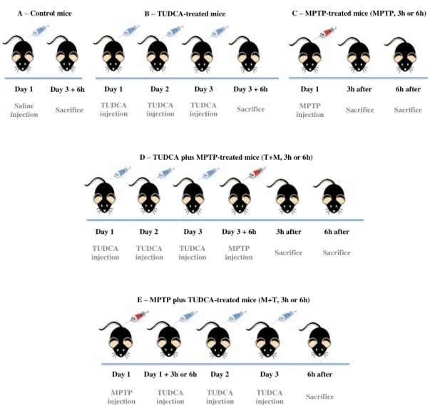
Figure II.1 – Experimental scheme of C57BL/6 male mice treatment course ... 25
Figure II.2 – Simplified scheme of SH-SY5Y cells treatment course ... 26
III. Results ... 33
Figure III.1 – TUDCA increases Nrf2 expression in mice striatum and midbrain ... 34
Figure III.2 – TUDCA increases the expression of Nrf2 downstream target enzymes in mice striatum and midbrain ... 36
Figure III.3 – DJ-1 expression is modulated by TUDCA in mice striatum and midbrain ... 38
Figure III.4 – TUDCA increases Gpx activity in mice striatum and midbrain ... 39
Figure III.5 – TUDCA modulates mRNA levels of the different cytoprotective enzymes in mice striatum ... 40
Figure III.6 – Role of TUDCA on ROS generation in mice striatum and midbrain ... ..41
Figure III.7 – TUDCA prevents MPP+-induced ROS formation in SH-SY5Y cells ... 42
Figure III.8 – TUDCA attenuates lipid peroxidation in SH-SY5Y cells ... 43
Figure III.9 – Nrf2 translocation to the nucleus is increased by TUDCA in SH-SY5Y cells ... 44
Figure III.10 – HO-1 mRNA levels are increased by TUDCA in SH-SY5Y cells ... 46
IV. Discussion ... 47
xix | P a g e
INDEX OF TABLES
I. Introduction ... 1
Table I.1 – Clinical features of Parkinson’s disease ... 3 Table I.2 – Schemes of MPTP administration in mice ... 19 Table I.3 – Key features of the MPTP mouse model in PD ... 19 II. Materials and Methods ... 23
Table II.1 – Primary antibodies used for Western blot and immunocytochemistry ... 24
Table II.2 – Secondary antibodies used for Western blot and immunocytochemistry ... 24
xxi | P a g e ABBREVIATIONS 6-OHDA ADP ARE ATP BBB BSA BTB cDNA CNS Cys DA DAT DCF-DA DGR DNA ETC FAD FADH2 FBS Gpx GSH GUDCA
H2O2 HNE HO HPRT i.p IVR JNK Keap1 LAA 6-hydroxydopamine Adenosine diphosphate Antioxidant response element Adenosine triphosphate Blood-brain barrier Bovine serum albumin
Broad complex, tramtrack, bric-a-brac domain Complementary DNA
Central nervous system Cysteine
Dopamine
Dopamine transporter
2’, 7’-dichlorofluorescein diacetate Double glycine repeat domain Deoxyribonucleic acid Electron transport chain Flavin adenine dinucleotide
Flavin adenine dinucleotide (reduced form) Fetal bovine serum
Glutathione peroxidase Glutathione Glycoursodeoxycholic acid Hydrogen peroxide 4-hydroxyl-2-nonenal Heme oxygenase
Hypoxanthine-guanine phosphoribosyl transferase Intra-peritoneally
Intervening region c-Jun N-terminal kinase
xxii | P a g e LB LRRK2 Maf MAO-B MEM MPDP+ MPP+ MPPP MPTP mRNA mtDNA NAD+ NADH NADPH NEAA Neh NQOs Nrf2
O2 -PARK7 PBS PD PFA PINK1 POLG1 Prx PVDF qRT-PCR Rbx1 RNA ROS SDS Lewy bodies
Leucine-rich repeat kinase 2 Musculo-aponeurotic fibrosarcoma Monoamine oxidase B
Minimum essential medium
1-methyl-4-phenyl-2,3-dihydropyridium 1-methyl-4-phenylpyridinium 1-methyl-4-phenyl-4-propionpiperidine 1-methyl-4-phenyl-1,2,3,6-tetrahydropyridine Messenger RNA Mitochondrial DNA
Nicotinamide adenine dinucleotide
Nicotinamide adenine dinucleotide (reduced form) Nicotinamide adenine dinucleotide phosphate Non-essential amino acids
Nrf2-ECH homologies
NADPH: quinone oxireductases
Nuclear factor erythroid 2 related factor 2 Superoxide anion
Parkinson protein 7 Phosphate buffered saline Parkinson’s disease Paraformaldehyde
Phosphatase and tensin (PTEN)-induced putative kinase1 Polymerase γ 1
Peroxiredoxins Polyvinyl difluoride
Quantitative real time polymerase chain reaction Ring-box protein 1
xxiii | P a g e
SDS-PAGE
SNpc
SOD
TBS-T
TFAM
Trx
TUDCA
UDCA
VMAT
Sodium dodecyl sulphate-polyacrilamide gel electrophoresis
Substantia nigra pars compacta
Superoxide dismutase
Tris-buffered saline-Tween 20
Mitochondrial transcriptional factor A Thioredoxins
Tauroursodeoxycholic acid Ursodeoxycholic acid
1 | P a g e
I. Introduction
1. Parkinson’s disease
Parkinson’s disease (PD), is the second most common neurodegenerative disorder, after Alzheimer’s disease, and it was first reported by James Parkinson, in 1817 (Parkinson, 1817). PD is a severe progressive neurological disorder characterized, not only by the loss of dopaminergic neurons in
the substantia nigra pars compacta (SNpc) and the consequent depletion of the neurotransmitter
dopamine (DA) in the striatum, but also by the presence of intracytoplasmic inclusions of aggregated
proteins designated by Lewy bodies (LB), formed mainly by α-synuclein and ubiquitin (Przedborski,
2005; Nagatsu and Sawada, 2006; Thomas and Beal, 2007).
Although the cause of PD remains elusive, several lines of evidence implicate that dopaminergic cell loss is associated with different mechanisms of cell damage, all of which may be interconnected. In fact, mitochondrial dysfunction and the consequent oxidative stress, inflammation, protein aggregation, excitotoxicity, impairment of both ubiquitin system and calcium homeostasis, and finally apoptosis have all been reported in PD patients’ brains (Jenner, 1999; Lev et al., 2003; Keane et al., 2011).
Despite the intensive research in PD field, it still remains unclear whether the disease results from either environmental factors, genetic causes or a combination of both. Therefore, PD is considered to have a multifactorial etiology, including genetic factors (designated by familial PD), in about 5% of PD cases, and environmental factors (referred to as sporadic PD), in the remaining 95% of PD cases (Kurth and Kurth, 1999; Lev et al., 2003; Martins et al., 2013). The familial form of PD comprises the
autosomal dominant forms, involving possibly gain-of-function mutations in α-synuclein and
leucine-rich repeat kinase 2 (LRRK2) genes, and the autosomal recessive forms, involving presumably
loss-of-function mutations in parkin, phosphatase and tensin (PTEN)-induced putative kinase 1 (PINK1) and
DJ-1 genes (Krüger, 2004; Lesage and Brice, 2012). In turn, the sporadic form of PD assumes that the
progressive nigral cell loss, characteristic of the disease, results from either chronic or limited exposure to environmental dopaminergic neurotoxins (Dauer and Przedborski, 2003). Remarkably, in the last few
2 | P a g e
Introduction
years, sporadic PD has gained a large genetic influence in its etiopathogenesis (Moon and Paek, 2015). For instance, numerous studies have demonstrated that several polymorphisms are responsible for
conferring increased susceptibility to sporadic PD (Vilar et al., 2007; Lesage and Brice, 2012; De Rosa
et al., 2015). Therefore, all of those factors account for the multifactorial etiology of this complex
disease.
1.1. Clinical and neuropathological features of PD
The prevalence of PD is approximately 0.3% of the entire population, affecting about 8 to 18 out of 100.000 people per year (Massano and Bahtia, 2012). PD affects more than 1% of people older than 60 years, and since PD incidence increases with age, it is estimated an increase of 3% in the population
over 80 years (Massano and Bahtia, 2012; Andalib et al., 2014). In addition, the mean age of onset of
the disease is roughly 60 years; however in about 10% of PD cases, the onset occurs earlier, between 20 and 50 years of age, being classified as young onset (Dexter and Jenner, 2013). Interestingly, Schrag
and collaborators (2000), among others, demonstratedthat PD affects more men than women, probably
due to the protective effects of estrogen (Dluzen, 2000).
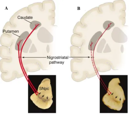
The main neuropathological feature of PD is the loss of midbrain dopaminergic neurons within SNpc. These dopaminergic neurons, whose cell bodies are located in SNpc, send projections to the caudate and putamen nucleus, in the striatum, creating the nigrostriatal pathway (Figure I.1), which is
essential for a normal motor function and voluntary movement control (Speciale, 2002; Martins et al.,
2013). Therefore, the loss of these nigral neurons, normally enriched in neuromelanin, results in complex changes in the brain’s motor system and also in a profound striatal dopamine loss and SNpc
depigmentation (Dauer and Przedborski, 2003; Smeyne and Jackson-Lewis, 2005) (Figure I.1 – B).
Together, all these alterations cause the motor deficits characteristics of PD.
Unfortunately, it is well known, that relevant clinical symptoms do not fully develop until there is a loss of about 60% of SNpc cells and 70% of dopamine response (Smeyne and Jackson-Lewis, 2005). Thus, the main symptoms of the disease can be divided in two categories: i) the motor symptoms, including tremor which occurs at rest but decreases with voluntary movement, rigidity (stiffness), slowness of movement known as bradykinesia, and postural instability; and ii) the non-motor symptoms, including depression and cognitive decline (Dickson, 2012; Massano and Bahtia, 2012; Dexter and Jenner, 2013). Other important motor and non-motor symptoms are described in Table I.1.
3 | P a g e
Introduction
available tests, during the patient’s life. Therefore, the final diagnosis of this disease is only confirmed
through brain autopsy (Massano and Bahtia, 2012).
Table I.1 –Clinical features of Parkinson’s disease. Sources: Dauer and Przedborski, Dickson, 2012; Massano and Bahtia, 2012; Dexter and Jenner, 2013
Motor Symptoms Non-motor Symptoms
Resting tremors
Depression Postural instability
Drooling
Rigidity (increased resistance to passive movement)
Cognitive decline Bradykinesia (slowness of movement)
Hypokinesia (reduction in movement amplitude) Akinesia (absence of normal unconscious movements)
Sleep disturbance Hypomimia (absence of normal facial expression)
Hypophonia (decreased voice volume) Micrographia (decreased size of handwriting)
Dementia Decreased speed of handwriting
A B
Figure I.1 –Schematic representation of Parkinson’s disease neuropathology. A –Normal nigrostriatal
pathway.This nigrostriatal pathway is composed of midbrain dopaminergic neurons, whose cell bodies are
located in substantia nigra pars compacta (SNpc) (indicated by the arrows). These neurons send projections,
represented by the thick red lines, to the striatum (caudate and putamen nucleus). In the picture it is evident the normal pigmentation of SNpc, produced by neuromelanin within the dopaminergic neurons. B – Diseased
nigrostriatal pathway. The degeneration of the nigrostriatal pathway is marked by a sharp loss of the
dopaminergic neurons that project to the striatum, represented by the dashed thin red line. It is also possible to see the characteristic depigmentation, caused by the loss of the dark-brown pigment, neuromelanin, in the SNpc.
4 | P a g e
Introduction
1.2. Mitochondrial dysfunction and oxidative stress in PD
Mitochondria are considered unique and important cellular organelles, since they have their own deoxyribonucleic acid (DNA), and function to produce cellular energy, in the form of adenosine
triphosphate (ATP), through a mechanism called oxidative phosphorylation (Keane et al., 2011).
Besides their main function as energy producers, mitochondria also play important roles in the regulation
of cell death via apoptosis, and are implicated in the control of cell growth and division (Keane et al.,
2011; Moon and Paek, 2015).
Mitochondrial DNA (mtDNA) is also known by its increased vulnerability to damages, probably due to a less efficient DNA repair mechanism, and also due to an absence of a protective histone coating
(Schapira et al., 1990; Schapira, 1994; Winklhofer and Haass, 2010). Since mtDNA is located close to
the electron transport chain (ETC) it is particularly susceptible to suffer damage from free radicals
produced during oxidative phosphorylation (Keane et al., 2011). Protein mutations, caused by oxidative
stress, lead to respiratory chain dysfunctions and/or alterations on the processes of mitochondrial replication, transcription or translation (Finsterer, 2006). Importantly, several mutations in mtDNA have been linked with PD, namely in the mitochondrial transcriptional factor A (TFAM), a regulator of mtDNA transcription, in the mitochondrial DNA polymerase γ 1 (POLG1), an enzyme involved in the
synthesis of mtDNA, and in genes that encode for proteins that constitute the ETC (Ekstrand et al., 2007;
Keane et al., 2011;Moon and Paek, 2015).
To accomplish the generation of cellular energy, the transport of electrons from reduced
nicotinamide adenine dinucleotide (NADH) or reduced flavin adenine dinucleotide (FADH2) oxidation,
are passed along the complexes of ETC, located in the inner mitochondrial membrane, until they reduce oxygen to water at complex IV (Winklhofer and Haass, 2010). This transport of electrons, generates proton movement from the mitochondrial matrix to the intermembrane space, creating an electrochemical gradient resulting in ATP production from adenosine diphosphate (ADP), through the
ATP synthase (complex V) (Winklhofer and Haass, 2010; Keane et al., 2011) (Figure I.2).
During the process of oxidative phosphorylation, electrons can leak from the ETC, specifically from complex I and III, and react with molecular oxygen to form reactive oxygen species (ROS) such
as superoxide anion (O2-), hydrogen peroxide (H2O2), and nitric oxide that can produce oxidative damage
by reacting with DNA, lipids and proteins (Kirkinezos and Moraes, 2001). Under normal physiological conditions, the cell is endowed with a free radical scavenging system, capable of clearing ROS from
mitochondria, preventing the damage of cellular and mitochondrial structures (Betarbet et al., 2002).
These scavenging systems include the antioxidant, glutathione (GSH), and the enzymes glutathione
peroxidase (Gpx) and superoxide dismutase (SOD) (Betarbet et al., 2002).
5 | P a g e
Introduction
brain contains a relatively low level of antioxidant enzymes, compared to other tissues, as well as high amounts of phospholipids, which are vulnerable to oxidative changes (Dias et al., 2013; Gaki and
Papavassiliou, 2014). Therefore, mitochondrial dysfunction, characterized by mitochondrial complex I impairment, together with the consequent rise in the leakage of electrons from the ETC, results in an increase in ROS generation, which may overwhelm the endogenous antioxidant mechanisms of the cell
(Betarbet et al., 2002; Moon and Paek, 2015). Importantly, it has been well documented that several
oxidative stress-related changes have been detected in PD patients’ brains (Jenner, 1998). Specifically,
different postmortem analysis revealed decreased GSH levels in SNpc (Perry et al., 1982; Perry and
Yong, 1986), increased levels of byproducts of lipid peroxidation, like 4-hydroxyl-2-nonenal (HNE), as described by Yoritaka and colleagues (1996), carbonyl modifications of soluble proteins, described by
Floor and Wetzel (1998), and also DNA and ribonucleic acid (RNA) oxidation products (Alam et al.,
1997; Zhang et al., 1999).
In addition, the characteristic neuronal cell death in PD may occur through severe oxidative mtDNA damage, protein oxidation and lipid peroxidation, and also through redox signaling pathways perturbation, due to an increase in mitochondrial ROS formation and/or defective ROS scavengers
(Winklhofer and Haass, 2010; Keane et al., 2011). Although the exact mechanisms leading to neuronal
death in PD remain elusive, it is believed that besides oxidative stress, apoptosis, p53, c-Jun N-terminal
kinase (JNK), as well as inflammation contribute to the process (Lev et al., 2003;Winklhofer and Haass,
2010; Castro-Caldas et al., 2012a).
6 | P a g e
Introduction
1.2.1. Role of DJ-1 in oxidative stress
DJ-1, a small homodimeric protein, ubiquitously expressed and encoded by Parkinson protein 7
(PARK7) gene, was initially identified as an oncogene, by Nagakubo and co-workers (1997), with crucial roles in cancer and male infertility (Thomas and Beal, 2007). Loss of function mutations in DJ-1 lead to autosomal recessive early-onset of familial PD, although the underlying mechanisms remain
unknown (Bonifati et al., 2003). Importantly, several studies involving mouse models lacking DJ-1
demonstrated that the absence of this protein results in age-dependent motor deficits, hypokinesia,
dopaminergic dysfunction and increased susceptibility to oxidative insults (Chen et al., 2005;Goldberg
et al., 2005; Kim et al., 2005), revealing the neuroprotective role of DJ-1 in PD. Moreover, DJ-1 is
considered a multifunctional protein that encompasses functions, such as chaperone, antioxidant, autophagy modulator, and transcriptional regulator (Im et al., 2012; Milani et al., 2013).
Under normal physiological conditions, DJ-1 has a predominantly cytoplasmic localization, however, under oxidative stress situations, this protein can be recruited either to the mitochondria or to the nucleus, where it functions as a ROS scavenger, by undergoing self-oxidation, oxidizing in particular Figure I.2 – Simplified scheme of the electron transport chain. The electrons (e-) generated by the
conversion of NADH to NAD+ (C I) or FADH
2 to FAD (C II), are passed through ubiquinone (Q), complex
III (C III), cytochrome c (Cyt c) and complex IV (C IV), where the electrons are used to reduce oxygen (O2)
to water (H2O). This electron transportation along the ETC, generates proton movement (H+) creating an
electrochemical gradient that culminates in ATP formation from ADP, by ATP-synthase. ADP – Adenosine diphosphate; ATP – Adenosine triphosphate; C I – Complex I; C II – Complex II; ETC – Electron transport chain; FAD – Flavin adenine dinucleotide; FADH2 – Reduced flavin adenine dinucleotide; NAD+ –
7 | P a g e
Introduction
its cysteine residue 106 (Canet-Avilés et al., 2004; Milani et al., 2013). Notably, it was showed by Choi
and colleagues (2006) that this oxidized form of DJ-1 is present in sporadic PD patients’ brains. Additionally, it is well documented that the overexpression of this protein allows cell protection, against
oxidative stress, induced either by H2O2 or neurotoxin-based models of PD (Im et al., 2012), which will
be further described in this thesis.
Summarizing, DJ-1 acts as a sensor of cellular redox status and therefore, it is considered a signaling molecule that responds to oxidative stress. Taken together the previous information, it is possible to conclude that DJ-1 has an essential role in cell protection, preventing ROS-mediated damage.
2. The Nrf2-Keap1 signaling pathway
To maintain a proper physiological redox balance, cells are endowed with a wide variety of endogenous antioxidant enzymes to lessen the levels of ROS production and the consequent oxidative stress (de Vries et al., 2008; Tufekci et al., 2011). Crucially, the expression of several of these
cytoprotective enzymes is activated, upon ROS exposure, by the transcription factor, nuclear factor erythroid 2 related factor 2 (Nrf2), the master regulator of cellular redox status (de Vries et al., 2008).
Nrf2 belongs to the basic leucine zipper transcription factor family, which is characterized by the requirement of a heterodimeric formation, with small musculo-aponeurotic fibrosarcoma (Maf) proteins, for DNA binding, as described by Itoh and collaborators (1997). Structurally, Nrf2 is formed by six functional domains, each one of them with specific functions, designated by Nrf2-ECH homologies (Neh1-6) (Tong et al., 2006).
Kelch-like ECH associated protein 1 (Keap1), in turn, is constituted by three functional domains, well documented by Tong and co-workers (2006), designated by Broad complex, Tramtrack, and Bric-a-brac (BTB) domain, an intervening region (IVR) and a Kelch domain, also designated by double glycine repeat (DGR) domain. Keap1 is an endogenous negative regulator of the Nrf2 pathway by forming a Keap1/Nrf2 complex, holding the transcription factor in the cytosol, with actin filaments, thus preventing it to function as a transcription factor in the nucleus (Tufeckci et al., 2011; Williamson et al.,
2012). To form this cytoplasmic complex, Keap1 has to form a homodimer, wherein each dimer binds
one molecule of Nrf2, in the DLG and ETGE motifs, via its two DGR domains (Tong et al., 2007). In
addition, Nrf2 regulation involves interactions between the conserved motifs DLG (week affinity for Keap1) and ETGE (high affinity for Keap1), within the Neh2 domain (responsible for cellular stress
response regulation), and also the DGR domain on Keap1 (Tufekci et al., 2011).
Under normal physiological conditions, the cytoplasmic Keap1/Nrf2 complex is connected, by the
BTB domain in Keap1, to a functional E3 ubiquitin ligase complex (Ring-box protein 1 – Rbx1) through
8 | P a g e
Introduction
ligase complex is responsible for the poly-ubiquitination and the consequent rapid degradation of Nrf2,
by the 26S proteasome (Zhang et al., 2004;Williamson et al., 2012).
On the other hand, under oxidative stress situations, the high reactive cysteine residues of Keap1 (Cys273 and Cys288 in the IVR domain; Cys151 in the BTB domain) are oxidized, affecting Keap1 conformation and causing its dissociation from the DLG motif of Nrf2, preventing its degradation
(Zhang and Hannink, 2003; Yamamoto et al., 2008;Williamson et al., 2012). These events allow Nrf2
stabilization and subsequent translocation into the nucleus, where Nrf2 forms a heterodimer with a Maf protein, through its Neh1 domain and subsequently binds the antioxidant response element (ARE), located in the promoter or enhancer regions of antioxidant and cytoprotective genes, activating their
expression (Williamson et al., 2012; Zhang et al., 2013). The schematic representation of Nrf2
regulation by Keap1, under normal conditions and oxidative stress, is illustrated in Figure I.3.
It is noteworthy that once the cellular redox homeostasis is restored, Nrf2 is transported out of the
nucleus to the cytoplasm, where it is poly-ubiquitinated and subsequently degraded (de Vries et al.,
9 | P a g e
Introduction
Figure I.3 – Illustrative representation of Nrf2 regulation by Keap1. Under normal physiological conditions (A), Keap1 forms a dimer that binds to Nrf2, in the DLG and ETGE motifs, through its two DGR domains. The formation of Keap1/Nrf2 complex, retains Nrf2 in the cytoplasm preventing its translocation to the nucleus and therefore the activation of antioxidant enzymes. In addition, since Nrf2 is not activated, the complex Keap1/Nrf2 binds to Rbx1, through the adaptor protein Cullin3. The formation of the complex Rbx1/Cullin3/Keap1/Nrf2 permits Nrf2 ubiquitination and proteasomal degradation. Under oxidative stress situations (B), cysteine residues of Keap1 are oxidized, upon ROS exposure, allowing the dissociation of Nrf2 from the complex Rbx1/Cullin3/Keap1. This dissociation allows Nrf2 stabilization and nuclear translocation, where it binds to Maf protein and subsequently to ARE, initiating the transcription of antioxidant and cytoprotective genes. ARE – Antioxidant response element; BTB – Broad complex, tramtrack, and bric-a-brac domain; Cys – Cysteine; DGR – Double glycine repeat domain; IVR – Intervening region; Keap1 – Kelch-like ECH associated protein 1; Maf – Musculo-aponeurotic fibrosarcoma; Neh2 – Nrf2-ECH homology 2; Nrf2 – Nuclear factor erythroid 2 related factor 2; Rbx1 – Ring-box protein 1; ROS – Reactive oxygen species; Ub – Ubiquitin. Adapted from Zhang et al. (2013).
26S Proteasome Nrf2 degradation C ull in 3 Ub Ub Ub Rbx1 DL G Neh2 E T G E Ub A Nucleus ARE Maf
Nrf2 Antioxidant enzymes
10 | P a g e
Introduction
2.1. Role of DJ-1 in Nrf2 regulation
As previously described in section “Role of DJ-1 in oxidative stress”, the redox-sensitive protein,
DJ-1, is responsible for the activation of antioxidant defenses, upon exposure to oxidative stress. Interestingly, it was recently demonstrated that the antioxidant response of DJ-1 may also result from
the activation of the Nrf2 pathway (Im et al., 2012). The evidence of the existence of this link between
DJ-1 and Nrf2 emerged from studies involving primary cell lines, from both human and mouse species, and also DJ-1-deficient patients. Studies regarding primary cell lines demonstrated that knockdown and knockout of DJ-1 caused a decrease in Nrf2 expression and stability, respectively, along with an increase of Nrf2 degradation and consequently, a decrease in the expression of downstream antioxidant enzymes (Clements et al., 2006; Im et al., 2012). Another study carried out by Gan and colleagues (2010)
demonstrated that messenger RNA (mRNA) levels of Nrf2 were decreased in DJ-1 knockout mice, when compared with wild-type mice. On the other hand, studies involving DJ-1 overexpression demonstrated a significant increase in Nrf2 stabilization and subsequently, diminished Nrf2 ubiquitination (Clements
et al., 2006). Moreover, DJ-1-deficient patients showed diminished expression of the cytoprotective
genes, accompanied by an increase in the oxidative stress levels (Zhang et al., 2013).
Together, these studies suggest that DJ-1 stabilizes Nrf2, either by disrupting Keap1/Nrf2 complex and/or by preventing its interaction with Keap1, thus reducing Nrf2 ubiquitination and consequent degradation.
2.2. Involvement of Nrf2 dysregulation in the pathogenesis of PD
In the last few years, several studies, including postmortem studies from PD patients’ brains, and
studies involving toxin-based animal models, have implicated the involvement of Nrf2 dysregulation in the pathogenesis of PD (Tufekci et al., 2011). In fact, studies regarding postmortem data from PD
patients’ brains revealed that in the nucleus of SNpc neurons, Nrf2, as well as its downstream targets
levels are enhanced, suggesting an increased activation of this transcription factor (Ramsey et al., 2007;
Wang et al., 2014). Importantly, these increments observed may be a compensatory response of the cell
to increase the levels of antioxidant cytoprotective enzymes, in response to oxidative toxicity (Zhang et
al., 2013). Additionally, it was showed, in studies using neurotoxin-based animal models of PD that
Nrf2 knockout mice displayed increased susceptibility to different neurotoxins, decreased levels of
dopamine transporters (DAT) in the striatum, and increased dopaminergic neurons depletion (Burton et
al., 2006; Jakel et al., 2007;Chen et al., 2009).
Taking together these observations, it can be suggested that Nrf2 is a promising candidate to limit oxidative stress-mediated damage, and therefore it could be used as a target for therapeutic strategies in
11 | P a g e
Introduction
2.3. Downstream targets of Nrf2
The activation of the Nrf2 pathway induces the transcription of several endogenous antioxidant,
detoxification, GSH synthesis enzymes, heat shock proteins, among others (Trachootham et al., 2008;
de Vries et al., 2008). Between the different Nrf2 downstream targets, the most important enzymes,
whose expression is activated by this transcription factor are SOD, catalase, Gpx, peroxiredoxins (Prx), nicotinamide adenine dinucleotide phosphate (NADPH): quinone oxidoreductases (NQOs), GSH and
its synthesis enzymes, heme oxygenases (HO) and thioredoxins (Trx) (de Vries et al., 2008). Here we
12 | P a g e
Introduction
Cul3
Rbx1
Nrf2 Keap1
ROS
DJ-1
Oxidative Damage
ROS
ROS
Caspase activation
Apoptosis
Nigral cell death
ARE
Maf
Mitochondria
Nrf2
Antioxidant enzymes Detoxification enzymes
Heat shock proteins GSH system
Antioxidant defense mechanisms
Cytc release
Figure I.4 – Schematic representation of the neuroprotective role of Nrf2 and DJ-1 in Parkinson’s disease. Mitochondrial dysfunction, characterized by mitochondrial complex I impairment which in turn results in higher levels of ROS formation, has been deeply implicated in PD. Under normal physiological conditions, the master regulator of cellular redox status, Nrf2, is maintained in the cytoplasm through its interaction with the complex Keap1/Cul3/Rbx1. Therefore, if not activated, Nrf2 is ubiquitinated and consequently degraded. Upon ROS exposure, Keap1 is oxidized and its conformation is affected, allowing Nrf2 dissociation and subsequent translocation to the nucleus. In the nucleus, Nrf2 binds to a Maf protein forming a complex which in turn binds to ARE, located in the regulatory regions of antioxidant genes, thus activating their expression. The antioxidant proteins, including for instance SOD, Gpx, and the heat shock proteins HO, act quickly in the cell reducing the levels of free radicals, by degradation or conversion, generating more powerful antioxidants, thus reducing cell damage induced by oxidative stress. Finally, DJ-1, upon ROS exposure, has the ability to undergo self-oxidation, being recruited either to the mitochondria or to the nucleus, functioning as a ROS scavenger preventing, once again, oxidative damage. Another important role of DJ-1 is related to an increase in Nrf2 stabilization, by disrupting and/or preventing Keap1/Nrf2 complex, reducing Nrf2 ubiquitination and degradation, thus increasing its nuclear translocation. Thereby, DJ-1 is also responsible for causing the activation of antioxidant genes, culminating in an increase in cellular defenses, being responsible for protecting the cells against apoptotic death, induced by ROS. ARE –
Antioxidant response element; Cul3 – Cullin3; Cyt c –Cytochrome c;Gpx – Glutathione peroxidase; GSH –
Glutathione; HO – Heme oxygenase; Keap1 – Kelch-like ECH associated protein 1; Maf – Musculo-aponeurotic fibrosarcoma; Nrf2 – Nuclear factor erythroid 2 related factor 2; Rbx1 – Ring-box protein 1; ROS
13 | P a g e
Introduction
2.3.1. Superoxide dismutases
The first line of defense against oxidative stress is provided by the metal containing SODs, such as cytosolic cooper, zinc superoxide dismutase (Cu, Zn-SOD or SOD1), mitochondrial manganese superoxide dismutase (MnSOD or SOD2) and extracellular superoxide dismutase (SOD3) (Johnson and Giulivi, 2005; de Vries et al., 2008). These enzymes are responsible, in general, for catalyzing the
dismutation of the O2- to molecular oxygen and H2O2 (de Vries et al., 2008).
SOD1 is a cytoplasmic protein, mainly expressed in astrocytes and neurons (de Vries et al., 2008;
Johnson and Giulivi, 2005). SOD3, in turn, is found in the extracellular matrix in most tissues, including the central nervous system (CNS) (Flynn and Melov, 2014). Therefore, both SOD1 and SOD3 are responsible for reducing the levels of superoxide in the extracellular and cytosolic environment, thus
preventing CNS damage (de Vries et al., 2008; Flynn and Melov, 2014).
Finally, SOD2 is the most important isoform in the defense against oxidative stress (Flynn and Melov, 2014). This enzyme is mainly localized in neurons, within the mitochondrial matrix, which in turn is the major site of free radical generation, as described by Weisiger and Fridovich (1973). Consequently, SOD2 is characterized as a critical enzyme in the fight against mitochondrial dysfunction and oxidative stress, playing an important role in several neurodegenerative diseases, including PD (de
Vries et al., 2008; Flynn and Melov, 2014).
2.3.2. Glutathione system
The GSH system is one of the most important antioxidant systems in the cell. GSH, a powerful antioxidant, scavenges alone or with different enzymes, several oxidative species such as NO, O2-, hydroxyl radicals, peroxynitrites among others, thus providing protection to the cell (Smeyne and
Smeyne, 2013; Zhang et al., 2013).
The GSH system comprises numerous enzymes with specific functions. For instance, γ
-glutamylcysteine ligase and glutathione synthetase are the enzymes responsible for GSH synthesis. In turn, glutathione reductase is responsible for recycling GSH, by converting oxidized GSH into reduced GSH. In addition, the enzymes Gpx and glutathione s-transferases are responsible for catalyzing the transfer of GSH to its substrates (Zhang et al., 2013).
Glutathione peroxidases are a group of 8 enzymes (Gpx1 to Gpx8) that play a crucial role in
reducing H2O2 to water, as well as reducing the levels of oxidized lipids in the cell (Smeyne and Smeyne,
2013). Gpx1 is the most abundant member of the Gpx family and is characterized as a crucial antioxidant enzyme because it is responsible for preventing the detrimental accumulation of intracellular hydrogen
peroxide (Lubos et al., 2011). Importantly, this enzyme is found in both neurons and glial cells, either
in cytosol, nucleus, mitochondria or peroxisomal compartments (Trépanier et al., 1996; Power and
14 | P a g e
Introduction
Curiously, it was demonstrated by Wang and collaborators (2003) that the overexpression of these family members, under neurotoxic conditions, prevents neuron loss, and also hydrogen peroxide accumulation and lipid peroxidation.
2.3.3. Heme oxygenases
There are two isoforms of active HO, the inducible isoform, HO-1, and the constitutive isoform,
HO-2 (Zhang et al., 2013). These two isoforms belong to the family of heat shock proteins and are
responsible for protecting brain cells from oxidative stress (Wagener et al., 2003). HO enzymes are
responsible for catalyzing the first step of heme catabolism, in other words, these enzymes are responsible for the degradation of intracellular heme (present in oxidases and peroxidases) into
biliverdin, free iron and carbon monoxide (Wagener et al., 2003; Hung et al., 2008). Subsequently,
biliverdin is converted to bilirubin, by biliverdin reductase (Wagener et al., 2003). Crucially, both
biliverdin and bilirubin are powerful antioxidants that are capable to protect the brain from ischemic injury, as described by Deguchi and co-workers (2008), as well as perform anti-inflammatory actions
(Hung et al., 2008). Not only biliverdin and bilirubin play important roles in cell defense, but also does
carbon monoxide, for instance, this endogenous gaseous molecule plays essential roles in anti-apoptosis,
anti-inflammation, anti-proliferation and in neurotransmission actions (Hung et al., 2008).
Ryter and colleagues (2006) described that HO enzymes can be found in several cell membranes, such as endoplasmic reticulum, nucleus and plasma membrane. HO-1 is considered a cellular stress response protein that is uniquely and rapidly expressed under oxidative stress and other harmful stimuli
(Hung et al., 2008). HO-2, in turn, is expressed constitutively and does not respond to oxidative stress
(Hung et al., 2008). Significantly, under oxidative stress situations, the synthesis of HO-1, in both
neuronal and non-neuronal cells, increases. Thus, HO-1 performs a key role in stress response and therefore, it can be considered crucial in neuroprotection, firstly by degrading heme, and secondly by
being responsible for the production of powerful antioxidants (Hung et al., 2008).
3. Experimental models of PD
Despite the years of research, very little is known about why and how the neurodegenerative mechanism of PD starts and evolves. Even so, in the last years, remarkable advances in the etiology and pathogenesis of PD have been made, thanks to the experimental models of the disease. Moreover, these models can be divided in two major categories: genetic models, which do not present the classic degeneration of nigral neurons, and neurotoxin-based models, which produce selective neuronal death,
both in vitro and in vivo thus, being considered the most valuable and popular models in PD (Bové et
15 | P a g e
Introduction
mechanisms that lead to the development of the pathophysiology of PD, much more needs to be done to fully understand which mechanisms are actually responsible for the severe neurodegeneration, characteristic of the disease, and what are the causes that culminate in these mechanisms.
3.1. Neurotoxin models of PD
An optimal model of PD should encompass all of the clinical and pathological features of the disease. In fact, the greater the similarity between a model and PD, the higher the predictive validity for clinical efficacy, as mentioned by Emborg (2004). Therefore, the neurotoxins currently available, should comprise both non-dopaminergic and dopaminergic systems, together with non-motor and motor symptoms, to be considered ideal models of PD (Tieu, 2011). Unfortunately, it is well known that, none of the available substances reproduces completely all the clinical and pathological features of the disease.
Currently, we can find among the neurotoxic chemicals used to induce dopaminergic neurodegeneration, 6-hydroxydopamine (6-OHDA), paraquat, rotenone and
1-methyl-4-phenyl-1,2,3,6-tetrahydropyridine (MPTP) (Bové et al., 2005). Despite all the neurotoxins available, 6-OHDA and
MPTP are the best characterized and most widely used agents, not only to study the molecular mechanisms leading to the neuropathology of the disease, but also to be used in the development of therapeutic strategies (Emborg, 2004). Nevertheless, the MPTP model, which does not exactly reproduces all the neuropathological features of PD (absence of LB, for instance), is clearly the most widely used neurotoxin as an experimental model of PD, due to the great similarity that individuals intoxicated with this substance, present with PD patients (Speciale, 2002; Dauer and Przedborski, 2003). The discovery of the MPTP model occurred in California, in the early 1980's, when several drug
users showed severe motor symptoms similar to those observed in PD (Langston et al., 1983). Further
investigations revealed that these patients had injected a "street" preparation of 1-methyl-4-phenyl-4-propionpiperidine (MPPP), an analog of the narcotic meperidine, contaminated with MPTP (Langston
et al., 1983). Later, it was discovered that the substance responsible for the severe clinical
manifestations, observed in these patients, was MPTP (Langston et al., 1983). Crucially, postmortem
studies in some of these patients revealed, like in PD, the loss of nigrostriatal structures (Langston et al.,
1999).
Since the discovery of MPTP as an inducer of Parkinsonism, a massive progress has been made in the discovery of the mechanisms underlying cell death in PD (Tieu, 2011). Importantly, studies using this neurotoxic model have led to propose the environmental toxicity, as a potential cause in sporadic PD, and the mitochondrial dysfunction and the consequent oxidative stress as a possible pathogenic
16 | P a g e
Introduction
this widely known model, for testing therapeutic approaches, trying to delay or even prevent the
degeneration of dopaminergic neurons (Le Couteur, 1999; Matthews et al., 1999).
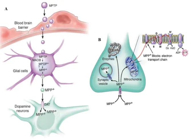
3.1.1. MPTP mechanism of action
MPTP is a highly lipophilic molecule that after systemic administration, easily crosses the blood-brain barrier (BBB), entering the blood-brain (Emborg, 2004). Once inside the blood-brain, the pro-toxin MPTP is
metabolized to 1-methyl-4-phenyl-2,3-dihydropyridium (MPDP+), by the enzyme monoamine oxidase
B (MAO-B), within non-dopaminergic neurons, like glial cells (Dauer and Przedborski, 2003). After
that, MPDP+ is converted to the active toxic metabolite, 1-methyl-4-phenylpyridinium (MPP+), possibly
by spontaneous oxidation (Dauer and Przedborski, 2003). Most importantly, it was demonstrated in several studies that MAO-B inhibition prevented both clinical and neuropathological evidence associated to MPTP and therefore, this enzyme is considered obligatory for the MPTP-induced neurotoxicity (Chiba et al., 1984; Langston et al., 1984).
Since MPP+ is a polar compound, it cannot freely exit from glial cells and enter the dopaminergic
neurons (Smeyne and Jackson-Lewis, 2005). Therefore, as described by Javitch and co-workers (1985) MPP+ is taken up into dopaminergic neurons, selectively, through its high affinity for the plasma membrane DAT. It is also essential to underline that the relevance of DAT in the MPTP neurotoxic mechanism, is proved in the studies of Javitch and colleagues (1985) and Bezard and collaborators (1999), where the blocking of DAT with antagonists or the ablation of DAT expression, respectively, results in the prevention of the neurotoxicity induced by MPTP. Accordingly, Donovan and co-workers (1999) demonstrated in their study, involving increased DAT levels in the brain of transgenic mice, that these animals are more susceptible to MPTP toxicity. Together, these studies reveal that the presence of DAT is obligatory for the neurotoxicity induced by MPTP. The schematic representation of the MPTP
metabolism is shown in Figure I.5 – A.
Once inside the dopaminergic neurons (Figure I.5 – B), MPP+ can follow three different pathways:
i) it can be sequestered into synaptic vesicles, by the action of the proton-dependent vesicular monoamine transporter (VMAT); ii) it can enter into the mitochondria, through the inner membrane, by a mechanism actively driven by the membrane electrical gradient, where it interferes with mitochondrial respiration, by blocking complex I (NADH dehydrogenase or NADH ubiquinone oxireductase) of the ETC, firstly described by Nicklas and colleagues (1985); and iii) it can remain in the cytosol and interact with different cytosolic enzymes (Dauer and Przedborski, 2003). Importantly, the blockage of
mitochondrial complex I, by MPP+, results in increased ROS production, leading to oxidative stress
17 | P a g e
Introduction
(Watanabe et al., 2005). Together, all these situations, recapitulate the deleterious events observed in
PD patients’ brains, and culminate in neuronal death (Watanabe et al., 2005).
3.2. Animal models of PD
To be considered a good PD animal model some requisites have to be fulfilled, such as: i) have reproducible nigral damage; ii) the neurodegeneration of dopaminergic cells must be steady over time, without unprompted recuperation and iii) should provide an opportunity for the implementation of a neuroprotective strategy (Emborg, 2004). Besides that, an ideal animal model should resemble the clinical and pathological features of the disease, including therefore, the loss of neurons in SNpc and
A
B
Figure I.5 – Schematic illustration of MPTP metabolism and intracellular pathways. A – MPTP
metabolism. MPTP crosses the blood-brain barrier where is first metabolized to MPDP+ by glial MAO-B, and
then converted to its active metabolite MPP+, probably by spontaneous oxidation. Thereafter, MPP+ is released
into the extracellular space and taken up into dopaminergic neurons through dopamine transporters. B –
Intracellular pathways of MPP+. Once inside the dopaminergic neurons, MPP+ can move through several
cellular compartments: it can concentrate within the mitochondria, where it inhibits complex I of the mitochondrial electron transport chain; it can interact with cytosolic enzymes; and it can be sequestered into synaptic vesicles by VMAT. DAT – Dopamine transporter; MAO-B – Monoamine oxidase B; MPDP+–
1-methyl-4-phenyl-2,3-dihydropyridinium; MPP+– 1-methyl-4-phenylpyridinium; MPTP –
18 | P a g e
Introduction
the formation of LB, as well as, α-synuclein aggregation and the clinical symptoms that appear during
the progress of the disease (Potashkin et al., 2010). Once the requirements are achieved, they become
an extreme valuable tool, in the therapeutic field, allowing to predict the capacity of a particular substance to protect dopaminergic neurons, against severe damage, as well as uncover potential problems associated with the therapeutic use of the substance in question (Emborg, 2004).
Each animal model, currently available, presents specific advantages and disadvantages, for instance, rodents are the most widely used models to study the underlying mechanisms of PD, by comparison to larger animals such as, cats, dogs and non-human primates, due to the fact that rats and mice are extensively accessible, genetically manageable, they reproduce easily and in large scale, their cost are quite affordable, and they do not need large spaces nor complex feeding conditions (Emborg,
2004; Potashkin et al., 2010). It is also important to emphasize that despite the existence of considerable
studies using cats, dogs and non-human primates in PD, these models bring countless ethical issues and elevated costs and therefore, their utility has been limitated (Potashkin et al., 2010).
3.2.1.The neurotoxin MPTP in rodent models
Susceptibility to the neurotoxin MPTP varies, not only across species, but also across animal
strains, as reviewed by Betarbetand collaborators (2002). For example, it is currently known that rodents
are more resistant to MPTP than humans and primates, and among rodents, mice exhibit more
susceptibility to MPTP toxicity, when compared to rats (Betarbet et al., 2002). It is believed that the
cause of this resistance, verified in rats, is due to their lower intracerebral levels of MAO-B and for this reason, mice are the most widely used animal models of PD (Emborg, 2004). In addition, it has been shown that only specific strains of mice are sensitive to MPTP, and that mice gender, age and body weight affect MPTP sensibility and reproducibility of the characteristic damage (Emborg, 2004). Therefore, female mice, mice under 8 weeks and mice smaller than 25g are more resistant to MPTP and their lesions are more variable, when compared to male mice, mice older than 8 weeks and heavier than
25g (Emborg, 2004). Accordingly, Przedborski and co-workers (2001) described that an optimal
reproducibility of MPTP-lesioning is obtained in C57BL/6 male mice.
MPTP can be administered by a number of different ways, including oral, intracerebral, systemically or intracarotid artery injections (Emborg, 2004). The most common way is done by systemic administration, which in turn, can be done by subcutaneous, intraperitoneal, intravenous or
intramuscular injections (Betarbet et al., 2002). Lastly, MPTP schemes of administration can also follow
different routes. In fact, MPTP is usually administered to mice in three different schemes: acute, sub-acute or chronic administration (Przedborski and Vila, 2001; Emborg, 2004). The sub-acute scheme consists of four intraperitoneal injections of MPTP, with 2 h intervals between the injections, on the same day,